

Search Count: 13
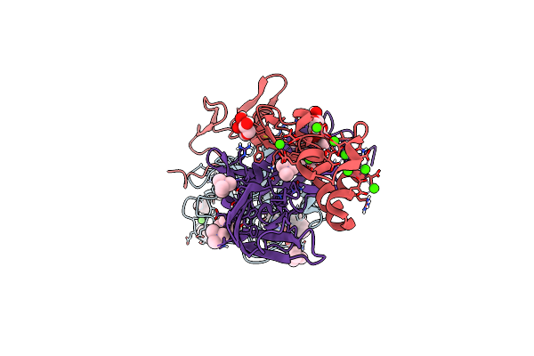 |
Crystal Structure Of The Super-Active Fviia Variant Vyt In Complex With Tissue Factor
Organism: Homo sapiens
Method: X-RAY DIFFRACTION Resolution:1.25 Å Release Date: 2019-12-11 Classification: HYDROLASE Ligands: CA, FUC, TMA, 0Z7 |
Organism: Homo sapiens
Method: X-RAY DIFFRACTION
Release Date: 2019-12-11
Ligands: CA, FUC, TMA, 0Z7
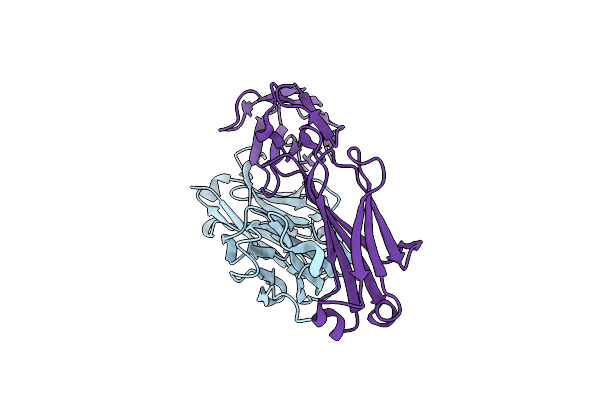 |
Crystal Structure Of The Fab Fragment Of Fviia Antibody Mab4F5
Organism: Mus musculus
Method: X-RAY DIFFRACTION Resolution:1.81 Å Release Date: 2019-06-19 Classification: IMMUNE SYSTEM |
Organism: Mus musculus
Method: X-RAY DIFFRACTION
Release Date: 2019-06-19
 |
X-Ray Structure Determined From Ex Vivo Charcot-Leyden Crystal
Organism: Homo sapiens
Method: X-RAY DIFFRACTION Resolution:2.30 Å Release Date: 2019-06-05 Classification: SUGAR BINDING PROTEIN Ligands: GOL |
Organism: Homo sapiens
Method: X-RAY DIFFRACTION
Release Date: 2019-06-05
Ligands: GOL
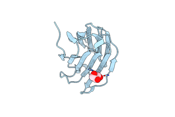 |
X-Ray Structure Determined From Recombinant Charcot-Leyden Crystal
Organism: Homo sapiens
Method: X-RAY DIFFRACTION Resolution:1.38 Å Release Date: 2019-06-05 Classification: SUGAR BINDING PROTEIN Ligands: GOL |
Organism: Homo sapiens
Method: X-RAY DIFFRACTION
Release Date: 2019-06-05
Ligands: GOL
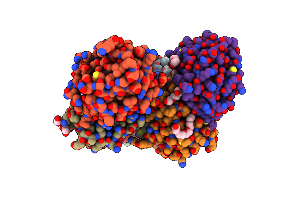 |
X-Ray Structure Of A Non-Autocrystallizing Galectin-10 Variant, Gal10-Tyr69Glu
Organism: Homo sapiens
Method: X-RAY DIFFRACTION Resolution:2.10 Å Release Date: 2019-06-05 Classification: SUGAR BINDING PROTEIN Ligands: PGE, PG4, P6G |
Organism: Homo sapiens
Method: X-RAY DIFFRACTION
Release Date: 2019-06-05
Ligands: PGE, PG4, P6G
 |
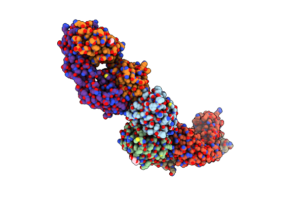
Structure Of Galectin-10 In Complex With The Fab Fragment Of A Charcot-Leyden Crystal Solubilizing Antibody, 6F5
Organism: Homo sapiens, Lama glama
Method: X-RAY DIFFRACTION Resolution:1.91 Å Release Date: 2019-06-05 Classification: SUGAR BINDING PROTEIN Ligands: CA, PGE |
Organism: Homo sapiens, Lama glama
Method: X-RAY DIFFRACTION
Release Date: 2019-06-05
Ligands: CA, PGE
 |
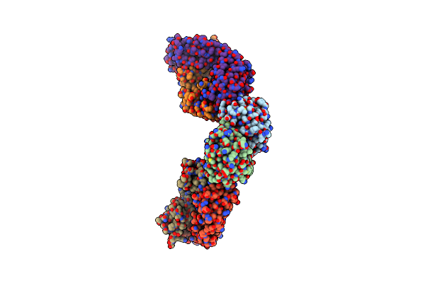
Structure Of Galectin-10 In Complex With The Fab Fragment Of A Charcot-Leyden Crystal Solubilizing Antibody, 1D11
Organism: Homo sapiens, Lama glama
Method: X-RAY DIFFRACTION Resolution:1.90 Å Release Date: 2019-06-05 Classification: SUGAR BINDING PROTEIN Ligands: PG4, ACT |
Organism: Homo sapiens, Lama glama
Method: X-RAY DIFFRACTION
Release Date: 2019-06-05
Ligands: PG4, ACT
 |
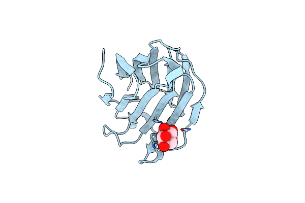
Structure Of Galectin-10 In Complex With The Fab Fragment Of A Charcot-Leyden Crystal Solubilizing Antibody, 4E8
Organism: Homo sapiens, Lama glama
Method: X-RAY DIFFRACTION Resolution:3.40 Å Release Date: 2019-06-05 Classification: SUGAR BINDING PROTEIN |
Organism: Homo sapiens, Lama glama
Method: X-RAY DIFFRACTION
Release Date: 2019-06-05
 |
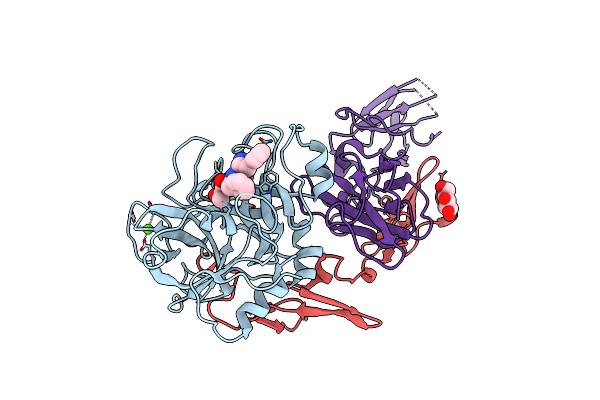
Galectin-10 Complexed With Ribose
Organism: Homo sapiens
Method: X-RAY DIFFRACTION Resolution:1.40 Å Release Date: 2019-06-05 Classification: IMMUNE SYSTEM Ligands: RIP |
Organism: Homo sapiens
Method: X-RAY DIFFRACTION
Release Date: 2019-06-05
Ligands: RIP
 |
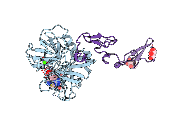
Crystal Structure Of Active Site Inhibited Coagulation Factor Viia Mutant In Complex With Soluble Tissue Factor
Organism: Homo sapiens
Method: X-RAY DIFFRACTION Resolution:2.20 Å Release Date: 2008-11-04 Classification: HYDROLASE/HYDROLASE INHIBITOR Ligands: GLC, FUC, CA, 0Z6 |
Organism: Homo sapiens
Method: X-RAY DIFFRACTION
Release Date: 2008-11-04
Ligands: GLC, FUC, CA, 0Z6
 |
Structure Of Human Factor Viia And Its Implications For The Triggering Of Blood Coagulation
Organism: Homo sapiens
Method: X-RAY DIFFRACTION Resolution:2.80 Å Release Date: 1999-08-01 Classification: hydrolase/hydrolase inhibitor Ligands: GLC, FUC, 0Z6, CA |
Organism: Homo sapiens
Method: X-RAY DIFFRACTION
Release Date: 1999-08-01
Ligands: GLC, FUC, 0Z6, CA
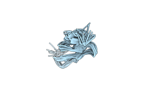 |
How An Epidermal Growth Factor (Egf)-Like Domain Binds Calcium-High Resolution Nmr Structure Of The Calcium Form Of The Nh2-Terminal Egf-Like Domain In Coagulation Factor X
Organism: Bos taurus
Method: SOLUTION NMR Release Date: 1994-05-31 Classification: COAGULATION FACTOR |
Organism: Bos taurus
Method: SOLUTION NMR
Release Date: 1994-05-31
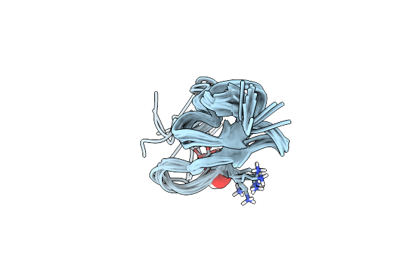 |
Three-Dimensional Structure Of The Apo Form Of The N-Terminal Egf-Like Module Of Blood Coagulation Factor X As Determined By Nmr Spectroscopy And Simulated Folding
Organism: Bos taurus
Method: SOLUTION NMR Release Date: 1994-01-31 Classification: COAGULATION FACTOR Ligands: OH |
Organism: Bos taurus
Method: SOLUTION NMR
Release Date: 1994-01-31
Ligands: OH