

Search Count: 36
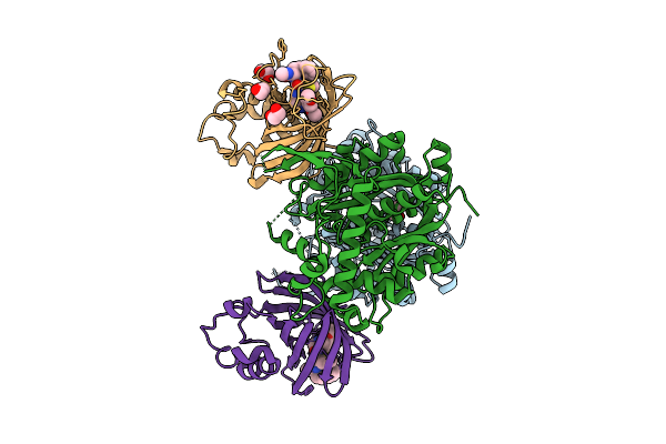 |
Gid4 In Complex With Cleo4-88 And Acaa1
Organism: Homo sapiens
Method: X-RAY DIFFRACTION Resolution:2.28 Å Release Date: 2026-04-15 Classification: PEPTIDE BINDING PROTEIN Ligands: L6U, EDO |
Organism: Homo sapiens
Method: X-RAY DIFFRACTION
Release Date: 2026-04-15
Ligands: L6U, EDO
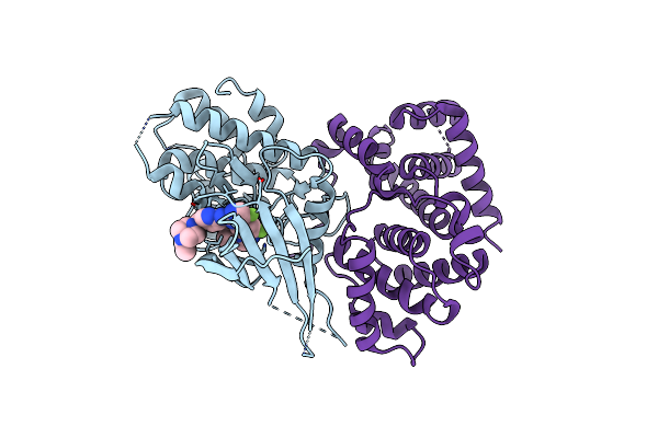 |
Structure Of Cdk4-Cyclin D3 Bound To Abemaciclib
Organism: Homo sapiens
Method: X-RAY DIFFRACTION Resolution:2.51 Å Release Date: 2022-11-02 Classification: CELL CYCLE Ligands: 6ZV |
Organism: Homo sapiens
Method: X-RAY DIFFRACTION
Release Date: 2022-11-02
Ligands: 6ZV
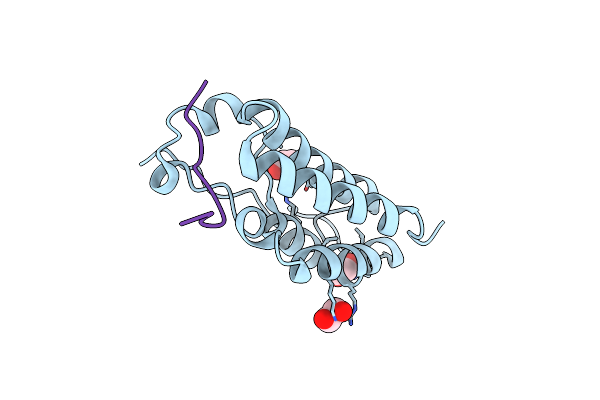 |
Crystal Structure Of The First Bromodomain Of Human Brd4 In Complex With An Acetylated Atrx Peptide (K1030Ac/K1033Ac)
Organism: Homo sapiens
Method: X-RAY DIFFRACTION Resolution:1.40 Å Release Date: 2018-11-28 Classification: TRANSCRIPTION Ligands: EDO |
Organism: Homo sapiens
Method: X-RAY DIFFRACTION
Release Date: 2018-11-28
Ligands: EDO
 |
Crystal Structure Of The First Bromodomain Of Human Brd4 In Complex With An Acetylated E2F1 Peptide (K117Ac/K120Ac)
Organism: Homo sapiens
Method: X-RAY DIFFRACTION Resolution:1.30 Å Release Date: 2018-11-28 Classification: TRANSCRIPTION Ligands: EDO |
Organism: Homo sapiens
Method: X-RAY DIFFRACTION
Release Date: 2018-11-28
Ligands: EDO
 |
Crystal Structure Of The First Bromodomain Of Human Brd4 In Complex With An Acetylated Gata1 Peptide (K312Ac/K315Ac)
Organism: Homo sapiens
Method: X-RAY DIFFRACTION Resolution:1.40 Å Release Date: 2018-11-28 Classification: TRANSCRIPTION Ligands: EDO |
Organism: Homo sapiens
Method: X-RAY DIFFRACTION
Release Date: 2018-11-28
Ligands: EDO
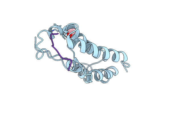 |
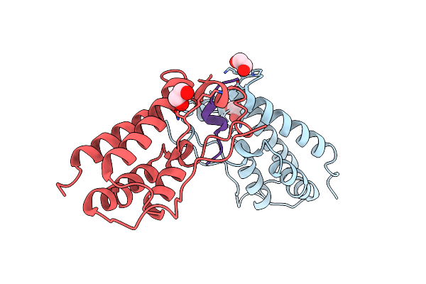
Crystal Structure Of The First Bromodomain Of Human Brd4 In Complex With An Acetylated Polr2A Peptide (K775Ac/K778Ac)
Organism: Homo sapiens
Method: X-RAY DIFFRACTION Resolution:1.25 Å Release Date: 2018-11-28 Classification: TRANSCRIPTION Ligands: EDO |
Organism: Homo sapiens
Method: X-RAY DIFFRACTION
Release Date: 2018-11-28
Ligands: EDO
 |
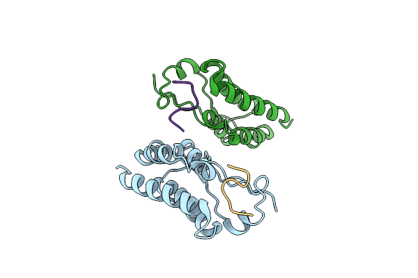
Crystal Structure Of The First Bromodomain Of Human Brd4 In Complex With An Acetylated Sirt7 Peptide (K272Ac/K275Ac)
Organism: Homo sapiens
Method: X-RAY DIFFRACTION Resolution:1.48 Å Release Date: 2018-11-28 Classification: TRANSCRIPTION Ligands: EDO |
Organism: Homo sapiens
Method: X-RAY DIFFRACTION
Release Date: 2018-11-28
Ligands: EDO
 |
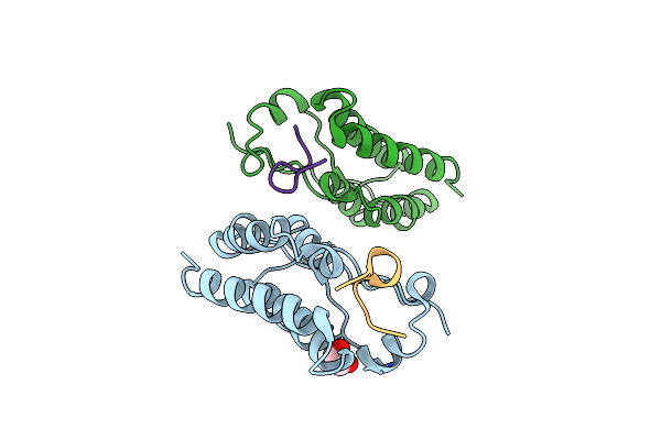
Crystal Structure Of The First Bromodomain Of Human Brd4 In Complex With A Diacetylated Histone 4 Peptide (H3K9Ac/K14Ac)
Organism: Homo sapiens
Method: X-RAY DIFFRACTION Resolution:2.22 Å Release Date: 2018-05-16 Classification: TRANSCRIPTION |
Organism: Homo sapiens
Method: X-RAY DIFFRACTION
Release Date: 2018-05-16
 |
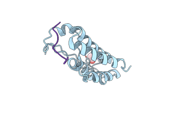
Crystal Structure Of The First Bromodomain Of Human Brd4 In Complex With A Diacetylated Histone 4 Peptide (H3K9Ac/K14Ac)
Organism: Homo sapiens
Method: X-RAY DIFFRACTION Resolution:1.82 Å Release Date: 2018-05-16 Classification: TRANSCRIPTION Ligands: EDO |
Organism: Homo sapiens
Method: X-RAY DIFFRACTION
Release Date: 2018-05-16
Ligands: EDO
 |
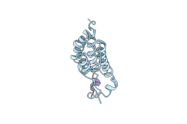
Crystal Structure Of The First Bromodomain Of Human Brd4 In Complex With A Diacetylated Top2A Peptide (K1201Ac/K1204Ac)
Organism: Homo sapiens
Method: X-RAY DIFFRACTION Resolution:1.15 Å Release Date: 2018-05-16 Classification: TRANSCRIPTION Ligands: EDO |
Organism: Homo sapiens
Method: X-RAY DIFFRACTION
Release Date: 2018-05-16
Ligands: EDO
 |
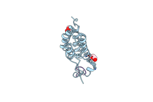
Crystal Structure Of The First Bromodomain Of Human Brd4 In Complex With An Acetylated Baz1B Peptide (K221Ac)
Organism: Homo sapiens
Method: X-RAY DIFFRACTION Resolution:1.15 Å Release Date: 2018-05-16 Classification: TRANSCRIPTION |
Organism: Homo sapiens
Method: X-RAY DIFFRACTION
Release Date: 2018-05-16
 |
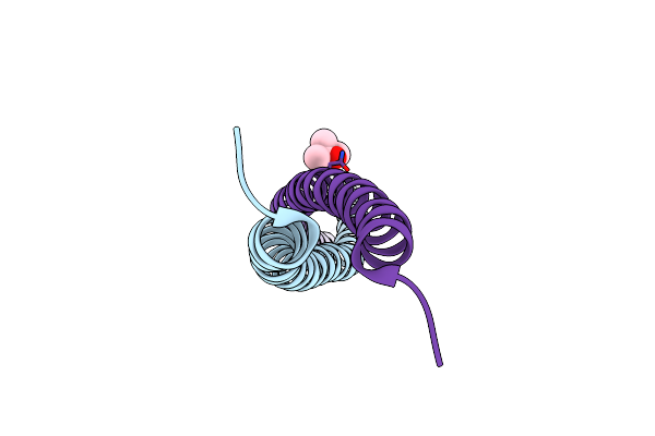
Crystal Structure Of The First Bromodomain Of Human Brd4 In Complex With An Acetylated Srpk1 Peptide (K585Ac)
Organism: Homo sapiens
Method: X-RAY DIFFRACTION Resolution:1.20 Å Release Date: 2018-05-16 Classification: TRANSCRIPTION Ligands: EDO |
Organism: Homo sapiens
Method: X-RAY DIFFRACTION
Release Date: 2018-05-16
Ligands: EDO
 |
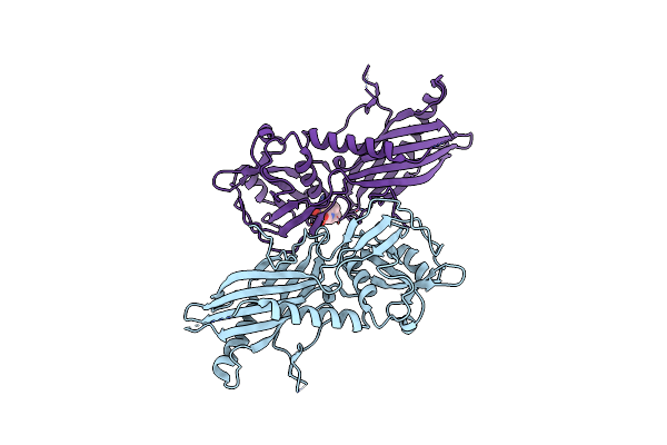
Human Cep85 - Coiled Coil Domain 4
Organism: Homo sapiens
Method: X-RAY DIFFRACTION Resolution:1.67 Å Release Date: 2018-04-18 Classification: PROTEIN BINDING Ligands: MPD |
Organism: Homo sapiens
Method: X-RAY DIFFRACTION
Release Date: 2018-04-18
Ligands: MPD
 |
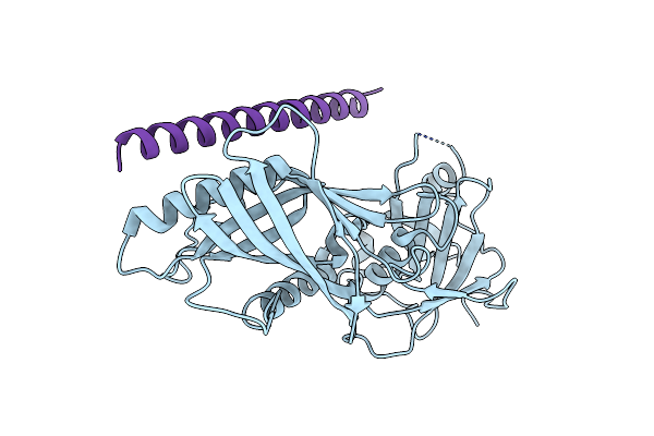
Trichoplax Adhaerens Stil N-Terminal Domain
Organism: Trichoplax adhaerens
Method: X-RAY DIFFRACTION Resolution:2.09 Å Release Date: 2018-04-18 Classification: PROTEIN BINDING Ligands: MES |
Organism: Trichoplax adhaerens
Method: X-RAY DIFFRACTION
Release Date: 2018-04-18
Ligands: MES
 |
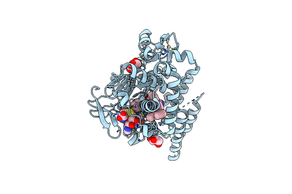
Complex Trichoplax Stil-Ntd:Human Cep85 Coiled Coil Domain 4
Organism: Trichoplax adhaerens, Homo sapiens
Method: X-RAY DIFFRACTION Resolution:4.60 Å Release Date: 2018-04-18 Classification: PROTEIN BINDING |
Organism: Trichoplax adhaerens, Homo sapiens
Method: X-RAY DIFFRACTION
Release Date: 2018-04-18
 |
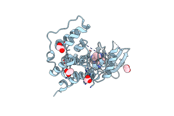
Crystal Structure Of Protein Lysine Methyltransferase Smyd2 In Complex With Lly-507, A Cell-Active, Potent And Selective Inhibitor
Organism: Homo sapiens
Method: X-RAY DIFFRACTION Resolution:1.63 Å Release Date: 2015-04-08 Classification: Transferase/transferase inhibitor Ligands: 3UJ, SAH, GOL, ZN |
Organism: Homo sapiens
Method: X-RAY DIFFRACTION
Release Date: 2015-04-08
Ligands: 3UJ, SAH, GOL, ZN
 |
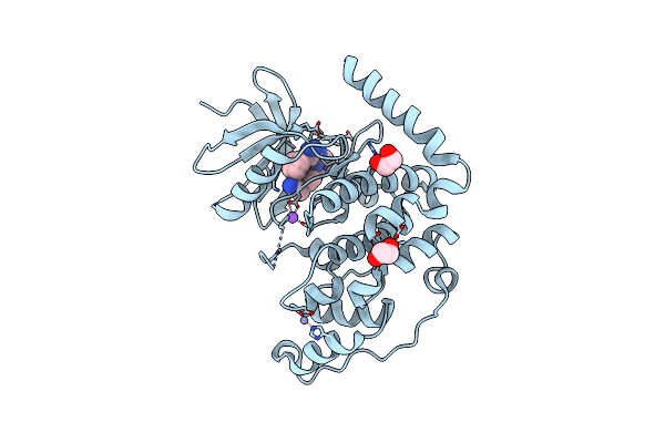
Crystal Structure Of The Human Cdkl5 Kinase Domain
Organism: Homo sapiens
Method: X-RAY DIFFRACTION Resolution:2.00 Å Release Date: 2013-05-08 Classification: TRANSFERASE |
Organism: Homo sapiens
Method: X-RAY DIFFRACTION
Release Date: 2013-05-08
 |
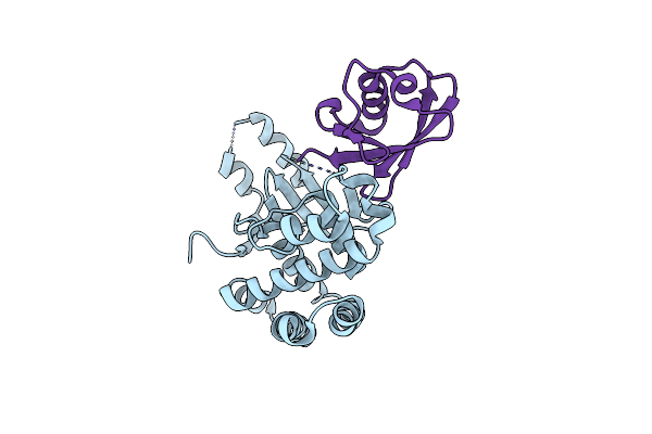
Crystal Structure Of The Human Cdkl3 Kinase Domain
Organism: Homo sapiens
Method: X-RAY DIFFRACTION Resolution:2.20 Å Release Date: 2013-03-20 Classification: TRANSFERASE Ligands: 38R, ZN, NA, EDO |
Organism: Homo sapiens
Method: X-RAY DIFFRACTION
Release Date: 2013-03-20
Ligands: 38R, ZN, NA, EDO
 |
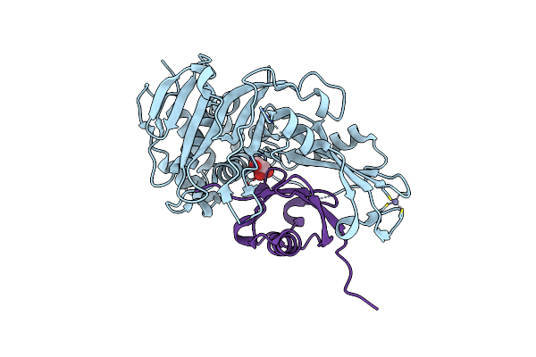
Crystal Structure Of Otub1 In Complex With Ubiquitin Variant
Organism: Homo sapiens
Method: X-RAY DIFFRACTION Resolution:2.49 Å Release Date: 2013-01-16 Classification: HYDROLASE |
Organism: Homo sapiens
Method: X-RAY DIFFRACTION
Release Date: 2013-01-16
 |
Crystal Structure Of Usp2 And A Mutant Form Of Ubiquitin
Organism: Homo sapiens
Method: X-RAY DIFFRACTION Resolution:2.10 Å Release Date: 2012-12-19 Classification: hydrolase/signaling protein Ligands: ZN, CL, GOL |
Organism: Homo sapiens
Method: X-RAY DIFFRACTION
Release Date: 2012-12-19
Ligands: ZN, CL, GOL