

Search Count: 118
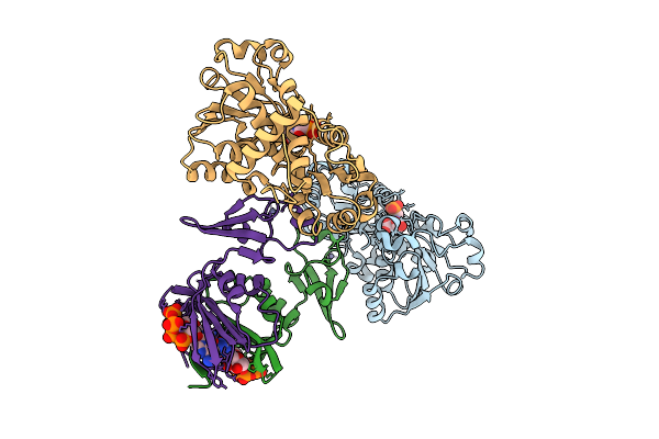 |
Crystal Structure Of E. Coli Aspartate Transcarbamoylase In The R-State Complexed With Pala, Atp, Gtp, And Mg2+
Organism: Escherichia coli
Method: X-RAY DIFFRACTION Resolution:2.19 Å Release Date: 2026-04-15 Classification: CYTOSOLIC PROTEIN Ligands: PAL, ZN, ATP, MG, GTP |
Organism: Escherichia coli
Method: X-RAY DIFFRACTION
Release Date: 2026-04-15
Ligands: PAL, ZN, ATP, MG, GTP
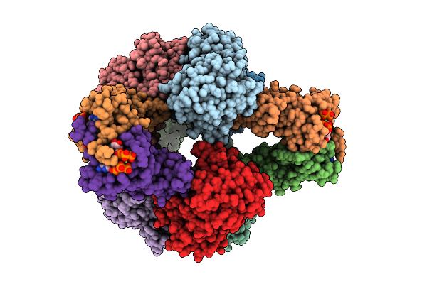 |
Crystal Structure Of E. Coli Aspartate Transcarbamoylase In The R-State Complexed With Cp, Succinate, Atp, And Mg2+
Organism: Escherichia coli
Method: X-RAY DIFFRACTION Resolution:3.00 Å Release Date: 2026-04-15 Classification: CYTOSOLIC PROTEIN Ligands: CP, SIN, ZN, ATP, MG |
Organism: Escherichia coli
Method: X-RAY DIFFRACTION
Release Date: 2026-04-15
Ligands: CP, SIN, ZN, ATP, MG
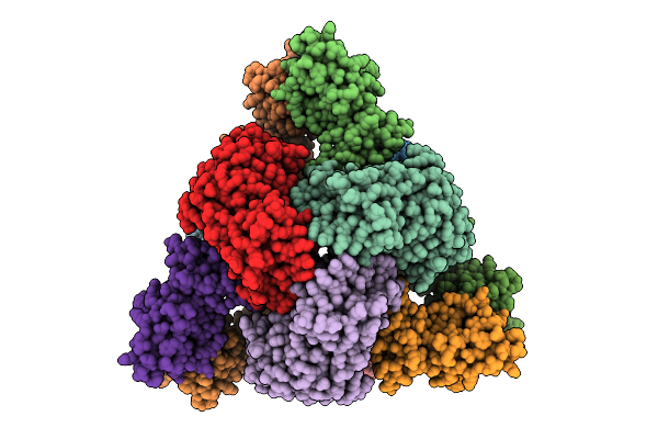 |
Cryo-Em Model Of E. Coli Aspartate Transcarbamoylase In The Ligand-Free T-State
Organism: Escherichia coli
Method: ELECTRON MICROSCOPY Release Date: 2026-04-15 Classification: CYTOSOLIC PROTEIN Ligands: ZN |
Organism: Escherichia coli
Method: ELECTRON MICROSCOPY
Release Date: 2026-04-15
Ligands: ZN
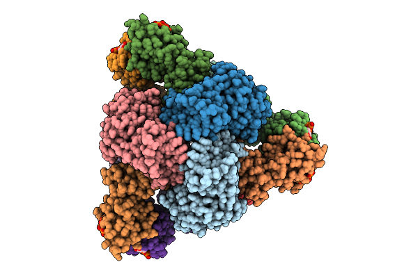 |
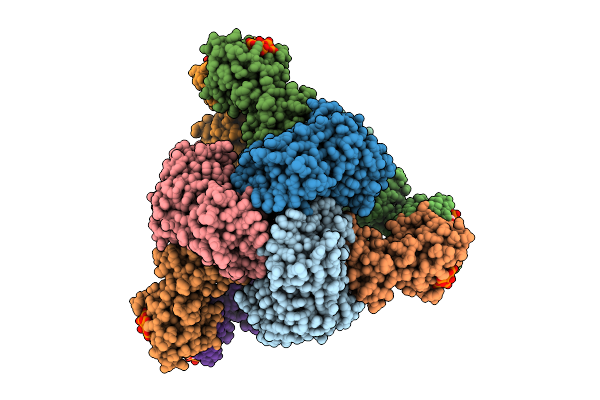
Cryo-Em Model Of E. Coli Aspartate Transcarbamoylase In The T-State Complexed With Cp, Ctp, Utp, And Mg2+
Organism: Escherichia coli
Method: ELECTRON MICROSCOPY Release Date: 2026-04-15 Classification: CYTOSOLIC PROTEIN Ligands: ZN, CTP, UTP, MG, CP |
Organism: Escherichia coli
Method: ELECTRON MICROSCOPY
Release Date: 2026-04-15
Ligands: ZN, CTP, UTP, MG, CP
 |
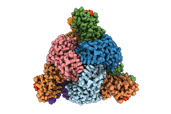
Cryo-Em Model Of E. Coli Aspartate Transcarbamoylase In The R-State Complexed With Cp, Succinate, Ctp, Utp, And Mg2+
Organism: Escherichia coli
Method: ELECTRON MICROSCOPY Release Date: 2026-04-15 Classification: CYTOSOLIC PROTEIN Ligands: CP, SIN, ZN, CTP, UTP, MG |
Organism: Escherichia coli
Method: ELECTRON MICROSCOPY
Release Date: 2026-04-15
Ligands: CP, SIN, ZN, CTP, UTP, MG
 |
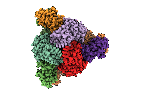
Cryo-Em Model Of E. Coli Aspartate Transcarbamoylase In The R-State Complexed With Cp, Succinate, Ctp, And Mg2+
Organism: Escherichia coli
Method: ELECTRON MICROSCOPY Release Date: 2026-04-15 Classification: CYTOSOLIC PROTEIN Ligands: CP, SIN, ZN, CTP, MG |
Organism: Escherichia coli
Method: ELECTRON MICROSCOPY
Release Date: 2026-04-15
Ligands: CP, SIN, ZN, CTP, MG
 |
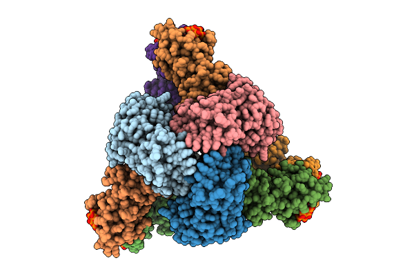
Cryo-Em Model Of E. Coli Aspartate Transcarbamoylase In The R-State Complexed With Cp And Succinate
Organism: Escherichia coli
Method: ELECTRON MICROSCOPY Release Date: 2026-04-15 Classification: CYTOSOLIC PROTEIN Ligands: CP, SIN, ZN |
Organism: Escherichia coli
Method: ELECTRON MICROSCOPY
Release Date: 2026-04-15
Ligands: CP, SIN, ZN
 |
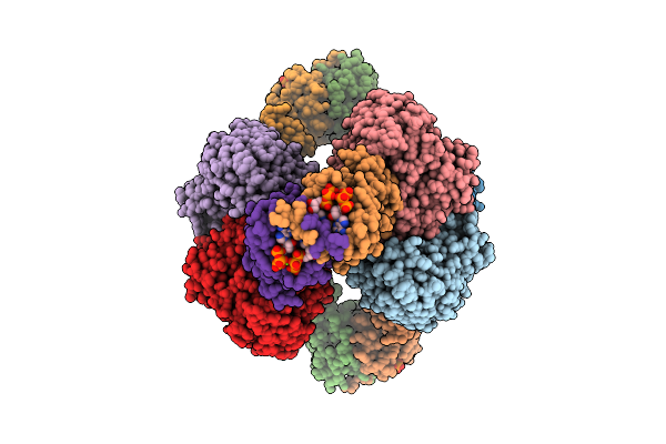
Cryo-Em Model Of E. Coli Aspartate Transcarbamoylase In The R-State Complexed With Cp, Succinate, Atp, And Mg2+
Organism: Escherichia coli
Method: ELECTRON MICROSCOPY Release Date: 2026-04-15 Classification: CYTOSOLIC PROTEIN Ligands: CP, SIN, ZN, ATP, MG |
Organism: Escherichia coli
Method: ELECTRON MICROSCOPY
Release Date: 2026-04-15
Ligands: CP, SIN, ZN, ATP, MG
 |
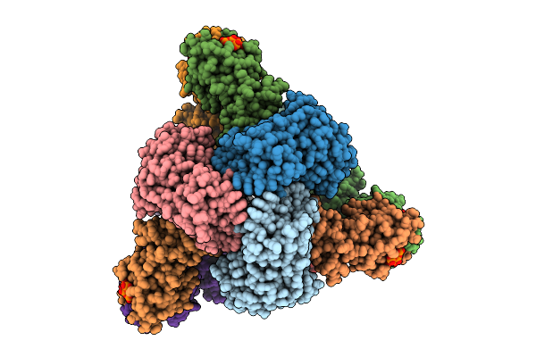
Cryo-Em Model Of E. Coli Aspartate Transcarbamoylase In The R-State Complexed With Cp, Succinate, Atp, Gtp, And Mg2+
Organism: Escherichia coli
Method: ELECTRON MICROSCOPY Release Date: 2026-04-15 Classification: CYTOSOLIC PROTEIN Ligands: CP, SIN, ZN, ATP, GTP, MG |
Organism: Escherichia coli
Method: ELECTRON MICROSCOPY
Release Date: 2026-04-15
Ligands: CP, SIN, ZN, ATP, GTP, MG
 |
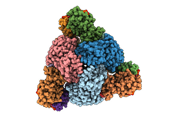
Cryo-Em Model Of E. Coli Aspartate Transcarbamoylase In An Expanded State Complexed With Cp, Atp, Gtp, And Mg2+
Organism: Escherichia coli
Method: ELECTRON MICROSCOPY Release Date: 2026-04-15 Classification: CYTOSOLIC PROTEIN Ligands: ZN, ATP, GTP, MG, CP |
Organism: Escherichia coli
Method: ELECTRON MICROSCOPY
Release Date: 2026-04-15
Ligands: ZN, ATP, GTP, MG, CP
 |
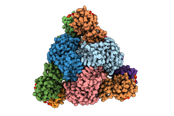
Cryo-Em Model Of E. Coli Aspartate Transcarbamoylase In The T-State Complexed With Cp, Atp, And Mg2+
Organism: Escherichia coli
Method: ELECTRON MICROSCOPY Release Date: 2026-04-15 Classification: CYTOSOLIC PROTEIN Ligands: ZN, ATP, MG, CP |
Organism: Escherichia coli
Method: ELECTRON MICROSCOPY
Release Date: 2026-04-15
Ligands: ZN, ATP, MG, CP
 |
Cryo-Em Model Of E. Coli Aspartate Transcarbamoylase In The T-State Complexed With Cp, Ctp, And Mg2+
Organism: Escherichia coli
Method: ELECTRON MICROSCOPY Release Date: 2026-04-01 Classification: CYTOSOLIC PROTEIN Ligands: ZN, CTP, MG, CP |
Organism: Escherichia coli
Method: ELECTRON MICROSCOPY
Release Date: 2026-04-01
Ligands: ZN, CTP, MG, CP
 |
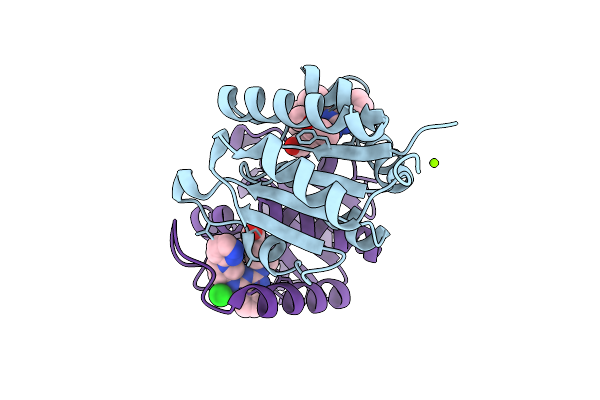
Crystal Structure Of Dnph1 Bound By Compound 2.
Organism: Homo sapiens
Method: X-RAY DIFFRACTION Resolution:1.60 Å Release Date: 2025-11-12 Classification: HYDROLASE Ligands: A1IZR |
Organism: Homo sapiens
Method: X-RAY DIFFRACTION
Release Date: 2025-11-12
Ligands: A1IZR
 |
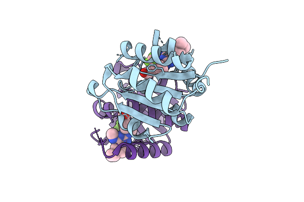
Crystal Structure Of Dnph1 Bound By Compound 39.
Organism: Homo sapiens
Method: X-RAY DIFFRACTION Resolution:1.22 Å Release Date: 2025-11-12 Classification: HYDROLASE Ligands: MG, A1IZW, CL |
Organism: Homo sapiens
Method: X-RAY DIFFRACTION
Release Date: 2025-11-12
Ligands: MG, A1IZW, CL
 |
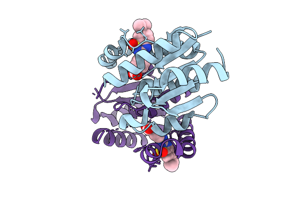
Crystal Structure Of Dnph1 Bound By Compound 31.
Organism: Homo sapiens
Method: X-RAY DIFFRACTION Resolution:1.21 Å Release Date: 2025-11-12 Classification: HYDROLASE Ligands: A1IZX |
Organism: Homo sapiens
Method: X-RAY DIFFRACTION
Release Date: 2025-11-12
Ligands: A1IZX
 |
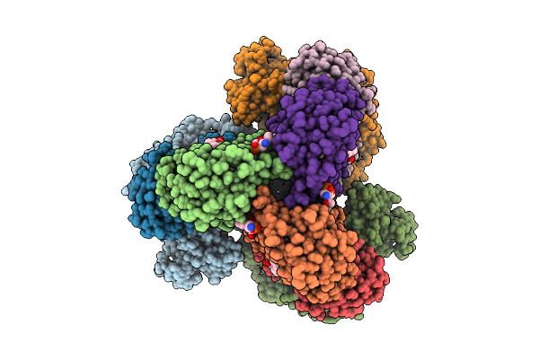
Crystal Structure Of Dnph1 Bound By Compound 5.
Organism: Homo sapiens
Method: X-RAY DIFFRACTION Resolution:1.49 Å Release Date: 2025-11-12 Classification: HYDROLASE Ligands: A1I0K |
Organism: Homo sapiens
Method: X-RAY DIFFRACTION
Release Date: 2025-11-12
Ligands: A1I0K
 |
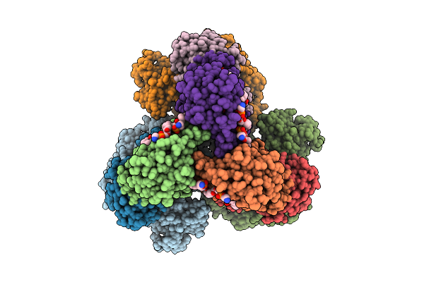
Cryo-Em Structure Of The Apo-Form Succinate Dehydrogenase From Chloroflexus Aurantiacus
Organism: Chloroflexus aurantiacus j-10-fl
Method: ELECTRON MICROSCOPY Release Date: 2025-11-12 Classification: MEMBRANE PROTEIN Ligands: FAD, SIN, SF4, FES, F3S, HEM, LMT, PGV, PEV |
Organism: Chloroflexus aurantiacus j-10-fl
Method: ELECTRON MICROSCOPY
Release Date: 2025-11-12
Ligands: FAD, SIN, SF4, FES, F3S, HEM, LMT, PGV, PEV
 |
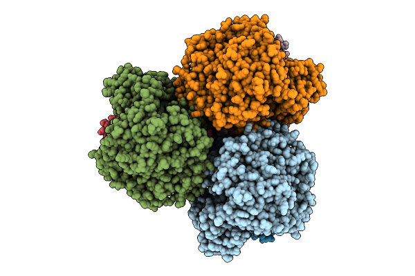
Cryo-Em Structure Of The Lipid-Bound Succiante Dehydrogenase From Chloroflexus Aurantiacus
Organism: Chloroflexus aurantiacus j-10-fl
Method: ELECTRON MICROSCOPY Release Date: 2025-11-12 Classification: MEMBRANE PROTEIN Ligands: FAD, SIN, SF4, FES, F3S, HEM, LMT, PGV, PEV |
Organism: Chloroflexus aurantiacus j-10-fl
Method: ELECTRON MICROSCOPY
Release Date: 2025-11-12
Ligands: FAD, SIN, SF4, FES, F3S, HEM, LMT, PGV, PEV
 |
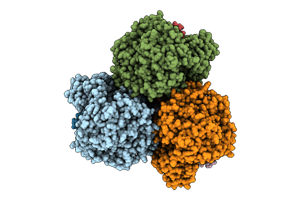
Cryo-Em Structure Of The Mk7-Bound Succinate Dehydrogenase From Chloroflexus Aurantiacus
Organism: Chloroflexus aurantiacus j-10-fl
Method: ELECTRON MICROSCOPY Release Date: 2025-11-12 Classification: MEMBRANE PROTEIN Ligands: FAD, SF4, FES, F3S, HEM, LMT, MQ7 |
Organism: Chloroflexus aurantiacus j-10-fl
Method: ELECTRON MICROSCOPY
Release Date: 2025-11-12
Ligands: FAD, SF4, FES, F3S, HEM, LMT, MQ7
 |
Cryo-Em Structure Of The Mk4-Bound Succinate Dehydrogenase From Chloroflexus Aurantiacus
Organism: Chloroflexus aurantiacus j-10-fl
Method: ELECTRON MICROSCOPY Release Date: 2025-11-12 Classification: MEMBRANE PROTEIN Ligands: FAD, SF4, FES, F3S, HEM, LMT, 1L3 |
Organism: Chloroflexus aurantiacus j-10-fl
Method: ELECTRON MICROSCOPY
Release Date: 2025-11-12
Ligands: FAD, SF4, FES, F3S, HEM, LMT, 1L3