

Search Count: 20
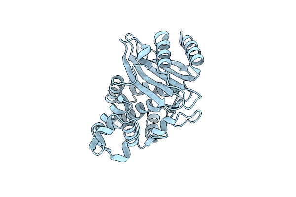 |
Crystal Structure Of Ctx-M-14 P167S/D240G Beta-Lactamase
Organism: Escherichia coli
Method: X-RAY DIFFRACTION Resolution:2.30 Å Release Date: 2020-04-22 Classification: HYDROLASE |
Organism: Escherichia coli
Method: X-RAY DIFFRACTION
Release Date: 2020-04-22
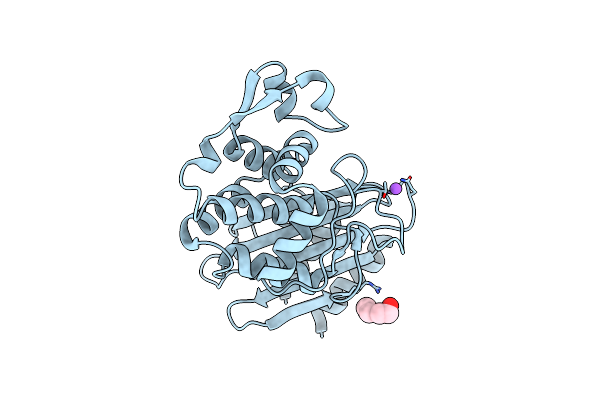 |
Crystal Structure Of Ctx-M-14 E166A/P167S/D240G Beta-Lactamase
Organism: Escherichia coli
Method: X-RAY DIFFRACTION Resolution:1.50 Å Release Date: 2020-04-22 Classification: HYDROLASE Ligands: PEG, NA |
Organism: Escherichia coli
Method: X-RAY DIFFRACTION
Release Date: 2020-04-22
Ligands: PEG, NA
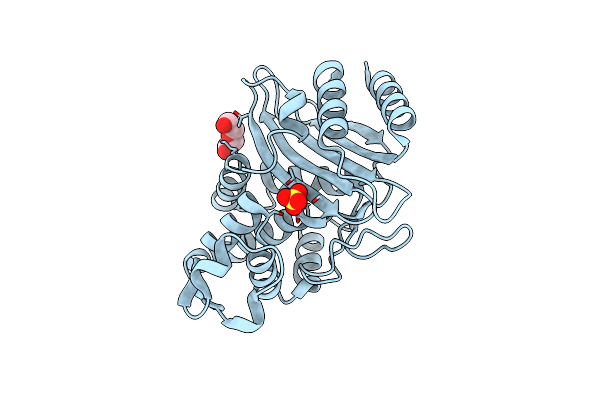 |
Crystal Structure Of Ctx-M-14 E166A/D240G Beta-Lactamase
Organism: Escherichia coli
Method: X-RAY DIFFRACTION Resolution:1.55 Å Release Date: 2020-04-22 Classification: HYDROLASE Ligands: SO4, PEG |
Organism: Escherichia coli
Method: X-RAY DIFFRACTION
Release Date: 2020-04-22
Ligands: SO4, PEG
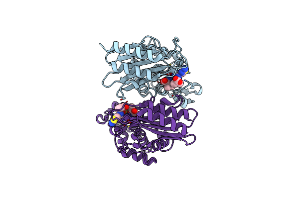 |
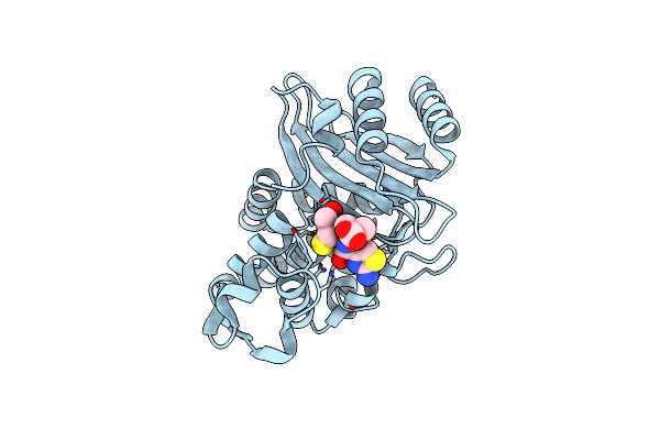
Crystal Structure Of Ctx-M-14 E166A/D240G Beta-Lactamase In Complex With Ceftazidime
Organism: Escherichia coli
Method: X-RAY DIFFRACTION Resolution:1.34 Å Release Date: 2020-04-22 Classification: HYDROLASE Ligands: CAZ |
Organism: Escherichia coli
Method: X-RAY DIFFRACTION
Release Date: 2020-04-22
Ligands: CAZ
 |
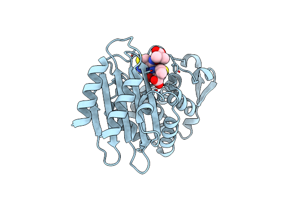
Crystal Structure Of Ctx-M-14 E166A/P167S/D240G Beta-Lactamase In Complex With Ceftazidime-1
Organism: Escherichia coli
Method: X-RAY DIFFRACTION Resolution:1.80 Å Release Date: 2020-04-22 Classification: HYDROLASE Ligands: CAZ |
Organism: Escherichia coli
Method: X-RAY DIFFRACTION
Release Date: 2020-04-22
Ligands: CAZ
 |
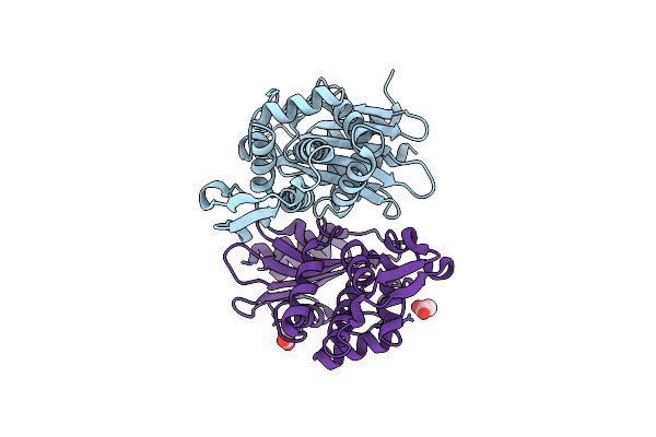
Crystal Structure Of Ctx-M-14 E166A/P167S/D240G Beta-Lactamase In Complex With Ceftazidime-2
Organism: Escherichia coli
Method: X-RAY DIFFRACTION Resolution:1.80 Å Release Date: 2020-04-22 Classification: HYDROLASE Ligands: CAZ |
Organism: Escherichia coli
Method: X-RAY DIFFRACTION
Release Date: 2020-04-22
Ligands: CAZ
 |
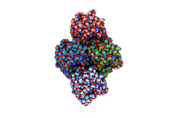
Ctx-M-14 N106S Mutant
Organism: Escherichia coli
Method: X-RAY DIFFRACTION Resolution:1.70 Å Release Date: 2018-10-10 Classification: HYDROLASE Ligands: ACT |
Organism: Escherichia coli
Method: X-RAY DIFFRACTION
Release Date: 2018-10-10
Ligands: ACT
 |
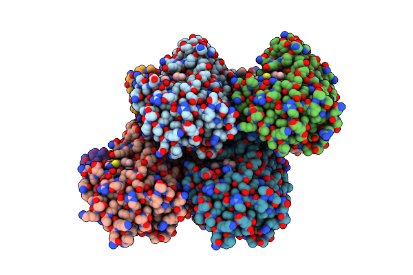
Ctx-M-14 N106S/D240G Mutant
Organism: Escherichia coli
Method: X-RAY DIFFRACTION Resolution:1.60 Å Release Date: 2018-10-10 Classification: HYDROLASE |
Organism: Escherichia coli
Method: X-RAY DIFFRACTION
Release Date: 2018-10-10
 |
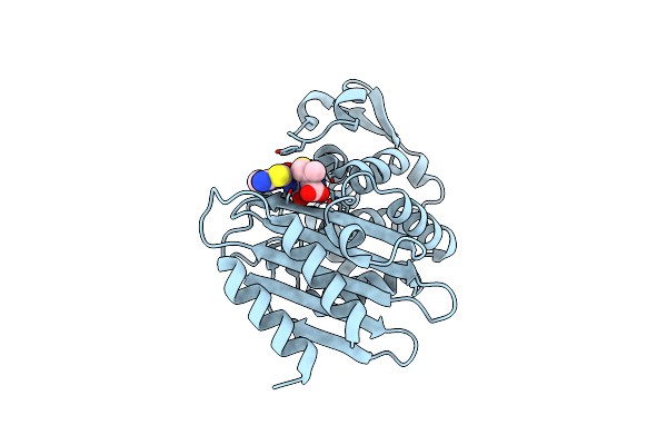
Crystal Structure Of Ctx-M-14 S70G/N106S Beta-Lactamase In Complex With Hydrolyzed Cefotaxime
Organism: Escherichia coli
Method: X-RAY DIFFRACTION Resolution:1.70 Å Release Date: 2018-10-10 Classification: hydrolase/antibiotic Ligands: CE4, GOL |
Organism: Escherichia coli
Method: X-RAY DIFFRACTION
Release Date: 2018-10-10
Ligands: CE4, GOL
 |
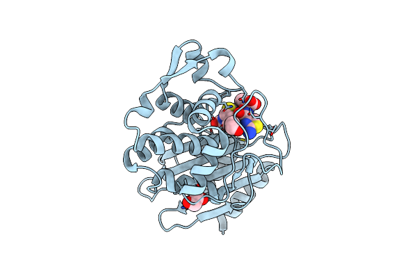
Crystal Structure Of Ctx-M-14 S70G/N106S/D240G Beta-Lactamase In Complex With Hydrolyzed Cefotaxime
Organism: Escherichia coli
Method: X-RAY DIFFRACTION Resolution:1.82 Å Release Date: 2018-10-10 Classification: hydrolase/antibiotic Ligands: CE4 |
Organism: Escherichia coli
Method: X-RAY DIFFRACTION
Release Date: 2018-10-10
Ligands: CE4
 |
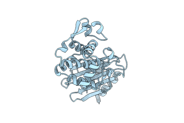
Ctx-M-14 P167S:E166A Mutant With Acylated Ceftazidime Molecule
Organism: Escherichia coli
Method: X-RAY DIFFRACTION Resolution:1.70 Å Release Date: 2017-06-28 Classification: HYDROLASE/ANTIBIOTIC Ligands: CAZ, EDO |
Organism: Escherichia coli
Method: X-RAY DIFFRACTION
Release Date: 2017-06-28
Ligands: CAZ, EDO
 |
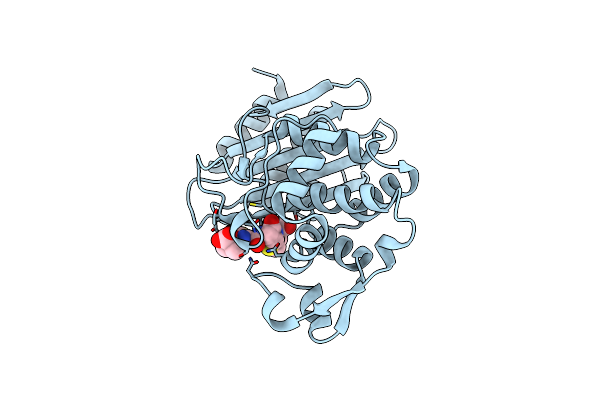
Ctx-M-14 P167S Apoenzyme
Organism: Escherichia coli
Method: X-RAY DIFFRACTION Resolution:1.70 Å Release Date: 2017-06-28 Classification: HYDROLASE |
Organism: Escherichia coli
Method: X-RAY DIFFRACTION
Release Date: 2017-06-28
 |
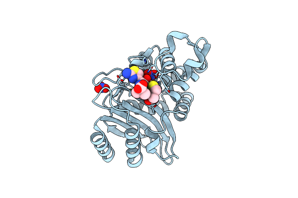
Ctx-M-14 P167S:S70G Mutant Enzyme Crystallized With Ceftazidime
Organism: Escherichia coli
Method: X-RAY DIFFRACTION Resolution:1.50 Å Release Date: 2017-06-28 Classification: HYDROLASE/ANTIBIOTIC Ligands: CAZ |
Organism: Escherichia coli
Method: X-RAY DIFFRACTION
Release Date: 2017-06-28
Ligands: CAZ
 |
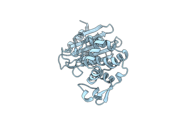
Ctx-M-14 E166A With Acylated Ceftazidime Molecule
Organism: Escherichia coli
Method: X-RAY DIFFRACTION Resolution:1.40 Å Release Date: 2017-06-28 Classification: HYDROLASE/ANTIBIOTIC Ligands: CAZ, NO3 |
Organism: Escherichia coli
Method: X-RAY DIFFRACTION
Release Date: 2017-06-28
Ligands: CAZ, NO3
 |
Ctx-M-14 P167S:E166A Mutant
Organism: Escherichia coli
Method: X-RAY DIFFRACTION Resolution:2.20 Å Release Date: 2017-06-28 Classification: HYDROLASE |
Organism: Escherichia coli
Method: X-RAY DIFFRACTION
Release Date: 2017-06-28
 |
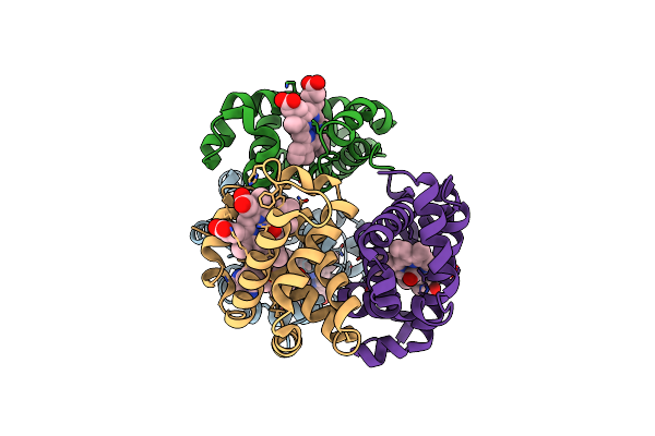
Crystal Structure Of Carbonmonoxy Hemoglobin S (Liganded Sickle Cell Hemoglobin) Complexed With Gbt Compound 31
Organism: Homo sapiens
Method: X-RAY DIFFRACTION Resolution:1.95 Å Release Date: 2017-02-22 Classification: OXYGEN TRANSPORT Ligands: HEM, CMO, 7SJ |
Organism: Homo sapiens
Method: X-RAY DIFFRACTION
Release Date: 2017-02-22
Ligands: HEM, CMO, 7SJ
 |
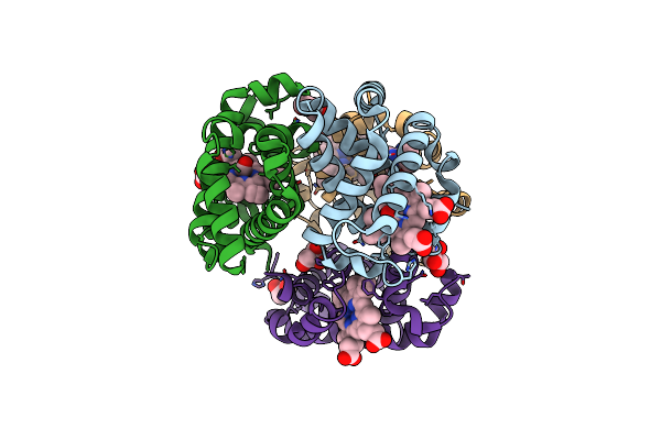
Crystal Structure Of Carbonmonoxy Hemoglobin S (Liganded Sickle Cell Hemoglobin) Complexed With Gbt Compound 6
Organism: Homo sapiens
Method: X-RAY DIFFRACTION Resolution:2.05 Å Release Date: 2017-02-22 Classification: OXYGEN TRANSPORT Ligands: HEM, CMO, 86M |
Organism: Homo sapiens
Method: X-RAY DIFFRACTION
Release Date: 2017-02-22
Ligands: HEM, CMO, 86M
 |
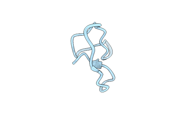
Crystal Structure Of Carbonmonoxy Hemoglobin S (Liganded Sickle Cell Hemoglobin) Complexed With Gbt440, Co-Crystallization Experiment
Organism: Homo sapiens
Method: X-RAY DIFFRACTION Resolution:1.80 Å Release Date: 2016-07-20 Classification: OXYGEN TRANSPORT Ligands: HEM, CMO, 5L7, GOL, UNL |
Organism: Homo sapiens
Method: X-RAY DIFFRACTION
Release Date: 2016-07-20
Ligands: HEM, CMO, 5L7, GOL, UNL
 |
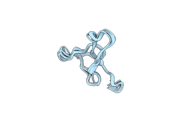
1H Assignments For Asip(93-126, P103A, P105A, P111A, Q115Y, S124Y)
Organism: Homo sapiens
Method: SOLUTION NMR Release Date: 2010-09-01 Classification: SIGNALING PROTEIN |
Organism: Homo sapiens
Method: SOLUTION NMR
Release Date: 2010-09-01
 |
Solution Structure Of Asip(80-132, P103A, P105A, Q115Y, S124Y)
Organism: Homo sapiens
Method: SOLUTION NMR Release Date: 2010-07-21 Classification: SIGNALING PROTEIN |
Organism: Homo sapiens
Method: SOLUTION NMR
Release Date: 2010-07-21