

Search Count: 44
 |
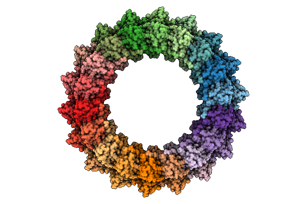
In Situ Cryo-Em Structure Of Outer Membrane Cap (Omc) Of The Legionella Dot/Icm T4Ss Machine
Organism: Legionella pneumophila subsp. pneumophila
Method: ELECTRON MICROSCOPY Release Date: 2025-09-17 Classification: PROTEIN TRANSPORT |
Organism: Legionella pneumophila subsp. pneumophila
Method: ELECTRON MICROSCOPY
Release Date: 2025-09-17
 |
In Situ Cryo-Em Structure Of Periplasmic Ring (Pr) Of The Legionella Dot/Icm T4Ss Machine.
Organism: Legionella pneumophila subsp. pneumophila
Method: ELECTRON MICROSCOPY Release Date: 2025-09-17 Classification: PROTEIN TRANSPORT |
Organism: Legionella pneumophila subsp. pneumophila
Method: ELECTRON MICROSCOPY
Release Date: 2025-09-17
 |
In-Situ Cryo-Em Structure Of Dome Of The Legionella Dot/Icm Machine
Organism: Legionella pneumophila subsp. pneumophila
Method: ELECTRON MICROSCOPY Release Date: 2025-09-17 Classification: PROTEIN TRANSPORT |
Organism: Legionella pneumophila subsp. pneumophila
Method: ELECTRON MICROSCOPY
Release Date: 2025-09-17
 |
In Situ Cryo-Em Structure Of Protochannel (Dota-Icmx) Of The Legionella Dot/Icm T4Ss Machine
Organism: Legionella pneumophila subsp. pneumophila
Method: ELECTRON MICROSCOPY Release Date: 2025-09-17 Classification: PROTEIN TRANSPORT |
Organism: Legionella pneumophila subsp. pneumophila
Method: ELECTRON MICROSCOPY
Release Date: 2025-09-17
 |
In Situ Cryo-Em Structure Of Pr And Dota-Icmx Of The Legionella Dot/Icm T4Ss Machine At C1 Symmetry
Organism: Legionella pneumophila subsp. pneumophila
Method: ELECTRON MICROSCOPY Release Date: 2025-09-17 Classification: PROTEIN TRANSPORT |
Organism: Legionella pneumophila subsp. pneumophila
Method: ELECTRON MICROSCOPY
Release Date: 2025-09-17
 |
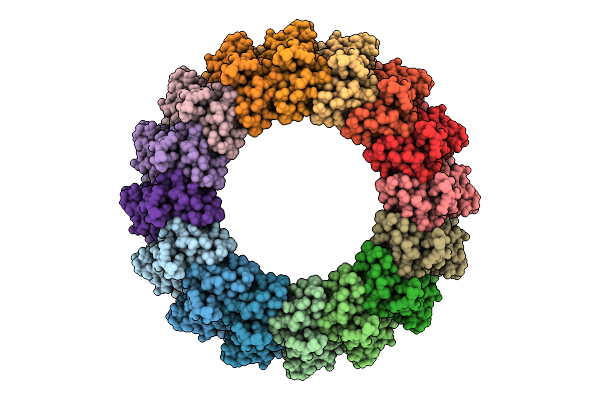
In Situ Cryo-Em Structure Of Porin I Of The Legionella Dot/Icm T4Ss Machine
Organism: Legionella pneumophila subsp. pneumophila
Method: ELECTRON MICROSCOPY Release Date: 2025-09-17 Classification: PROTEIN TRANSPORT |
Organism: Legionella pneumophila subsp. pneumophila
Method: ELECTRON MICROSCOPY
Release Date: 2025-09-17
 |
In Situ Cryo-Em Structure Of Porin Iii Of The Legionella Dot/Icm T4Ss Machine
Organism: Legionella pneumophila subsp. pneumophila
Method: ELECTRON MICROSCOPY Release Date: 2025-09-17 Classification: PROTEIN TRANSPORT |
Organism: Legionella pneumophila subsp. pneumophila
Method: ELECTRON MICROSCOPY
Release Date: 2025-09-17
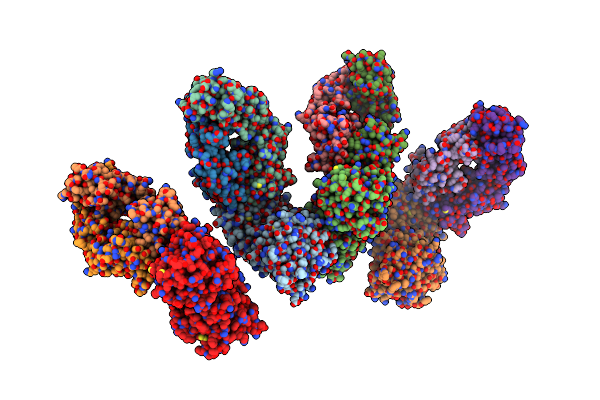 |
Crystal Structure Of N-Terminal Domain Of N-Methyl-D-Aspartate Receptor Subunit Nr1 In Complex With Patient-Derived Antibody
Organism: Homo sapiens
Method: X-RAY DIFFRACTION Resolution:3.50 Å Release Date: 2025-05-14 Classification: MEMBRANE PROTEIN |
Organism: Homo sapiens
Method: X-RAY DIFFRACTION
Release Date: 2025-05-14
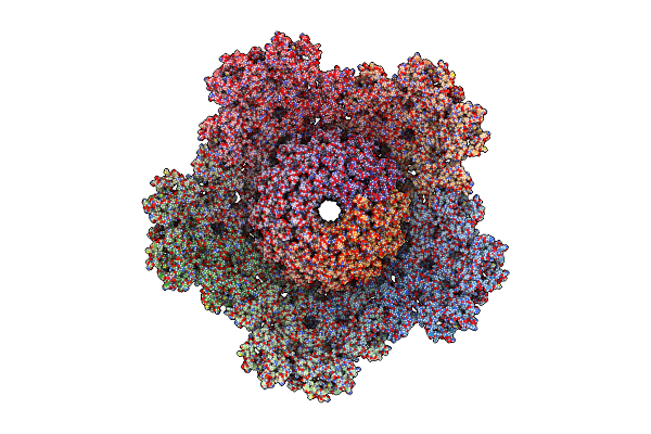 |
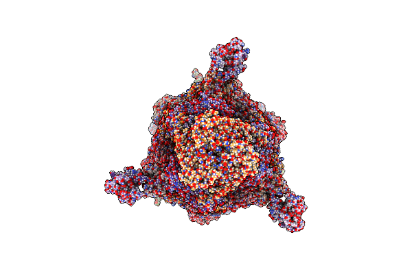
Mycobacteriophage Bxb1 C1 Capsid And Portal - Composite Map And Model
Organism: Mycobacterium phage bxb1
Method: ELECTRON MICROSCOPY Release Date: 2024-10-09 Classification: VIRAL PROTEIN |
Organism: Mycobacterium phage bxb1
Method: ELECTRON MICROSCOPY
Release Date: 2024-10-09
 |
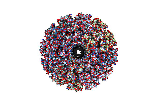
Mycobacteriophage Bxb1 Tail Tip - Composite Map And Model
Organism: Mycobacterium phage bxb1
Method: ELECTRON MICROSCOPY Release Date: 2024-09-25 Classification: VIRAL PROTEIN |
Organism: Mycobacterium phage bxb1
Method: ELECTRON MICROSCOPY
Release Date: 2024-09-25
 |
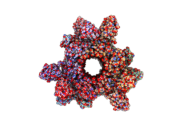
Mycobacteriophage Bxb1 Portal And Connector Assembly - Composite Map And Model
Organism: Mycobacterium phage bxb1
Method: ELECTRON MICROSCOPY Release Date: 2024-09-25 Classification: VIRAL PROTEIN |
Organism: Mycobacterium phage bxb1
Method: ELECTRON MICROSCOPY
Release Date: 2024-09-25
 |
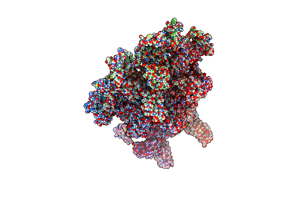
Mycobacteriophage Bxb1 Tail Tube Segment - Composite Map And Model
Organism: Mycobacterium phage bxb1
Method: ELECTRON MICROSCOPY Release Date: 2024-09-25 Classification: VIRAL PROTEIN |
Organism: Mycobacterium phage bxb1
Method: ELECTRON MICROSCOPY
Release Date: 2024-09-25
 |
Mycobacteriophage Bxb1 Capsid - Composite Map And Model
Organism: Mycobacterium phage bxb1
Method: ELECTRON MICROSCOPY Release Date: 2024-09-25 Classification: VIRAL PROTEIN |
Organism: Mycobacterium phage bxb1
Method: ELECTRON MICROSCOPY
Release Date: 2024-09-25
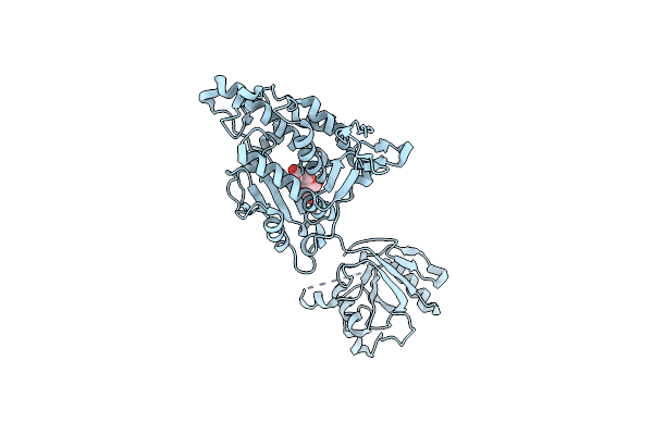 |
Functional Implication Of The Homotrimeric Multidomain Vacuolar Sorting Receptor 1 From Arabidopsis Thaliana
Organism: Arabidopsis thaliana
Method: X-RAY DIFFRACTION Resolution:3.50 Å Release Date: 2024-05-15 Classification: PROTEIN TRANSPORT |
Organism: Arabidopsis thaliana
Method: X-RAY DIFFRACTION
Release Date: 2024-05-15
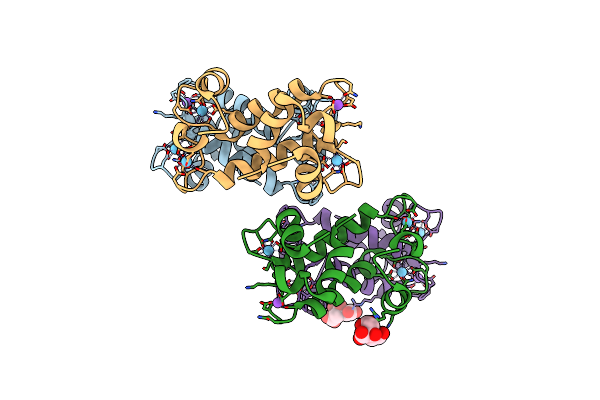 |
X-Ray Crystal Structure Of Hansschlegelia Quercus Lanmodulin (Lanm) With Lanthanum (Iii) Bound At Ph 7
Organism: Hansschlegelia quercus
Method: X-RAY DIFFRACTION Resolution:1.80 Å Release Date: 2023-06-07 Classification: METAL BINDING PROTEIN Ligands: LA, NA, CIT |
Organism: Hansschlegelia quercus
Method: X-RAY DIFFRACTION
Release Date: 2023-06-07
Ligands: LA, NA, CIT
 |
X-Ray Crystal Structure Of Hansschlegelia Quercus Lanmodulin (Lanm) With Dysprosium (Iii) Bound At Ph 7
Organism: Hansschlegelia quercus
Method: X-RAY DIFFRACTION Resolution:1.40 Å Release Date: 2023-06-07 Classification: METAL BINDING PROTEIN Ligands: DY |
Organism: Hansschlegelia quercus
Method: X-RAY DIFFRACTION
Release Date: 2023-06-07
Ligands: DY
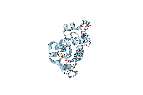 |
X-Ray Crystal Structure Of Methylorubrum Extorquens Am1 Lanmodulin (Lanm) With Neodymium (Iii) Bound At Ph 7
Organism: Methylorubrum extorquens am1
Method: X-RAY DIFFRACTION Resolution:1.01 Å Release Date: 2023-06-07 Classification: METAL BINDING PROTEIN Ligands: ND |
Organism: Methylorubrum extorquens am1
Method: X-RAY DIFFRACTION
Release Date: 2023-06-07
Ligands: ND
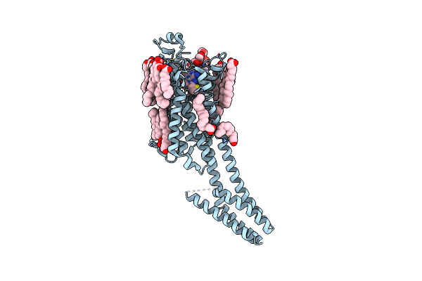 |
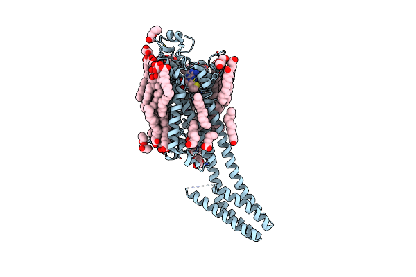
Crystal Structure Of A2Aar-Star2-S277-Bril In Complex With A Novel A2A Antagonist, Lj-4517
Organism: Homo sapiens, Escherichia coli
Method: X-RAY DIFFRACTION Resolution:2.80 Å Release Date: 2022-08-31 Classification: MEMBRANE PROTEIN Ligands: OLA, OLC, PEG, LJX, CLR |
Organism: Homo sapiens, Escherichia coli
Method: X-RAY DIFFRACTION
Release Date: 2022-08-31
Ligands: OLA, OLC, PEG, LJX, CLR
 |
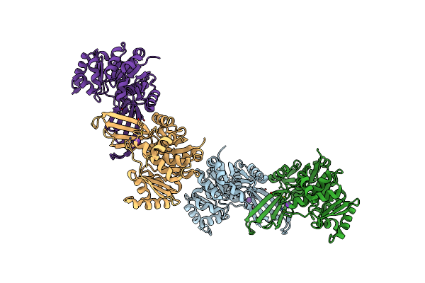
Crystal Structure Of A2Aar-Star2-Bril In Complex With A Novel A2A Antagonist, Lj-4517
Organism: Homo sapiens, Escherichia coli
Method: X-RAY DIFFRACTION Resolution:2.05 Å Release Date: 2022-08-31 Classification: MEMBRANE PROTEIN Ligands: PEG, LJX, NA, OLC, OLA, CLR, ETF |
Organism: Homo sapiens, Escherichia coli
Method: X-RAY DIFFRACTION
Release Date: 2022-08-31
Ligands: PEG, LJX, NA, OLC, OLA, CLR, ETF
 |
Unliganded Structure Of Pseudouridine Kinase (Puki) From Escherichia Coli Strain B
Organism: Escherichia coli
Method: X-RAY DIFFRACTION Resolution:2.15 Å Release Date: 2022-04-06 Classification: TRANSFERASE Ligands: K |
Organism: Escherichia coli
Method: X-RAY DIFFRACTION
Release Date: 2022-04-06
Ligands: K