

Search Count: 171
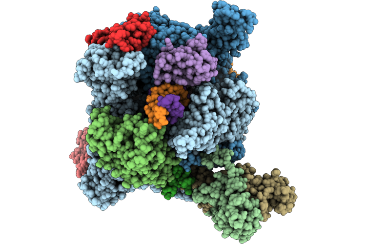 |
Structure Of Yeast Pol Ii In Complex With An Oxaliplatin-Icl Lesion At +7 Position.
Organism: Saccharomyces cerevisiae s288c, Synthetic construct
Method: ELECTRON MICROSCOPY Release Date: 2026-06-24 Classification: TRANSCRIPTION Ligands: ZN, MG, DNH, PT |
Organism: Saccharomyces cerevisiae s288c, Synthetic construct
Method: ELECTRON MICROSCOPY
Release Date: 2026-06-24
Ligands: ZN, MG, DNH, PT
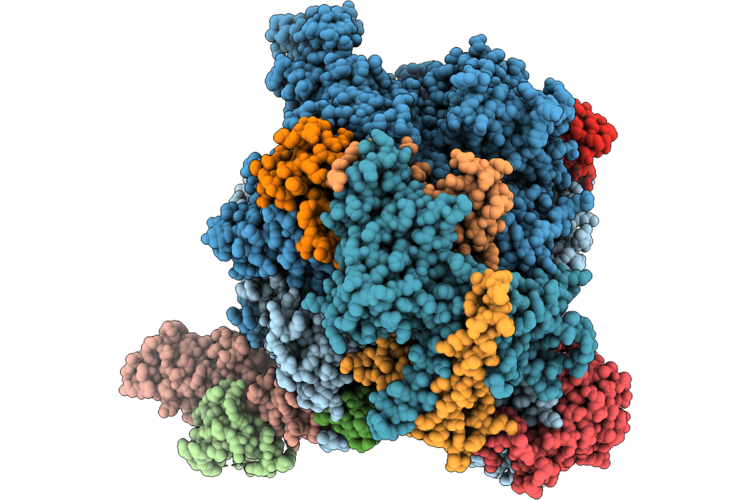 |
Structure Of Yeast Pol Ii In Complex With An Oxaliplatin-Icl Lesion At +2 Position.
Organism: Synthetic construct, Saccharomyces cerevisiae s288c
Method: ELECTRON MICROSCOPY Resolution:2.67 Å Release Date: 2026-06-24 Classification: TRANSCRIPTION Ligands: ZN, DNH, PT, MG |
Organism: Synthetic construct, Saccharomyces cerevisiae s288c
Method: ELECTRON MICROSCOPY
Release Date: 2026-06-24
Ligands: ZN, DNH, PT, MG
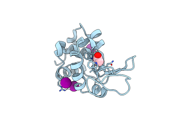 |
X-Ray Structure Of The Adduct Formed Upon Reaction Of The Diiodido Analogue Of Picoplatin With Lysozyme (Structure A)
Organism: Gallus gallus
Method: X-RAY DIFFRACTION Resolution:1.96 Å Release Date: 2025-05-14 Classification: HYDROLASE Ligands: NH3, ACT, CL, PT, IOD |
Organism: Gallus gallus
Method: X-RAY DIFFRACTION
Release Date: 2025-05-14
Ligands: NH3, ACT, CL, PT, IOD
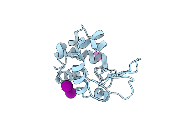 |
X-Ray Structure Of The Adduct Formed Upon Reaction Of The Diiodido Analogue Of Picoplatin With Lysozyme (Structure B)
Organism: Gallus gallus
Method: X-RAY DIFFRACTION Resolution:1.48 Å Release Date: 2025-05-14 Classification: HYDROLASE Ligands: PT, IOD |
Organism: Gallus gallus
Method: X-RAY DIFFRACTION
Release Date: 2025-05-14
Ligands: PT, IOD
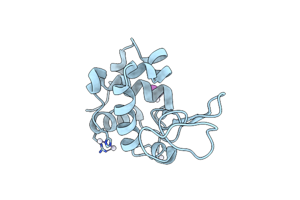 |
X-Structure Of The Adduct Formed Upon Reaction Of The Diiodido Analogue Of Picoplatin With Lysozyme (Structure C)
Organism: Gallus gallus
Method: X-RAY DIFFRACTION Resolution:2.25 Å Release Date: 2025-05-14 Classification: HYDROLASE Ligands: PT, NH3, IOD |
Organism: Gallus gallus
Method: X-RAY DIFFRACTION
Release Date: 2025-05-14
Ligands: PT, NH3, IOD
 |
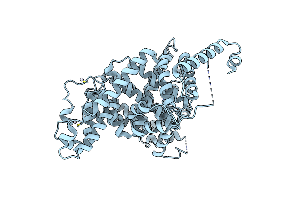
X-Ray Structure Of The Adduct Formed Upon Reaction Of The Diiodido Analogue Of Picoplatin With Ribonuclease A
Organism: Bos taurus
Method: X-RAY DIFFRACTION Resolution:1.77 Å Release Date: 2025-05-14 Classification: HYDROLASE Ligands: NH3, PT, IOD, CL |
Organism: Bos taurus
Method: X-RAY DIFFRACTION
Release Date: 2025-05-14
Ligands: NH3, PT, IOD, CL
 |
X-Ray Structure Of The Adduct Formed Upon Reaction Of The Diiodido Analogue Of Picoplatin With Human Serum Albumin
Organism: Homo sapiens
Method: X-RAY DIFFRACTION Resolution:3.90 Å Release Date: 2025-05-14 Classification: HYDROLASE Ligands: PT |
Organism: Homo sapiens
Method: X-RAY DIFFRACTION
Release Date: 2025-05-14
Ligands: PT
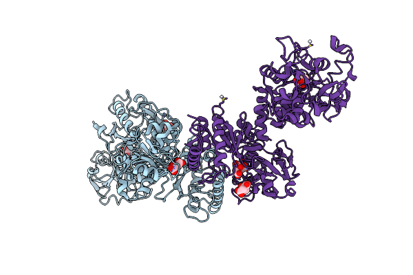 |
Crystal Structure Of The Adduct Between Human Serum Transferrin (Apo-Form) And Cisplatin
Organism: Homo sapiens
Method: X-RAY DIFFRACTION Resolution:3.52 Å Release Date: 2025-01-22 Classification: METAL TRANSPORT Ligands: CIT, PT, NH3 |
Organism: Homo sapiens
Method: X-RAY DIFFRACTION
Release Date: 2025-01-22
Ligands: CIT, PT, NH3
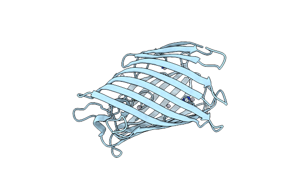 |
C2221 Crystal Structure Of Tama (Barrel Only) From Pseudomonas Aeruginosa At 3.15 Ang
Organism: Pseudomonas aeruginosa pao1
Method: X-RAY DIFFRACTION Resolution:3.15 Å Release Date: 2024-06-26 Classification: MEMBRANE PROTEIN Ligands: PT |
Organism: Pseudomonas aeruginosa pao1
Method: X-RAY DIFFRACTION
Release Date: 2024-06-26
Ligands: PT
 |
Crystal Structure Of Human Twisted Gastrulation Protein Homolog 1 (Twsg1) In Complex With Platinum
Organism: Homo sapiens
Method: X-RAY DIFFRACTION Resolution:3.61 Å Release Date: 2024-06-19 Classification: SIGNALING PROTEIN Ligands: PT |
Organism: Homo sapiens
Method: X-RAY DIFFRACTION
Release Date: 2024-06-19
Ligands: PT
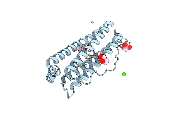 |
Ferritin Drug Carrier(Fdc) For Encapsulated Platinum (Iv) Prodrug For Esophageal Squamous Cell Carcinoma Targeted Therapy
Organism: Homo sapiens
Method: X-RAY DIFFRACTION Resolution:1.80 Å Release Date: 2024-05-29 Classification: TRANSPORT PROTEIN Ligands: PT, GOL, CA, CL, MG |
Organism: Homo sapiens
Method: X-RAY DIFFRACTION
Release Date: 2024-05-29
Ligands: PT, GOL, CA, CL, MG
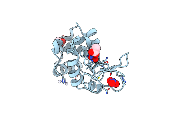 |
X-Ray Structure Of The Adduct Formed Upon Reaction Of Picoplatin With Lysozyme (Structure A)
Organism: Gallus gallus
Method: X-RAY DIFFRACTION Resolution:1.60 Å Release Date: 2024-05-22 Classification: HYDROLASE Ligands: NO3, ACT, NH3, PT |
Organism: Gallus gallus
Method: X-RAY DIFFRACTION
Release Date: 2024-05-22
Ligands: NO3, ACT, NH3, PT
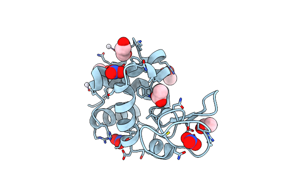 |
X-Ray Structure Of The Adduct Formed Upon Reaction Of Picoplatin With Lysozyme (Structure B)
Organism: Gallus gallus
Method: X-RAY DIFFRACTION Resolution:1.36 Å Release Date: 2024-05-22 Classification: HYDROLASE Ligands: NO3, GOL, ACT, PT |
Organism: Gallus gallus
Method: X-RAY DIFFRACTION
Release Date: 2024-05-22
Ligands: NO3, GOL, ACT, PT
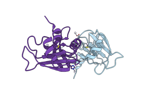 |
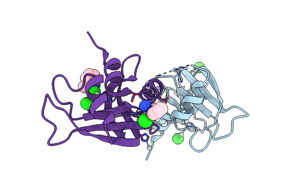
X-Ray Structure Of The Adduct Formed Upon Reaction Of Picoplatin With Bovine Pancreatic Ribonuclease (Structure C)
Organism: Bos taurus
Method: X-RAY DIFFRACTION Resolution:1.99 Å Release Date: 2024-05-22 Classification: RNA BINDING PROTEIN Ligands: NH3, PT |
Organism: Bos taurus
Method: X-RAY DIFFRACTION
Release Date: 2024-05-22
Ligands: NH3, PT
 |
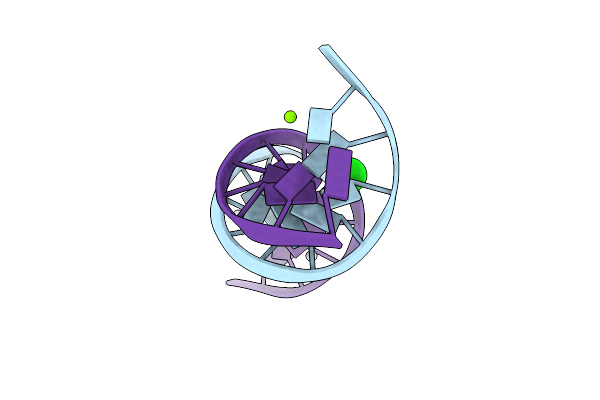
X-Ray Structure Of The Adduct Formed Upon Reaction Of Picoplatin With Bovine Pancreatic Ribonuclease (Structure D)
Organism: Gallus gallus
Method: X-RAY DIFFRACTION Resolution:1.76 Å Release Date: 2024-05-22 Classification: RNA BINDING PROTEIN Ligands: NH3, PT, CL, A1H58 |
Organism: Gallus gallus
Method: X-RAY DIFFRACTION
Release Date: 2024-05-22
Ligands: NH3, PT, CL, A1H58
 |
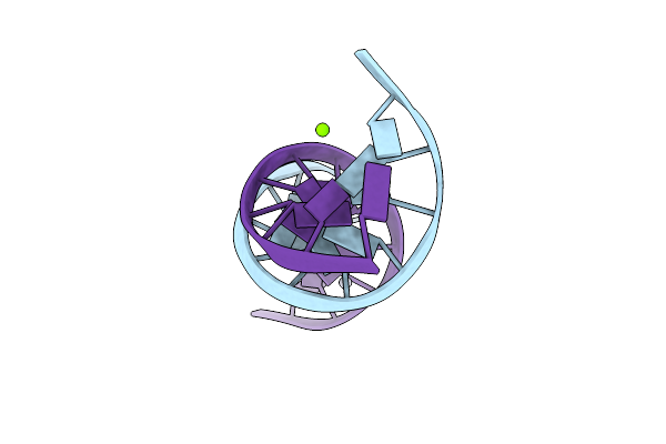
Crystal Structure Of Transplatin/B-Dna Adduct Obtained Upon 7 Days Of Soaking
Organism: Synthetic construct
Method: X-RAY DIFFRACTION Resolution:1.40 Å Release Date: 2024-02-28 Classification: DNA Ligands: MG, PT, NH3, CL |
Organism: Synthetic construct
Method: X-RAY DIFFRACTION
Release Date: 2024-02-28
Ligands: MG, PT, NH3, CL
 |
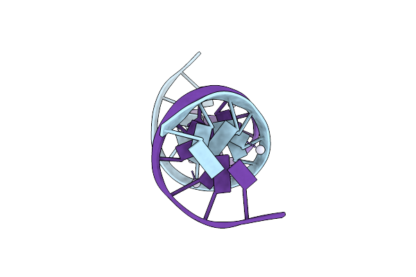
Crystal Structure Of Transplatin/B-Dna Adduct Obtained Upon 48 H Of Soaking
Organism: Dna molecule
Method: X-RAY DIFFRACTION Resolution:1.42 Å Release Date: 2024-02-07 Classification: DNA Ligands: MG, NH3, PT |
Organism: Dna molecule
Method: X-RAY DIFFRACTION
Release Date: 2024-02-07
Ligands: MG, NH3, PT
 |
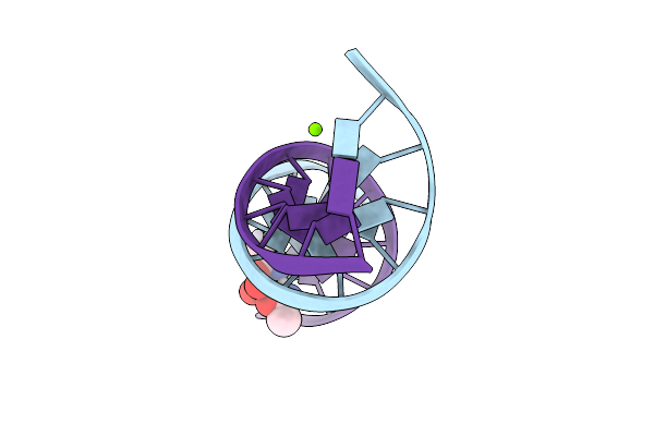
Crystal Structure Of Cisplatin/B-Dna Adduct
Organism: Synthetic construct
Method: X-RAY DIFFRACTION Resolution:2.31 Å Release Date: 2024-01-31 Classification: DNA Ligands: PT |
Organism: Synthetic construct
Method: X-RAY DIFFRACTION
Release Date: 2024-01-31
Ligands: PT
 |
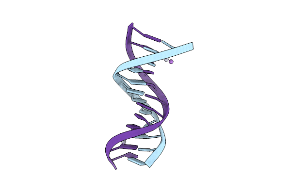
Crystal Structure Of Arsenoplatin-1/B-Dna Adduct Obtained Upon 4 H Of Soaking
Organism: Synthetic construct
Method: X-RAY DIFFRACTION Resolution:1.52 Å Release Date: 2024-01-31 Classification: DNA Ligands: PT, MG, A6R |
Organism: Synthetic construct
Method: X-RAY DIFFRACTION
Release Date: 2024-01-31
Ligands: PT, MG, A6R
 |
Crystal Structure Of Arsenoplatin-1/B-Dna Adduct Obtained Upon 48 H Of Soaking
Organism: Synthetic construct
Method: X-RAY DIFFRACTION Resolution:2.51 Å Release Date: 2024-01-31 Classification: DNA Ligands: PT, A6R |
Organism: Synthetic construct
Method: X-RAY DIFFRACTION
Release Date: 2024-01-31
Ligands: PT, A6R