
Search Count: 390
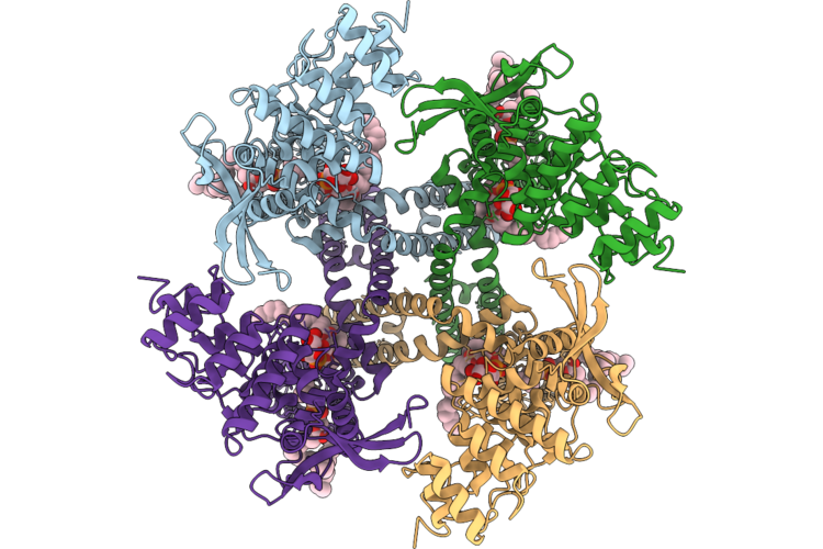 |
Organism: Homo sapiens
Method: ELECTRON MICROSCOPY Release Date: 2026-06-03 Classification: MEMBRANE PROTEIN Ligands: 8IJ, POV |
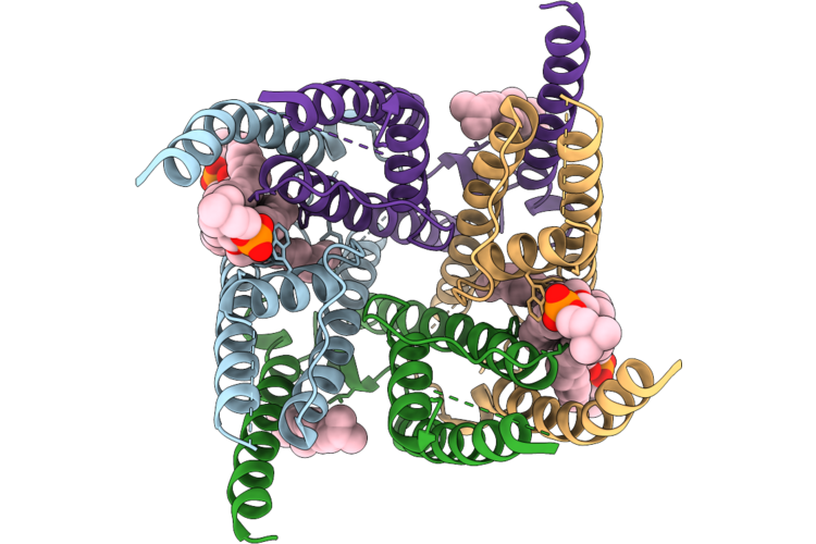 |
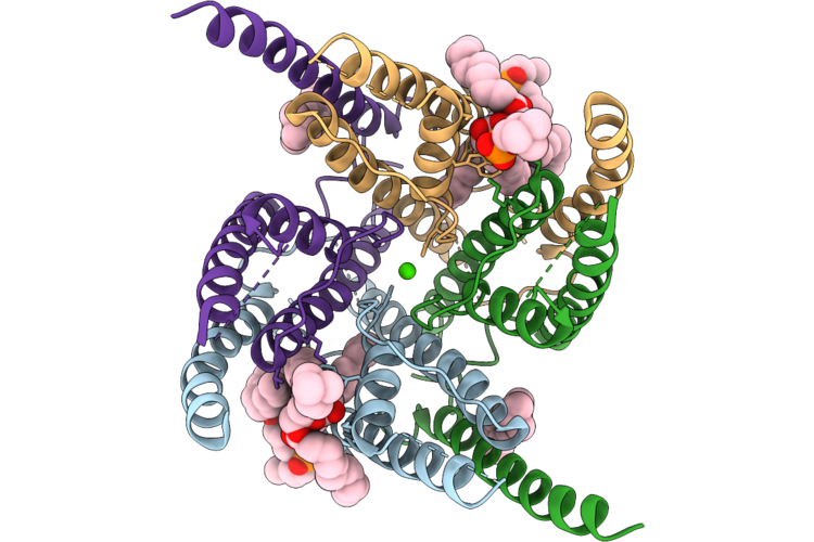
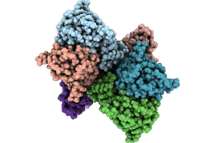
Organism: Rattus norvegicus
Method: ELECTRON MICROSCOPY Resolution:3.01 Å Release Date: 2026-05-06 Classification: MEMBRANE PROTEIN Ligands: POV, CLR |
 |
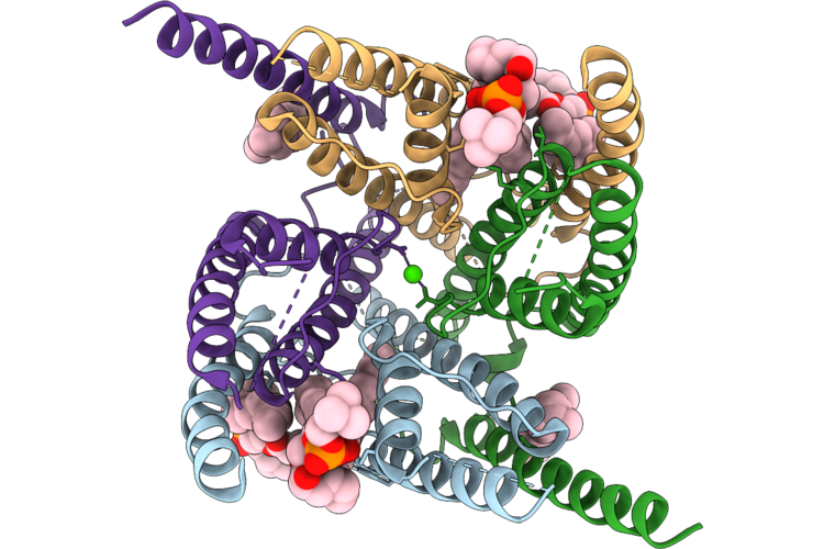
Organism: Rattus norvegicus
Method: ELECTRON MICROSCOPY Resolution:3.21 Å Release Date: 2026-05-06 Classification: MEMBRANE PROTEIN Ligands: POV, MG |
 |
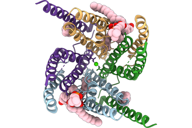
Organism: Rattus norvegicus
Method: ELECTRON MICROSCOPY Resolution:3.15 Å Release Date: 2026-05-06 Classification: MEMBRANE PROTEIN Ligands: POV, MG |
 |
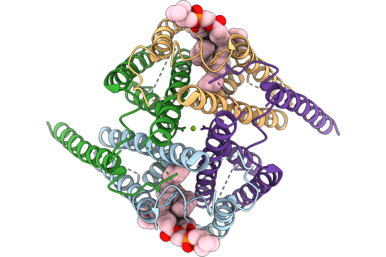
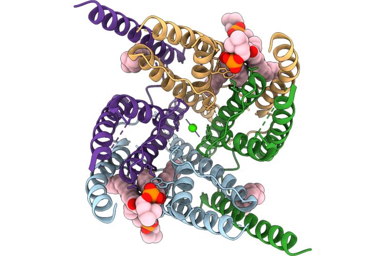
Organism: Rattus norvegicus
Method: ELECTRON MICROSCOPY Resolution:2.81 Å Release Date: 2026-05-06 Classification: MEMBRANE PROTEIN Ligands: POV, CLR, CA |
 |
Organism: Rattus norvegicus
Method: ELECTRON MICROSCOPY Resolution:2.97 Å Release Date: 2026-05-06 Classification: MEMBRANE PROTEIN Ligands: POV, CLR, CA |
 |
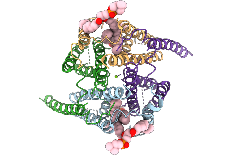
Organism: Rattus norvegicus
Method: ELECTRON MICROSCOPY Resolution:2.76 Å Release Date: 2026-05-06 Classification: MEMBRANE PROTEIN Ligands: CA, POV, CLR |
 |
Organism: Rattus norvegicus
Method: ELECTRON MICROSCOPY Resolution:2.69 Å Release Date: 2026-05-06 Classification: MEMBRANE PROTEIN Ligands: POV, CLR, CA |
 |
Organism: Mus musculus
Method: ELECTRON MICROSCOPY Resolution:3.13 Å Release Date: 2026-04-29 Classification: SIGNALING PROTEIN Ligands: PLM, OLC, POV |
 |
Organism: Mus musculus
Method: ELECTRON MICROSCOPY Resolution:2.80 Å Release Date: 2026-04-29 Classification: SIGNALING PROTEIN Ligands: PLM, OLC, POV |
 |
Organism: Mus musculus
Method: ELECTRON MICROSCOPY Resolution:3.04 Å Release Date: 2026-04-29 Classification: SIGNALING PROTEIN Ligands: PLM, OLC, POV |
 |
Organism: Mus musculus
Method: ELECTRON MICROSCOPY Resolution:2.87 Å Release Date: 2026-04-29 Classification: SIGNALING PROTEIN Ligands: PLM, OLC, POV |
 |
Organism: Mus musculus
Method: ELECTRON MICROSCOPY Resolution:3.36 Å Release Date: 2026-04-29 Classification: SIGNALING PROTEIN Ligands: PLM, OLC, POV |
 |
Organism: Mus musculus
Method: ELECTRON MICROSCOPY Resolution:3.08 Å Release Date: 2026-04-29 Classification: SIGNALING PROTEIN Ligands: PLM, OLC, POV |
 |
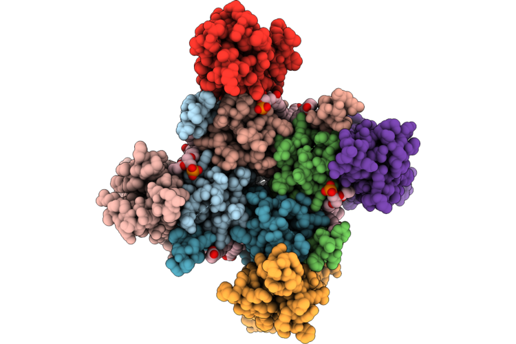
The Lbd-Tmd Structure Of Native Mouse Ampar With 2 Tarps 2 Cnihs And Prrt1/Syndig4
Organism: Mus musculus
Method: ELECTRON MICROSCOPY Resolution:3.36 Å Release Date: 2026-04-29 Classification: SIGNALING PROTEIN Ligands: PLM, OLC, POV |
 |
The Tmd Structure Of Native Mouse Ampar With 2 Tarps 2 Cnihs And Prrt1/Syndig4
Organism: Mus musculus
Method: ELECTRON MICROSCOPY Resolution:3.04 Å Release Date: 2026-04-29 Classification: SIGNALING PROTEIN Ligands: PLM, OLC, POV |
 |
The Lbd-Tmd Structure Of Native Mouse Ampar With 3 Tarps 1 Cnih And Prrt1/Syndig4
Organism: Mus musculus
Method: ELECTRON MICROSCOPY Resolution:3.75 Å Release Date: 2026-04-29 Classification: SIGNALING PROTEIN Ligands: PLM, OLC, POV |
 |
Organism: Mus musculus
Method: ELECTRON MICROSCOPY Resolution:3.45 Å Release Date: 2026-04-29 Classification: SIGNALING PROTEIN Ligands: PLM, OLC, POV |
 |
Organism: Mus musculus
Method: ELECTRON MICROSCOPY Resolution:3.33 Å Release Date: 2026-04-29 Classification: SIGNALING PROTEIN Ligands: PLM, OLC, POV |
 |
Organism: Escherichia coli k-12
Method: ELECTRON MICROSCOPY Release Date: 2026-04-22 Classification: OXIDOREDUCTASE Ligands: HEM, A1JN4, MQ9, LPP, UQ8, OXY, PGT, POV |