
Search Count: 4,550
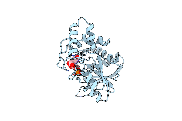 |
Organism: Salmonella typhimurium
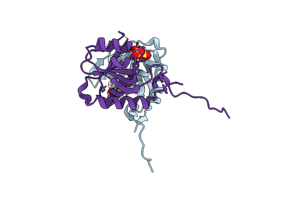
Method: X-RAY DIFFRACTION Resolution:2.60 Å Release Date: 1994-05-31 Classification: PHOSPHORIBOSYLTRANSFERASE Ligands: OMP |
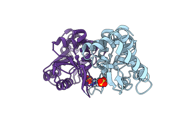 |
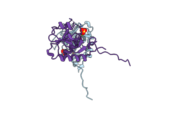
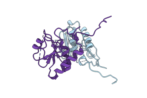
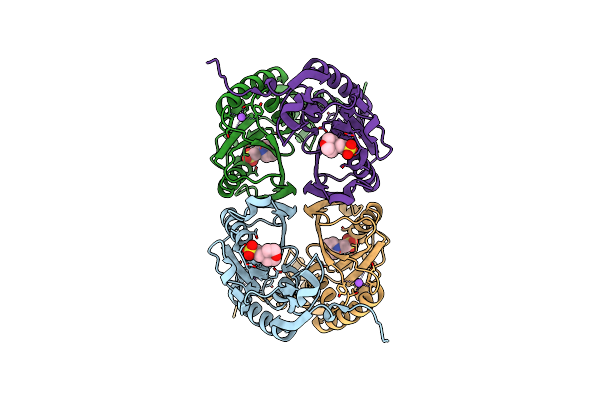
A Flexible Loop At The Dimer Interface Is A Part Of The Active Site Of The Adjacent Monomer Of Escherichia Coli Orotate Phosphoribosyltransferase
Organism: Escherichia coli
Method: X-RAY DIFFRACTION Resolution:2.40 Å Release Date: 1996-04-03 Classification: PHOSPHORIBOSYLTRANSFERASE Ligands: SO4 |
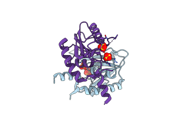 |
Helicobacter Pylori Xanthine-Guanine-Hypoxanthine Phosphoribosyltransferase
Organism: Helicobacter pylori
Method: X-RAY DIFFRACTION Resolution:1.47 Å Release Date: 2021-05-05 Classification: TRANSFERASE Ligands: SO4 |
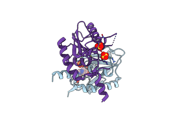 |
Helicobacter Pylori Xanthine-Guanine-Hypoxanthine Phosphoribosyltransferase
Organism: Helicobacter pylori
Method: X-RAY DIFFRACTION Resolution:1.97 Å Release Date: 2021-05-05 Classification: TRANSFERASE Ligands: WG7, SO4 |
 |
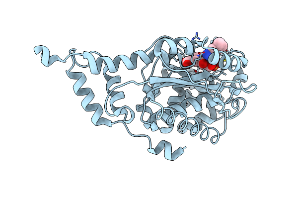
Organism: Escherichia coli
Method: X-RAY DIFFRACTION Resolution:1.80 Å Release Date: 1997-05-15 Classification: PHOSPHORIBOSYLTRANSFERASE Ligands: SO4, MG |
 |
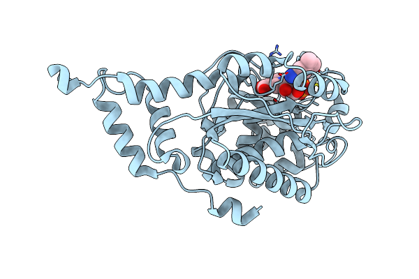
Organism: Escherichia coli
Method: X-RAY DIFFRACTION Resolution:2.60 Å Release Date: 1998-11-11 Classification: PHOSPHORIBOSYLTRANSFERASE Ligands: BO3, 5GP |
 |
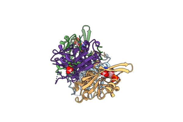
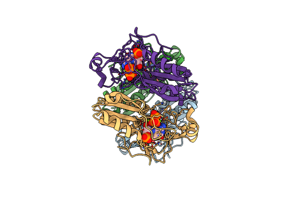
Organism: Escherichia coli
Method: X-RAY DIFFRACTION Resolution:2.00 Å Release Date: 1998-11-11 Classification: PHOSPHORIBOSYLTRANSFERASE Ligands: BO3, MG, PCP, GUN |
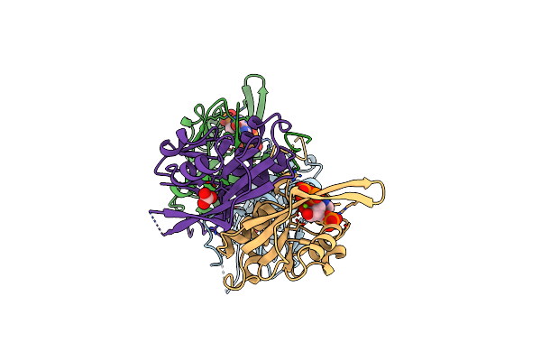 |
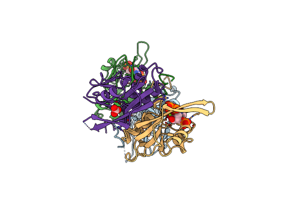
Organism: Escherichia coli
Method: X-RAY DIFFRACTION Resolution:2.00 Å Release Date: 1998-11-11 Classification: PHOSPHORIBOSYLTRANSFERASE Ligands: BO3, MG, PCP, XAN |
 |
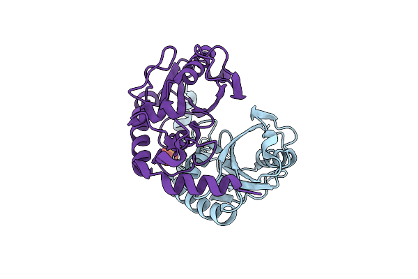
Organism: Escherichia coli
Method: X-RAY DIFFRACTION Resolution:2.25 Å Release Date: 1998-06-17 Classification: PHOSPHORIBOSYLTRANSFERASE |
 |
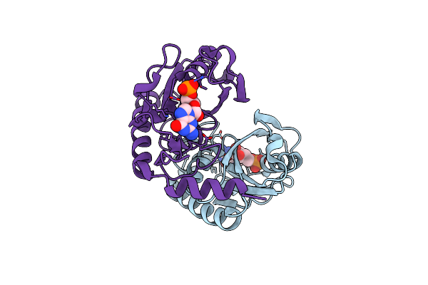
Organism: Homo sapiens
Method: X-RAY DIFFRACTION Resolution:2.00 Å Release Date: 1999-06-22 Classification: PHOSPHORIBOSYLTRANSFERASE Ligands: MG, IMU, POP |
 |
Crystal Structure Of Apo Hypoxanthine-Guanine Phosphoribosyltransferase From Legionella Pneumophila
Organism: Legionella pneumophila subsp. pneumophila strain philadelphia 1
Method: X-RAY DIFFRACTION Resolution:2.40 Å Release Date: 2016-03-30 Classification: TRANSFERASE Ligands: PO4 |
 |
Crystal Structure Of Hypoxanthine-Guanine Phosphoribosyltransferase Complexed With Gmp From Legionella Pneumophila
Organism: Legionella pneumophila subsp. pneumophila atcc 43290
Method: X-RAY DIFFRACTION Resolution:2.71 Å Release Date: 2016-03-30 Classification: TRANSFERASE Ligands: 5GP |
 |
1.95 Angstrom Crystal Structure Of Complex Of Hypoxanthine-Guanine Phosphoribosyltransferase From Bacillus Anthracis With 2-(N-Morpholino)Ethanesulfonic Acid (Mes)
Organism: Bacillus anthracis str. 'ames ancestor'
Method: X-RAY DIFFRACTION Resolution:1.95 Å Release Date: 2009-06-23 Classification: TRANSFERASE Ligands: MES, NA |
 |
Crystal Structure Of E136F Mutant Of Xanthine-Guanine Phosphoribosyltransferase From Yersinia Pestis
Organism: Yersinia pestis
Method: X-RAY DIFFRACTION Resolution:1.70 Å Release Date: 2019-09-11 Classification: TRANSFERASE Ligands: CL, GOL |
 |
Crystal Structure Of Xanthine-Guanine Phosphoribosyltransferase (Xgprt) From Yersinia Pestis In P21212 Space Group With Sulphate Ions In The Active Site
Organism: Yersinia pestis
Method: X-RAY DIFFRACTION Resolution:1.10 Å Release Date: 2019-09-25 Classification: TRANSFERASE Ligands: SO4 |
 |
The Crystal Structure Of Cobt Complexed With 4,5-Dimethyl-1,2-Phenylenediamine And Nicotinate Mononucleotide
Organism: Salmonella enterica
Method: X-RAY DIFFRACTION Resolution:2.10 Å Release Date: 2002-09-07 Classification: TRANSFERASE Ligands: 150, NCN |
 |
Organism: Salmonella enterica
Method: X-RAY DIFFRACTION Resolution:2.00 Å Release Date: 2002-09-07 Classification: TRANSFERASE Ligands: PO4, 2AF, NIO |
 |
Organism: Salmonella enterica
Method: X-RAY DIFFRACTION Resolution:1.90 Å Release Date: 2002-09-07 Classification: TRANSFERASE Ligands: PO4, IMD |
 |
Crystal Structure Of Cobt Complexed With N7-(5'-Phosphoribosyl)-2-Aminopurine And Nicotinate
Organism: Salmonella enterica
Method: X-RAY DIFFRACTION Resolution:2.00 Å Release Date: 2002-09-07 Classification: TRANSFERASE Ligands: 7RA, NIO |
 |
Crystal Structure Of Cobt Complexed With 3,4-Dimethylaniline And Nicotinate Mononucleotide
Organism: Salmonella enterica
Method: X-RAY DIFFRACTION Resolution:2.20 Å Release Date: 2002-09-07 Classification: TRANSFERASE Ligands: 34A, NCN |