

Search Count: 21
 |
Biochemical And Structural Characterization Of A Novel 4-Hydroxyphenylacetate-3-Monooxygenase From Geobacillus Mahadii Geo-05
Organism: Geobacillus mahadia
Method: X-RAY DIFFRACTION Resolution:2.40 Å Release Date: 2025-09-10 Classification: HYDROLASE Ligands: PEU |
Organism: Geobacillus mahadia
Method: X-RAY DIFFRACTION
Release Date: 2025-09-10
Ligands: PEU
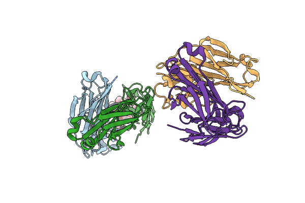 |
Humanized Anti-Peg H6.3 Fab In Complex With Peg
Organism: Homo sapiens
Method: X-RAY DIFFRACTION Resolution:2.26 Å Release Date: 2025-02-12 Classification: IMMUNE SYSTEM Ligands: PEU, IPA |
Organism: Homo sapiens
Method: X-RAY DIFFRACTION
Release Date: 2025-02-12
Ligands: PEU, IPA
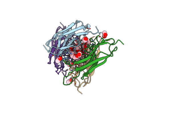 |
Structure Of The Sensor Domain (Short Construct) Of The Anti-Sigma Factor Rsgi4 In Pseudobacteroides Cellulosolvens
Organism: Pseudobacteroides cellulosolvens atcc 35603 = dsm 2933
Method: X-RAY DIFFRACTION Resolution:1.50 Å Release Date: 2021-06-30 Classification: TRANSCRIPTION Ligands: ACT, PEU |
Organism: Pseudobacteroides cellulosolvens atcc 35603 = dsm 2933
Method: X-RAY DIFFRACTION
Release Date: 2021-06-30
Ligands: ACT, PEU
 |
Anti-Peg Antibody 6-3 Fab Fragment In Complex With Peg
Organism: Mus musculus
Method: X-RAY DIFFRACTION Resolution:2.42 Å Release Date: 2020-09-09 Classification: IMMUNE SYSTEM Ligands: EDO, PEU |
Organism: Mus musculus
Method: X-RAY DIFFRACTION
Release Date: 2020-09-09
Ligands: EDO, PEU
 |
Crystal Structure Of Peg-Bound Sh3 Domain Of Myosin Ib From Entamoeba Histolytica
Organism: Entamoeba histolytica
Method: X-RAY DIFFRACTION Resolution:1.78 Å Release Date: 2017-08-16 Classification: CONTRACTILE PROTEIN Ligands: 1PE, PEU, PG6, SO4 |
Organism: Entamoeba histolytica
Method: X-RAY DIFFRACTION
Release Date: 2017-08-16
Ligands: 1PE, PEU, PG6, SO4
 |
Crystal Structure Of A Corynascus Thermopiles (Myceliophthora Fergusii) Carbohydrate Esterase Family 2 (Ce2) Enzyme Plus Carbohydrate Binding Domain (Cbd)
Organism: Chaetomium olivicolor
Method: X-RAY DIFFRACTION Resolution:1.94 Å Release Date: 2015-02-11 Classification: HYDROLASE Ligands: GOL, PEU |
Organism: Chaetomium olivicolor
Method: X-RAY DIFFRACTION
Release Date: 2015-02-11
Ligands: GOL, PEU
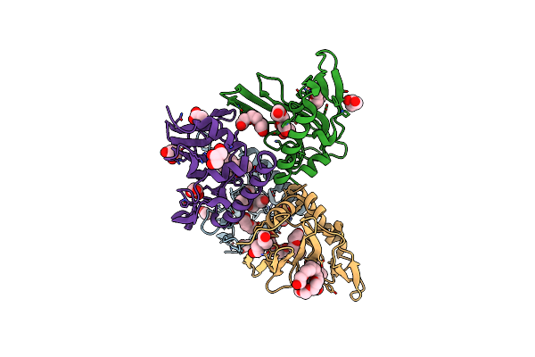 |
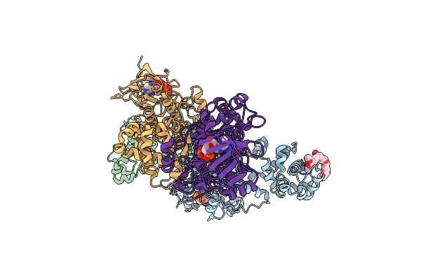
Crystal Structure Of A Cystatin-Like Protein (Baccac_01506) From Bacteroides Caccae Atcc 43185 At 1.69 A Resolution
Organism: Bacteroides caccae
Method: X-RAY DIFFRACTION Resolution:1.69 Å Release Date: 2014-10-01 Classification: STRUCTURAL GENOMICS, UNKNOWN FUNCTION Ligands: ACT, PEU, CL, GOL |
Organism: Bacteroides caccae
Method: X-RAY DIFFRACTION
Release Date: 2014-10-01
Ligands: ACT, PEU, CL, GOL
 |
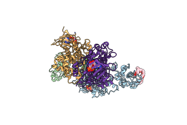
Crystal Structure Of Murine Ire1 In Complex With Oicr464 Inhibitor
Organism: Mus musculus
Method: X-RAY DIFFRACTION Resolution:3.00 Å Release Date: 2014-09-03 Classification: TRANSFERASE,HYDROLASE/INHIBITOR Ligands: 31K, ADP, PEU, MG |
Organism: Mus musculus
Method: X-RAY DIFFRACTION
Release Date: 2014-09-03
Ligands: 31K, ADP, PEU, MG
 |
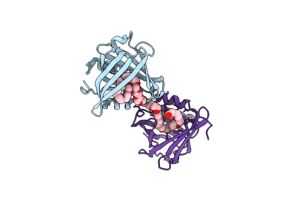
Crystal Structure Of Murine Ire1 In Complex With Oicr573 Inhibitor
Organism: Mus musculus
Method: X-RAY DIFFRACTION Resolution:3.40 Å Release Date: 2014-09-03 Classification: TRANSFERASE,HYDROLASE/INHIBITOR Ligands: ADP, MG, 31L, PEU |
Organism: Mus musculus
Method: X-RAY DIFFRACTION
Release Date: 2014-09-03
Ligands: ADP, MG, 31L, PEU
 |
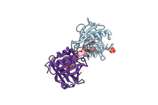
Three-Dimensional Structure Of The C65A Mutant Of Human Lipocalin-Type Prostaglandin D Synthase Holo-Form Second Space Group
Organism: Homo sapiens
Method: X-RAY DIFFRACTION Resolution:1.55 Å Release Date: 2014-08-06 Classification: ISOMERASE Ligands: PEU |
Organism: Homo sapiens
Method: X-RAY DIFFRACTION
Release Date: 2014-08-06
Ligands: PEU
 |
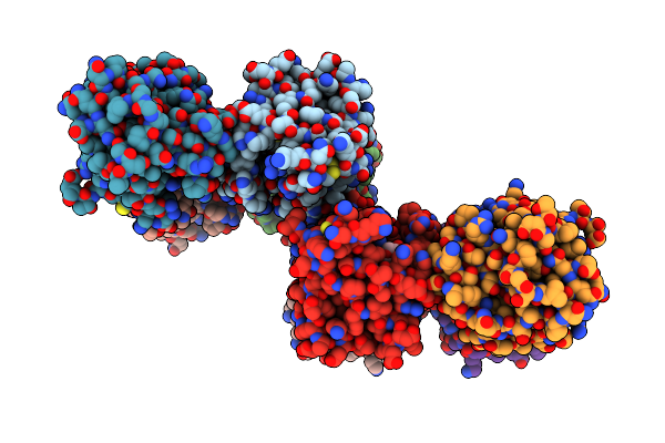
Three-Dimensional Structure Of The C65A-K59A Double Mutant Of Human Lipocalin-Type Prostaglandin D Synthase Holo-Form
Organism: Homo sapiens
Method: X-RAY DIFFRACTION Resolution:1.60 Å Release Date: 2014-08-06 Classification: ISOMERASE Ligands: SO4, PEU |
Organism: Homo sapiens
Method: X-RAY DIFFRACTION
Release Date: 2014-08-06
Ligands: SO4, PEU
 |
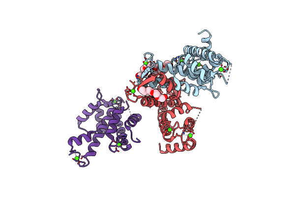
Three-Dimensional Structure Of The C65A-K59A Double Mutant Of Human Lipocalin-Type Prostaglandin D Synthase Holo, Second Crystal Form
Organism: Homo sapiens
Method: X-RAY DIFFRACTION Resolution:1.80 Å Release Date: 2014-08-06 Classification: ISOMERASE Ligands: PEU |
Organism: Homo sapiens
Method: X-RAY DIFFRACTION
Release Date: 2014-08-06
Ligands: PEU
 |
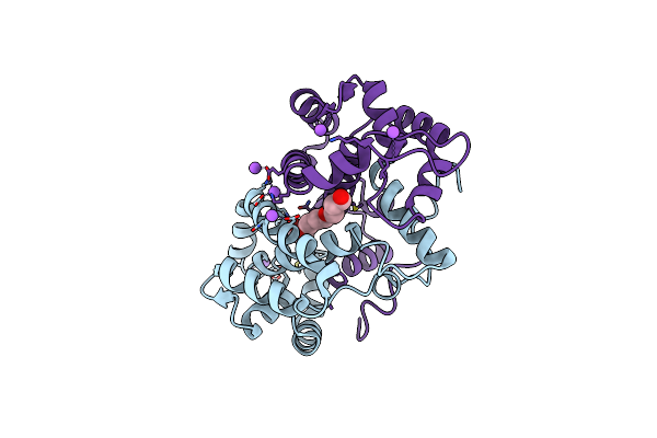
Crystal Structure Of Calpain-3 Penta-Ef-Hand Domain
Organism: Homo sapiens
Method: X-RAY DIFFRACTION Resolution:2.45 Å Release Date: 2014-05-28 Classification: HYDROLASE Ligands: CA, PEU |
Organism: Homo sapiens
Method: X-RAY DIFFRACTION
Release Date: 2014-05-28
Ligands: CA, PEU
 |
Crystal Structure Of Odorant Binding Protein 48 From Anopheles Gambiae (Agamobp48) With Peg
Organism: Anopheles gambiae
Method: X-RAY DIFFRACTION Resolution:2.25 Å Release Date: 2013-10-16 Classification: TRANSPORT PROTEIN Ligands: PEU, NA |
Organism: Anopheles gambiae
Method: X-RAY DIFFRACTION
Release Date: 2013-10-16
Ligands: PEU, NA
 |
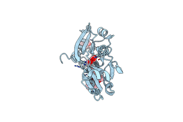
Bovine Trypsin Variant X(Tripleglu217Ile227) In Complex With Small Molecule Inhibitor
Organism: Bos taurus
Method: X-RAY DIFFRACTION Resolution:1.70 Å Release Date: 2011-12-21 Classification: HYDROLASE/HYDROLASE INHIBITOR Ligands: CA, PMJ, SO4, PEU |
Organism: Bos taurus
Method: X-RAY DIFFRACTION
Release Date: 2011-12-21
Ligands: CA, PMJ, SO4, PEU
 |
Dntr Inducer Binding Domain In Complex With Salicylate. Trigonal Crystal Form
Organism: Burkholderia sp.
Method: X-RAY DIFFRACTION Resolution:1.85 Å Release Date: 2011-07-20 Classification: TRANSCRIPTION REGULATOR Ligands: SAL, PEU |
Organism: Burkholderia sp.
Method: X-RAY DIFFRACTION
Release Date: 2011-07-20
Ligands: SAL, PEU
 |
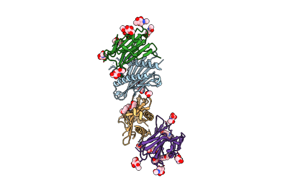
Crystal Structure Of Odorant Binding Protein 1 From Anopheles Gambiae (Agamobp1) With Deet (N,N-Diethyl-Meta-Toluamide) And Peg
Organism: Anopheles gambiae
Method: X-RAY DIFFRACTION Resolution:1.60 Å Release Date: 2011-06-08 Classification: TRANSPORT PROTEIN Ligands: MG, MOH, DE3, PEU, CL |
Organism: Anopheles gambiae
Method: X-RAY DIFFRACTION
Release Date: 2011-06-08
Ligands: MG, MOH, DE3, PEU, CL
 |
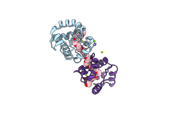
Structure Of The Hcmv Ul16-Micb Complex Elucidates Select Binding Of A Viral Immunoevasin To Diverse Nkg2D Ligands
Organism: Homo sapiens, Human herpesvirus 5 strain ad169
Method: X-RAY DIFFRACTION Resolution:1.80 Å Release Date: 2010-02-02 Classification: IMMUNE SYSTEM/VIRAL PROTEIN Ligands: PEU, ACT, NAG |
Organism: Homo sapiens, Human herpesvirus 5 strain ad169
Method: X-RAY DIFFRACTION
Release Date: 2010-02-02
Ligands: PEU, ACT, NAG
 |
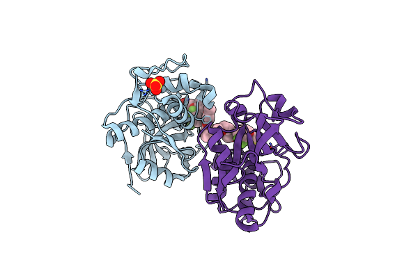
Crystal Structure Of Odorant Binding Protein 1 (Aaegobp1) From Aedes Aegypti
Organism: Aedes aegypti
Method: X-RAY DIFFRACTION Resolution:1.85 Å Release Date: 2009-12-08 Classification: ODORANT BINDING PROTEIN Ligands: MG, PEU, CL |
Organism: Aedes aegypti
Method: X-RAY DIFFRACTION
Release Date: 2009-12-08
Ligands: MG, PEU, CL
 |
The Structure Of Cathepsin S With A Novel 2-Arylphenoxyacetaldehyde Inhibitor Derived By The Substrate Activity Screening (Sas) Method
Organism: Homo sapiens
Method: X-RAY DIFFRACTION Resolution:1.60 Å Release Date: 2007-05-22 Classification: HYDROLASE Ligands: SO4, TF5, PEU |
Organism: Homo sapiens
Method: X-RAY DIFFRACTION
Release Date: 2007-05-22
Ligands: SO4, TF5, PEU