
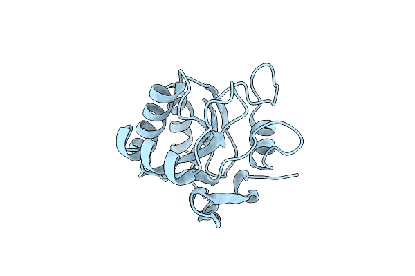
Search Count: 1,329
 |
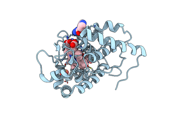
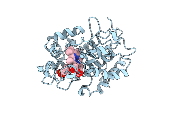
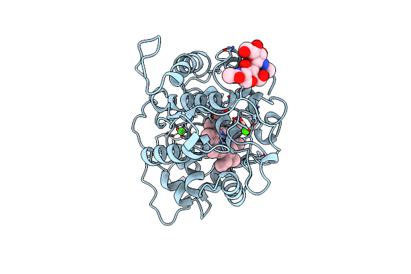
Crystal Structure Of Recombinant Ascorbate Peroxidase
Organism: Pisum sativum
Method: X-RAY DIFFRACTION Resolution:2.20 Å Release Date: 1996-03-08 Classification: PEROXIDASE Ligands: K, HEM |
Organism: Pisum sativum
Method: X-RAY DIFFRACTION
Release Date: 1996-03-08
Ligands: K, HEM
 |
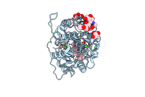
Crystal Structure Of Fungal Versatile Peroxidase From Pleurotus Eryngii Septuple Mutant E37K, H39R, V160A, T184M, Q202L, D213A & G330R
Organism: Pleurotus eryngii
Method: X-RAY DIFFRACTION Resolution:1.79 Å Release Date: 2016-07-13 Classification: OXIDOREDUCTASE Ligands: CA, HEM, SO4 |
Organism: Pleurotus eryngii
Method: X-RAY DIFFRACTION
Release Date: 2016-07-13
Ligands: CA, HEM, SO4
 |
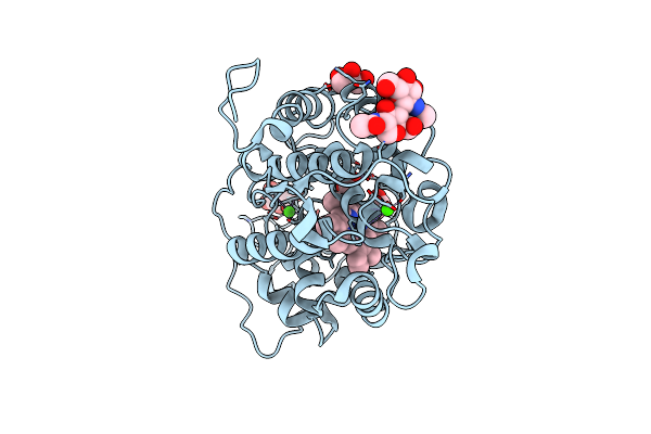
Crystal Structure Analysis Of Fungal Versatile Peroxidase From Pleurotus Eryngii. Mutant Vpi. Mutated Residues D69S, T70D, S86E, D146T, Q202L, H232E, Q239R And S301K.
Organism: Pleurotus eryngii
Method: X-RAY DIFFRACTION Resolution:2.19 Å Release Date: 2015-11-04 Classification: OXIDOREDUCTASE Ligands: CA, HEM, MG |
Organism: Pleurotus eryngii
Method: X-RAY DIFFRACTION
Release Date: 2015-11-04
Ligands: CA, HEM, MG
 |
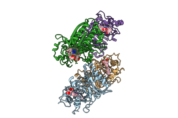
Crystal Structure Analysis Of Fungal Versatile Peroxidase From Pleurotus Eryngii. Mutant Vpi-Br. Mutated Residues T2K, D69S, T70D, S86E, A131K, D146T, Q202L, Q219K, H232E, Q239R, L288R, S301K, A308R, A309K And A314R.
Organism: Pleurotus eryngii
Method: X-RAY DIFFRACTION Resolution:1.10 Å Release Date: 2015-11-04 Classification: OXIDOREDUCTASE Ligands: CA, HEM |
Organism: Pleurotus eryngii
Method: X-RAY DIFFRACTION
Release Date: 2015-11-04
Ligands: CA, HEM
 |
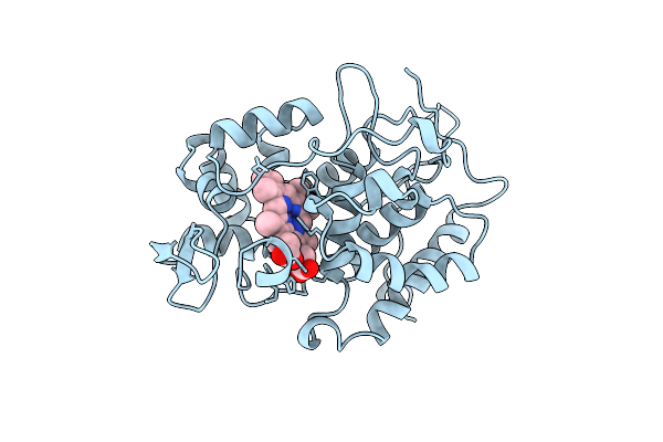
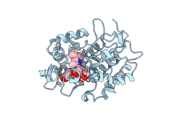
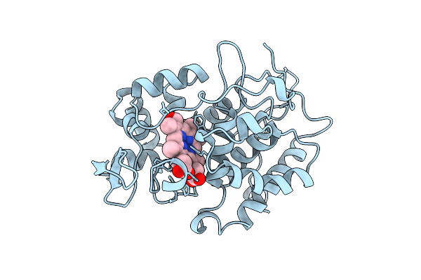
Structure Of Isoniazid (Inh) Bound To Cytosolic Soybean Ascorbate Peroxidase Mutant W41A
Organism: Glycine max
Method: X-RAY DIFFRACTION Resolution:1.20 Å Release Date: 2007-12-04 Classification: OXIDOREDUCTASE Ligands: HEM, SO4, ISZ |
Organism: Glycine max
Method: X-RAY DIFFRACTION
Release Date: 2007-12-04
Ligands: HEM, SO4, ISZ
 |
Crystal Structure Of Trypanosoma Cruzi Peroxidase
Organism: Trypanosoma cruzi
Method: X-RAY DIFFRACTION Resolution:1.76 Å Release Date: 2022-06-22 Classification: OXIDOREDUCTASE Ligands: HEM, SO4, GOL, NA, OXY, ACT |
Organism: Trypanosoma cruzi
Method: X-RAY DIFFRACTION
Release Date: 2022-06-22
Ligands: HEM, SO4, GOL, NA, OXY, ACT
 |
Crystal Structure Of A Type Ii Tryparedoxin-Dependant Peroxidase From Trypanosoma Brucei
Organism: Trypanosoma brucei
Method: X-RAY DIFFRACTION Resolution:2.10 Å Release Date: 2008-06-17 Classification: OXIDOREDUCTASE |
Organism: Trypanosoma brucei
Method: X-RAY DIFFRACTION
Release Date: 2008-06-17
 |
Interaction Between Proximal And Distals Regions Of Cytochrome C Peroxidase
Organism: Saccharomyces cerevisiae
Method: X-RAY DIFFRACTION Resolution:2.00 Å Release Date: 1998-10-21 Classification: PEROXIDASE Ligands: CL, HEM, MES |
Organism: Saccharomyces cerevisiae
Method: X-RAY DIFFRACTION
Release Date: 1998-10-21
Ligands: CL, HEM, MES
 |
Interaction Between Proximal And Distals Regions Of Cytochrome C Peroxidase
Organism: Saccharomyces cerevisiae
Method: X-RAY DIFFRACTION Resolution:2.40 Å Release Date: 1998-10-21 Classification: PEROXIDASE Ligands: HEM |
Organism: Saccharomyces cerevisiae
Method: X-RAY DIFFRACTION
Release Date: 1998-10-21
Ligands: HEM
 |
Interaction Between Proximal And Distals Regions Of Cytochrome C Peroxidase
Organism: Saccharomyces cerevisiae
Method: X-RAY DIFFRACTION Resolution:2.16 Å Release Date: 1998-10-21 Classification: PEROXIDASE Ligands: HEM, MES |
Organism: Saccharomyces cerevisiae
Method: X-RAY DIFFRACTION
Release Date: 1998-10-21
Ligands: HEM, MES
 |
Interaction Between Proximal And Distals Regions Of Cytochrome C Peroxidase
Organism: Saccharomyces cerevisiae
Method: X-RAY DIFFRACTION Resolution:2.20 Å Release Date: 1998-10-21 Classification: PEROXIDASE Ligands: HEM, MES |
Organism: Saccharomyces cerevisiae
Method: X-RAY DIFFRACTION
Release Date: 1998-10-21
Ligands: HEM, MES
 |
Glutathione Peroxidase-Type Tryparedoxin Peroxidase, Oxidized Form
Organism: Trypanosoma brucei
Method: X-RAY DIFFRACTION Resolution:1.41 Å Release Date: 2008-08-05 Classification: OXIDOREDUCTASE |
Organism: Trypanosoma brucei
Method: X-RAY DIFFRACTION
Release Date: 2008-08-05
 |
Effect Of Unnatural Heme Substitution On Kinetics Of Electron Transfer In Cytochrome C Peroxidase
Organism: Saccharomyces cerevisiae
Method: X-RAY DIFFRACTION Resolution:2.20 Å Release Date: 1998-10-21 Classification: PEROXIDASE Ligands: HEM |
Organism: Saccharomyces cerevisiae
Method: X-RAY DIFFRACTION
Release Date: 1998-10-21
Ligands: HEM
 |
Effect Of Unnatural Heme Substitution On Kinetics Of Electron Transfer In Cytochrome C Peroxidase
Organism: Saccharomyces cerevisiae
Method: X-RAY DIFFRACTION Resolution:2.20 Å Release Date: 1998-10-21 Classification: PEROXIDASE Ligands: CCH |
Organism: Saccharomyces cerevisiae
Method: X-RAY DIFFRACTION
Release Date: 1998-10-21
Ligands: CCH
 |
Crystal Structure Of Versatile Peroxidase
Organism: Pleurotus eryngii
Method: X-RAY DIFFRACTION Resolution:1.33 Å Release Date: 2005-11-02 Classification: OXIDOREDUCTASE Ligands: HEM, ZN, MN, CAC, CA |
Organism: Pleurotus eryngii
Method: X-RAY DIFFRACTION
Release Date: 2005-11-02
Ligands: HEM, ZN, MN, CAC, CA
 |
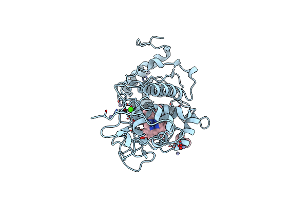
Manganese Peroxidase Substrate Binding Site Mutant D179N
Organism: Phanerochaete chrysosporium
Method: X-RAY DIFFRACTION Resolution:2.00 Å Release Date: 1997-09-04 Classification: PEROXIDASE Ligands: CA, HEM |
Organism: Phanerochaete chrysosporium
Method: X-RAY DIFFRACTION
Release Date: 1997-09-04
Ligands: CA, HEM
 |
Manganese Peroxidase Substrate Binding Site Mutant E35Q, D179N
Organism: Phanerochaete chrysosporium
Method: X-RAY DIFFRACTION Resolution:2.00 Å Release Date: 1997-09-04 Classification: PEROXIDASE Ligands: CA, HEM |
Organism: Phanerochaete chrysosporium
Method: X-RAY DIFFRACTION
Release Date: 1997-09-04
Ligands: CA, HEM
 |
1.05 A Structure Of Manganese-Free Manganese Peroxidase
Organism: Phanerochaete chrysosporium
Method: X-RAY DIFFRACTION Resolution:1.05 Å Release Date: 2010-04-14 Classification: OXIDOREDUCTASE Ligands: MAN, CA, GOL, HEM |
Organism: Phanerochaete chrysosporium
Method: X-RAY DIFFRACTION
Release Date: 2010-04-14
Ligands: MAN, CA, GOL, HEM
 |
0.93 A Structure Of Manganese-Bound Manganese Peroxidase
Organism: Phanerochaete chrysosporium
Method: X-RAY DIFFRACTION Resolution:0.93 Å Release Date: 2010-04-14 Classification: OXIDOREDUCTASE Ligands: MAN, CA, MN, GOL, HEM |
Organism: Phanerochaete chrysosporium
Method: X-RAY DIFFRACTION
Release Date: 2010-04-14
Ligands: MAN, CA, MN, GOL, HEM
 |
Study On The Mechanism Of Dye Decolorizing Peroxidase Degradation Of Pvc
Organism: Klebsiella variicola
Method: X-RAY DIFFRACTION Resolution:2.57 Å Release Date: 2026-01-21 Classification: OXIDOREDUCTASE Ligands: HEM |
Organism: Klebsiella variicola
Method: X-RAY DIFFRACTION
Release Date: 2026-01-21
Ligands: HEM