

Search Count: 29
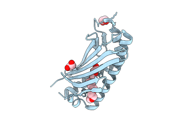 |
Crystal Structure Of Computationally Designed Protein Vtp-4
Organism: Synthetic construct
Method: X-RAY DIFFRACTION Resolution:1.60 Å Release Date: 2025-08-13 Classification: DE NOVO PROTEIN Ligands: EDO, P15 |
Organism: Synthetic construct
Method: X-RAY DIFFRACTION
Release Date: 2025-08-13
Ligands: EDO, P15
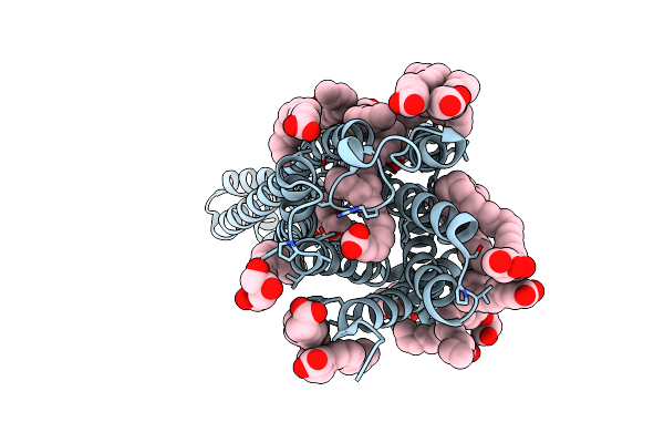 |
Crystal Structure Of Orphan G Protein-Coupled Receptor 6, Pseudoapo Form
Organism: Homo sapiens, Escherichia coli
Method: X-RAY DIFFRACTION Resolution:2.10 Å Release Date: 2024-12-04 Classification: MEMBRANE PROTEIN Ligands: OLC, P15, OLA, CLR |
Organism: Homo sapiens, Escherichia coli
Method: X-RAY DIFFRACTION
Release Date: 2024-12-04
Ligands: OLC, P15, OLA, CLR
 |
Structure Of Human Cysteine Desulfurase Nfs1 With L-Propargylglycine Bound To Active Site Plp In Complex With Isd11, Acp1 And Iscu2
Organism: Homo sapiens, Escherichia coli
Method: X-RAY DIFFRACTION Resolution:2.00 Å Release Date: 2024-09-11 Classification: TRANSFERASE Ligands: EDO, PLP, LPH, GOL, PEG, PG4, P15, DTT, PGE, EDT, 8Q1, MES, 1PE |
Organism: Homo sapiens, Escherichia coli
Method: X-RAY DIFFRACTION
Release Date: 2024-09-11
Ligands: EDO, PLP, LPH, GOL, PEG, PG4, P15, DTT, PGE, EDT, 8Q1, MES, 1PE
 |
Crystal Structure Of Anti-Mpeg H15-2B Fab W104Y Mutant
Organism: Homo sapiens
Method: X-RAY DIFFRACTION Resolution:2.42 Å Release Date: 2023-08-02 Classification: IMMUNE SYSTEM Ligands: P15 |
Organism: Homo sapiens
Method: X-RAY DIFFRACTION
Release Date: 2023-08-02
Ligands: P15
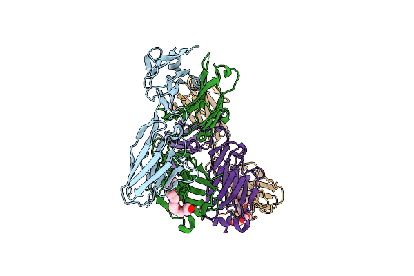 |
Crystal Structure Of Anti-Mpeg H15-2B Fab
Organism: Homo sapiens
Method: X-RAY DIFFRACTION Resolution:2.24 Å Release Date: 2023-03-01 Classification: IMMUNE SYSTEM Ligands: P15 |
Organism: Homo sapiens
Method: X-RAY DIFFRACTION
Release Date: 2023-03-01
Ligands: P15
 |
Crystal Structure Of Anti-Mpeg H15-2B Fab
Organism: Homo sapiens
Method: X-RAY DIFFRACTION Resolution:2.08 Å Release Date: 2022-08-10 Classification: IMMUNE SYSTEM Ligands: P15 |
Organism: Homo sapiens
Method: X-RAY DIFFRACTION
Release Date: 2022-08-10
Ligands: P15
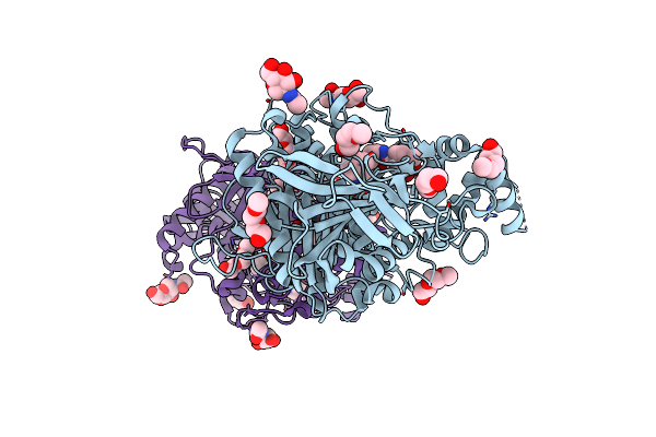 |
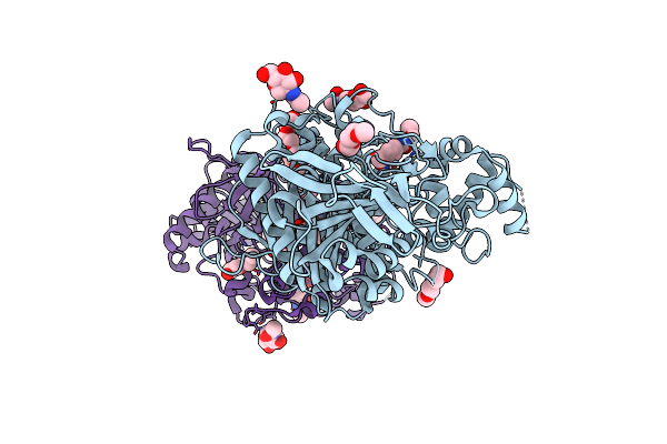
Mus Musculus Acetylcholinesterase In Complex With 2-((Hydroxyimino)Methyl)-1-(5-(4-Methyl-3-Nitrobenzamido)Pentyl)Pyridinium
Organism: Mus musculus
Method: X-RAY DIFFRACTION Resolution:2.50 Å Release Date: 2022-04-27 Classification: HYDROLASE Ligands: NAG, I1X, PG0, TOE, P15, CL |
Organism: Mus musculus
Method: X-RAY DIFFRACTION
Release Date: 2022-04-27
Ligands: NAG, I1X, PG0, TOE, P15, CL
 |
Structure Of Sarin Phosphonylated Acetylcholinesterase In Complex With 2-((Hydroxyimino)Methyl)-1-(5-(4-Methyl-3-Nitrobenzamido)Pentyl)Pyridinium
Organism: Mus musculus
Method: X-RAY DIFFRACTION Resolution:2.80 Å Release Date: 2022-04-27 Classification: HYDROLASE Ligands: I4H, NAG, PG0, P15, TOE |
Organism: Mus musculus
Method: X-RAY DIFFRACTION
Release Date: 2022-04-27
Ligands: I4H, NAG, PG0, P15, TOE
 |
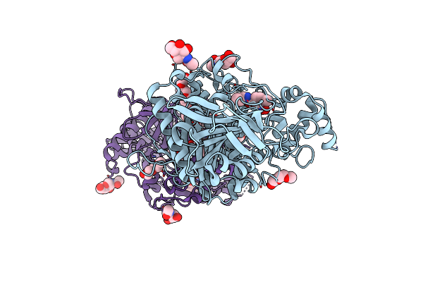
Vx-Inhibited Acetylcholinesterase In Complex With 2-((Hydroxyimino)Methyl)-1-(5-(4-Methyl-3-Nitrobenzamido)Pentyl)Pyridinium
Organism: Mus musculus
Method: X-RAY DIFFRACTION Resolution:2.40 Å Release Date: 2022-04-27 Classification: HYDROLASE Ligands: I1X, NAG, PG0, TOE, P15 |
Organism: Mus musculus
Method: X-RAY DIFFRACTION
Release Date: 2022-04-27
Ligands: I1X, NAG, PG0, TOE, P15
 |
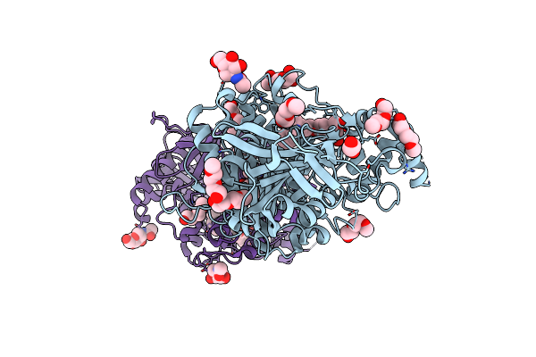
Rvx-Inhibited Acetylcholinesterase In Complex With 2-((Hydroxyimino)Methyl)-1-(5-(4-Methyl-3-Nitrobenzamido)Pentyl)Pyridinium
Organism: Mus musculus
Method: X-RAY DIFFRACTION Resolution:3.00 Å Release Date: 2022-04-27 Classification: HYDROLASE Ligands: I1X, NAG, PG0, TOE, P15 |
Organism: Mus musculus
Method: X-RAY DIFFRACTION
Release Date: 2022-04-27
Ligands: I1X, NAG, PG0, TOE, P15
 |
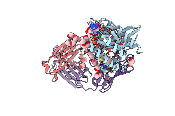
Mus Musculus Acetylcholinesterase In Complex With 7-[(4-{[Benzyl(Methyl)Amino]Methyl}Benzyl)Oxy]-4-(Hydroxymethyl)-2H-Chromen-2-One
Organism: Mus musculus
Method: X-RAY DIFFRACTION Resolution:2.60 Å Release Date: 2022-04-06 Classification: HYDROLASE Ligands: NAG, 5IK, EDO, PG0, TOE, P15 |
Organism: Mus musculus
Method: X-RAY DIFFRACTION
Release Date: 2022-04-06
Ligands: NAG, 5IK, EDO, PG0, TOE, P15
 |
Kras G12C In Complex With G-2897
Organism: Homo sapiens
Method: X-RAY DIFFRACTION Resolution:1.96 Å Release Date: 2022-03-16 Classification: HYDROLASE/Immune System Ligands: Z07, GDP, MG, ACT, CAD, P15, EDO, DMS |
Organism: Homo sapiens
Method: X-RAY DIFFRACTION
Release Date: 2022-03-16
Ligands: Z07, GDP, MG, ACT, CAD, P15, EDO, DMS
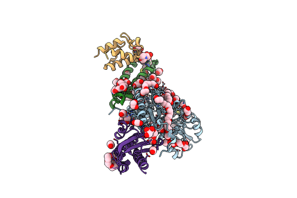 |
Structure Of The (Niau)2 Complex With N-Terminal Mutation Of Iscu2 Y35D At 2.5 A Resolution
Organism: Homo sapiens, Escherichia coli
Method: X-RAY DIFFRACTION Resolution:2.50 Å Release Date: 2021-08-25 Classification: TRANSFERASE Ligands: PLP, EDO, GOL, PG4, PEG, P15, DTT, PGE, EDT, 8Q1, MES, 1PE |
Organism: Homo sapiens, Escherichia coli
Method: X-RAY DIFFRACTION
Release Date: 2021-08-25
Ligands: PLP, EDO, GOL, PG4, PEG, P15, DTT, PGE, EDT, 8Q1, MES, 1PE
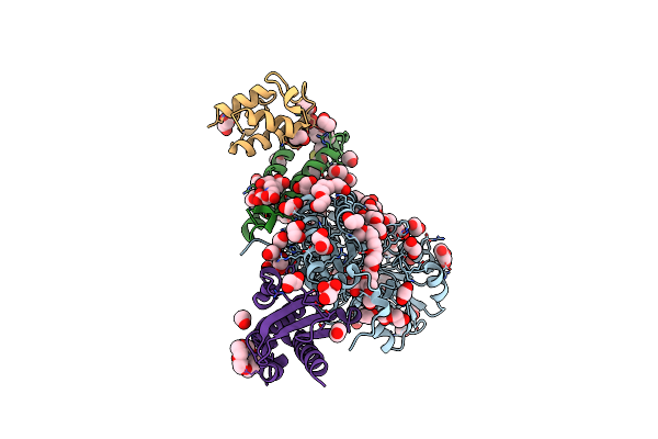 |
N-Terminal Mutation Of Iscu2 (L35H36) Traps Nfs1 Cys Loop In The Active Site Of Iscu2 Without Metal Present. Structure Of Human Mitochondrial Complex Nfs1-Iscu2(L35H36)-Isd11 With E.Coli Acp1 At 1.9 A Resolution (Niau)2.
Organism: Homo sapiens, Escherichia coli
Method: X-RAY DIFFRACTION Resolution:1.90 Å Release Date: 2020-05-13 Classification: TRANSFERASE Ligands: PLP, PEG, GOL, EDO, PG4, P15, PGE, EDT, 8Q1, 1PE |
Organism: Homo sapiens, Escherichia coli
Method: X-RAY DIFFRACTION
Release Date: 2020-05-13
Ligands: PLP, PEG, GOL, EDO, PG4, P15, PGE, EDT, 8Q1, 1PE
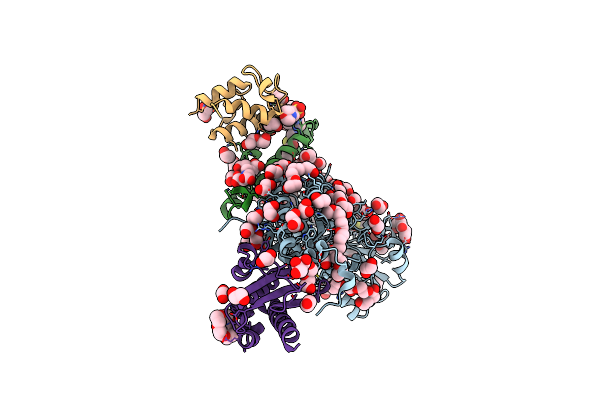 |
Structure Of Human Mitochondrial Complex Nfs1-Iscu2 (Wt)-Isd11 With E.Coli Acp1 At 1.8 A Resolution (Niau)2
Organism: Homo sapiens, Escherichia coli
Method: X-RAY DIFFRACTION Resolution:1.80 Å Release Date: 2020-03-18 Classification: TRANSFERASE Ligands: PLP, GOL, EDO, PEG, PG4, P15, DTT, PGE, EDT, 8Q1, MES, 1PE |
Organism: Homo sapiens, Escherichia coli
Method: X-RAY DIFFRACTION
Release Date: 2020-03-18
Ligands: PLP, GOL, EDO, PEG, PG4, P15, DTT, PGE, EDT, 8Q1, MES, 1PE
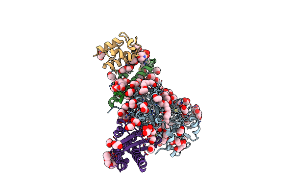 |
Structure Of The Human Mitochondrial Desulfurase Complex Nfs1-Iscu2(M140I)-Isd11 With E.Coli Acp1 At 1.57 A Resolution Showing Flexibility Of N Terminal End Of Iscu2
Organism: Homo sapiens, Escherichia coli
Method: X-RAY DIFFRACTION Resolution:1.57 Å Release Date: 2019-11-20 Classification: TRANSFERASE Ligands: PLP, GOL, EDO, PEG, PG4, P15, DTT, PGE, EDT, 8Q1, MES, 1PE |
Organism: Homo sapiens, Escherichia coli
Method: X-RAY DIFFRACTION
Release Date: 2019-11-20
Ligands: PLP, GOL, EDO, PEG, PG4, P15, DTT, PGE, EDT, 8Q1, MES, 1PE
 |
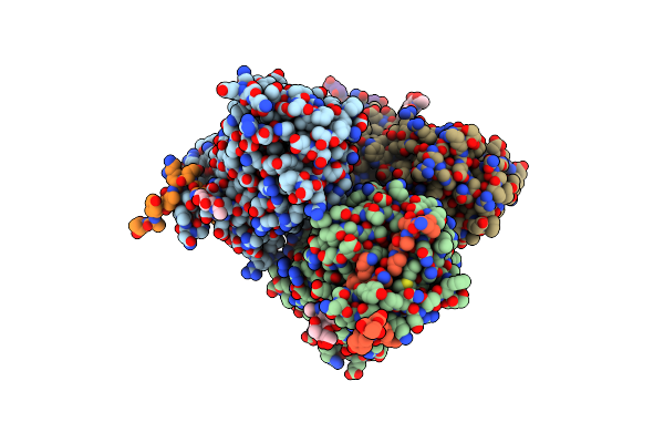
Molecular Features Of The Unc-45 Chaperone Critical For Binding And Folding Muscle Myosin
Organism: Caenorhabditis elegans
Method: X-RAY DIFFRACTION Resolution:1.88 Å Release Date: 2019-10-30 Classification: MOTOR PROTEIN Ligands: ADP, GOL, BU1, PGO, HEZ, P15 |
Organism: Caenorhabditis elegans
Method: X-RAY DIFFRACTION
Release Date: 2019-10-30
Ligands: ADP, GOL, BU1, PGO, HEZ, P15
 |
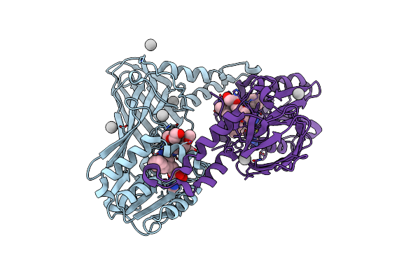
Structure Of The Clinically Used Myxomaviral Serine Protease Inhibitor 1 (Serp-1)
Organism: Myxoma virus (strain lausanne)
Method: X-RAY DIFFRACTION Resolution:2.50 Å Release Date: 2018-11-07 Classification: IMMUNOSUPPRESSANT Ligands: NAG, P15 |
Organism: Myxoma virus (strain lausanne)
Method: X-RAY DIFFRACTION
Release Date: 2018-11-07
Ligands: NAG, P15
 |
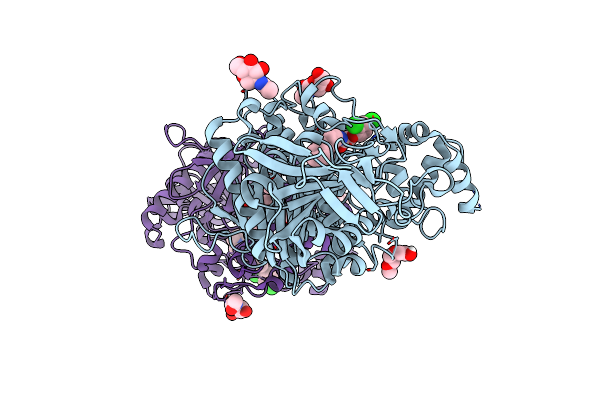
Crystal Structure Of Human Carm1 With (R)-Ski-72
Organism: Homo sapiens
Method: X-RAY DIFFRACTION Resolution:1.54 Å Release Date: 2018-07-25 Classification: TRANSFERASE/TRANSFERASE INHIBITOR Ligands: HDG, P15, UNX |
Organism: Homo sapiens
Method: X-RAY DIFFRACTION
Release Date: 2018-07-25
Ligands: HDG, P15, UNX
 |
Acetylcholinesterase In Complex With C7653
Organism: Mus musculus
Method: X-RAY DIFFRACTION Resolution:2.30 Å Release Date: 2016-03-02 Classification: HYDROLASE Ligands: NAG, GC8, PG0, P15 |
Organism: Mus musculus
Method: X-RAY DIFFRACTION
Release Date: 2016-03-02
Ligands: NAG, GC8, PG0, P15