

Search Count: 141
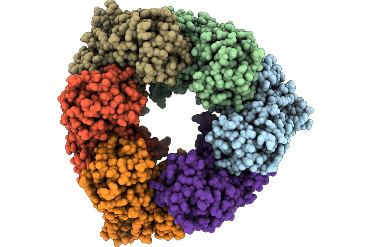 |
Single Particle Reconstruction Of Pilu From Vibrio Cholerae El Tor E7946, Form 1
Organism: Vibrio cholerae
Method: ELECTRON MICROSCOPY Release Date: 2026-05-20 Classification: MOTOR PROTEIN |
Organism: Vibrio cholerae
Method: ELECTRON MICROSCOPY
Release Date: 2026-05-20
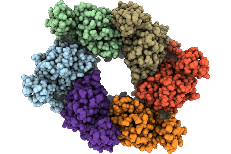 |
Single Particle Reconstruction Of Pilu From Vibrio Cholerae El Tor E7946, Form 2
Organism: Vibrio cholerae
Method: ELECTRON MICROSCOPY Release Date: 2026-05-20 Classification: MOTOR PROTEIN |
Organism: Vibrio cholerae
Method: ELECTRON MICROSCOPY
Release Date: 2026-05-20
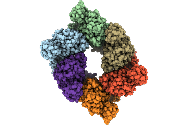 |
Single Particle Reconstruction Of Pilu From Vibrio Cholerae El Tor E7946, Form 3
Organism: Vibrio cholerae
Method: ELECTRON MICROSCOPY Release Date: 2026-05-20 Classification: MOTOR PROTEIN |
Organism: Vibrio cholerae
Method: ELECTRON MICROSCOPY
Release Date: 2026-05-20
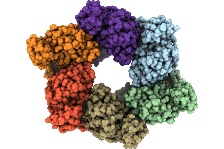 |
Single Particle Reconstruction Of Pilu From Vibrio Cholerae El Tor E7946, Form 4
Organism: Vibrio cholerae
Method: ELECTRON MICROSCOPY Release Date: 2026-05-20 Classification: MOTOR PROTEIN |
Organism: Vibrio cholerae
Method: ELECTRON MICROSCOPY
Release Date: 2026-05-20
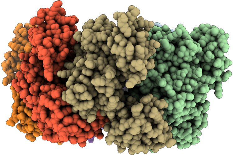 |
Crystal Structure Of Vibrio Cholerae Pilu, A Pilt-Dependent Retraction Atpase - Crystal Form 1.
Organism: Vibrio cholerae o1 biovar el tor str. n16961
Method: X-RAY DIFFRACTION Resolution:2.95 Å Release Date: 2026-05-20 Classification: MOTOR PROTEIN |
Organism: Vibrio cholerae o1 biovar el tor str. n16961
Method: X-RAY DIFFRACTION
Release Date: 2026-05-20
 |
Crystal Structure Of Vibrio Cholerae Pilu, A Pilt-Dependent Retraction Atpase - Crystal Form 2
Organism: Vibrio cholerae o1 biovar el tor str. n16961
Method: X-RAY DIFFRACTION Resolution:2.70 Å Release Date: 2026-05-20 Classification: MOTOR PROTEIN Ligands: SO4 |
Organism: Vibrio cholerae o1 biovar el tor str. n16961
Method: X-RAY DIFFRACTION
Release Date: 2026-05-20
Ligands: SO4
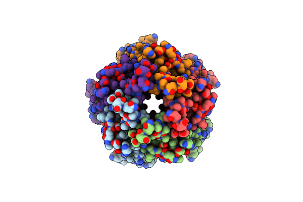 |
Tolq Inner Membrane Protein From Acinetobacter Baumannii
Organism: Acinetobacter baumannii
Method: ELECTRON MICROSCOPY Resolution:3.02 Å Release Date: 2025-01-22 Classification: MEMBRANE PROTEIN |
Organism: Acinetobacter baumannii
Method: ELECTRON MICROSCOPY
Release Date: 2025-01-22
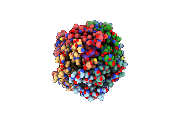 |
Tolq-Tolr Inner Membrane Protein Complex From Acinetobacter Baumannii
Organism: Acinetobacter baumannii
Method: ELECTRON MICROSCOPY Resolution:3.34 Å Release Date: 2025-01-15 Classification: MEMBRANE PROTEIN |
Organism: Acinetobacter baumannii
Method: ELECTRON MICROSCOPY
Release Date: 2025-01-15
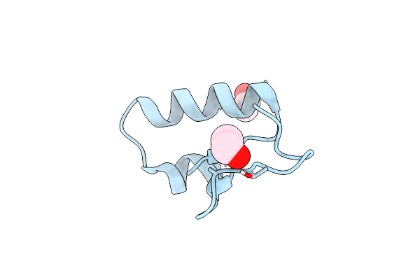 |
Solvent Organization In Ultrahigh-Resolution Protein Crystal Structure At Room Temperature
Organism: Crambe hispanica subsp. abyssinica
Method: X-RAY DIFFRACTION Resolution:0.70 Å Release Date: 2024-09-04 Classification: PLANT PROTEIN Ligands: EOH |
Organism: Crambe hispanica subsp. abyssinica
Method: X-RAY DIFFRACTION
Release Date: 2024-09-04
Ligands: EOH
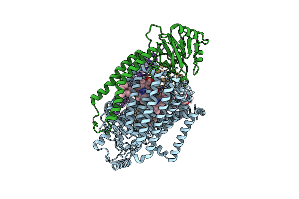 |
E. Coli Cytochrome Bo3 Ubiquinol Oxidase Monomer
Organism: Escherichia coli
Method: ELECTRON MICROSCOPY Release Date: 2022-11-30 Classification: PROTON TRANSPORT Ligands: HEM, HEO, CU, 3PE |
Organism: Escherichia coli
Method: ELECTRON MICROSCOPY
Release Date: 2022-11-30
Ligands: HEM, HEO, CU, 3PE
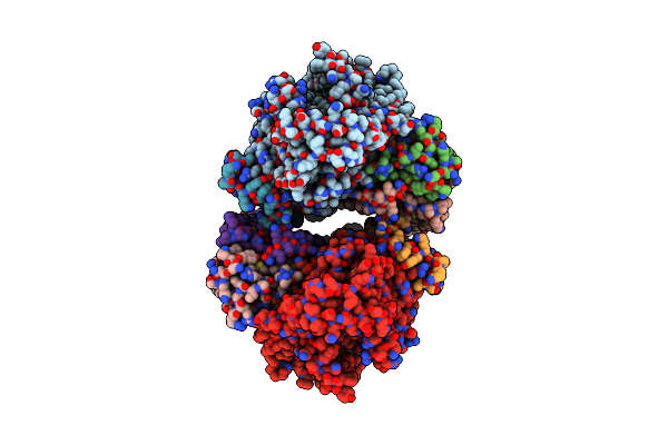 |
E. Coli Cytochrome Bo3 Ubiquinol Oxidase Dimer
Organism: Escherichia coli
Method: ELECTRON MICROSCOPY Resolution:3.46 Å Release Date: 2022-11-30 Classification: PROTON TRANSPORT Ligands: HEM, HEO, CU, 3PE |
Organism: Escherichia coli
Method: ELECTRON MICROSCOPY
Release Date: 2022-11-30
Ligands: HEM, HEO, CU, 3PE
 |
Cryoem Structure Of The Candida Albicans Aro1 Dimer
Organism: Candida albicans
Method: ELECTRON MICROSCOPY Release Date: 2022-05-04 Classification: BIOSYNTHETIC PROTEIN |
Organism: Candida albicans
Method: ELECTRON MICROSCOPY
Release Date: 2022-05-04
 |
Structure Of Dhqs/Epsps Dimer From Candida Albicans Aro1
Organism: Candida albicans
Method: ELECTRON MICROSCOPY Release Date: 2022-05-04 Classification: BIOSYNTHETIC PROTEIN |
Organism: Candida albicans
Method: ELECTRON MICROSCOPY
Release Date: 2022-05-04
 |
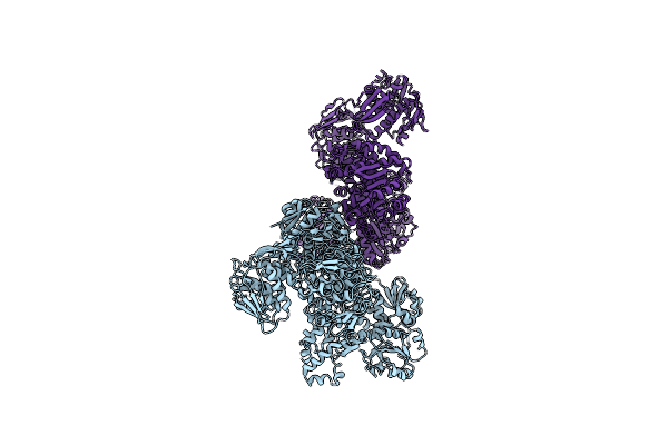
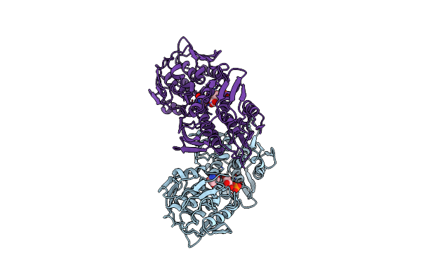
Structure Of The Sk/Dhqase/Dhsd Dimer From Candida Albicans Aro1
Organism: Candida albicans
Method: ELECTRON MICROSCOPY Release Date: 2022-05-04 Classification: BIOSYNTHETIC PROTEIN |
Organism: Candida albicans
Method: ELECTRON MICROSCOPY
Release Date: 2022-05-04
 |
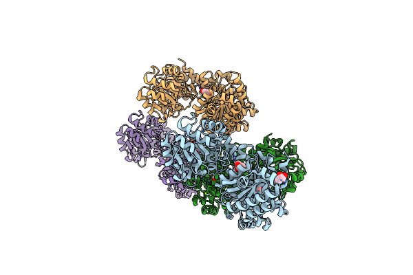
Crystal Structure Of The 5-Enolpyruvate-Shikimate-3-Phosphate Synthase (Epsps) Domain Of Aro1 From Candida Albicans In Complex With Shikimate-3-Phosphate
Organism: Candida albicans ca6
Method: X-RAY DIFFRACTION Resolution:1.85 Å Release Date: 2022-03-16 Classification: TRANSFERASE Ligands: TRS, S3P |
Organism: Candida albicans ca6
Method: X-RAY DIFFRACTION
Release Date: 2022-03-16
Ligands: TRS, S3P
 |
Crystal Structure Of The Shikimate Kinase + 3-Dehydroquinate Dehydratase + 3-Dehydroshikimate Dehydrogenase Domains Of Aro1 From Candida Albicans
Organism: Candida albicans ca6
Method: X-RAY DIFFRACTION Resolution:2.30 Å Release Date: 2022-03-16 Classification: TRANSFERASE,OXIDOREDUCTASE Ligands: MG, CL, GOL |
Organism: Candida albicans ca6
Method: X-RAY DIFFRACTION
Release Date: 2022-03-16
Ligands: MG, CL, GOL
 |
Crystal Structure Of The Peptidyl-Prolyl Cis-Trans Isomerase (Ppib) From Streptococcus Pyogenes.
Organism: Streptococcus pyogenes mgas5005
Method: X-RAY DIFFRACTION Resolution:1.28 Å Release Date: 2021-12-01 Classification: ISOMERASE |
Organism: Streptococcus pyogenes mgas5005
Method: X-RAY DIFFRACTION
Release Date: 2021-12-01
 |
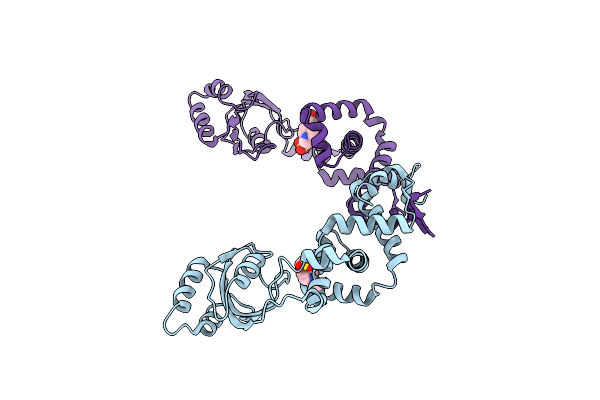
Crystal Structure Of Peptidyl-Prolyl Cis-Trans Isomerasefrom (Ppib) Streptococcus Pneumoniae R6
Organism: Streptococcus pneumoniae (strain atcc baa-255 / r6)
Method: X-RAY DIFFRACTION Resolution:1.88 Å Release Date: 2021-12-01 Classification: ISOMERASE Ligands: CL, EDO, MES |
Organism: Streptococcus pneumoniae (strain atcc baa-255 / r6)
Method: X-RAY DIFFRACTION
Release Date: 2021-12-01
Ligands: CL, EDO, MES
 |
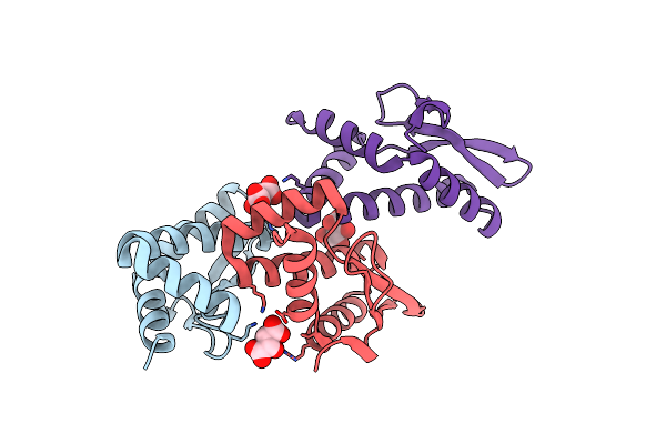
Crystal Structure Of Peptidylprolyl Isomerase Prsa From Streptococcus Mutans.
Organism: Streptococcus mutans
Method: X-RAY DIFFRACTION Resolution:3.15 Å Release Date: 2021-12-01 Classification: ISOMERASE Ligands: EPE, CL |
Organism: Streptococcus mutans
Method: X-RAY DIFFRACTION
Release Date: 2021-12-01
Ligands: EPE, CL
 |
Structure Of Human Respiratory Syncytial Virus Nonstructural Protein 2 (Ns2)
Organism: Human respiratory syncytial virus a
Method: X-RAY DIFFRACTION Resolution:2.82 Å Release Date: 2021-03-17 Classification: VIRAL PROTEIN Ligands: TAR, CL |
Organism: Human respiratory syncytial virus a
Method: X-RAY DIFFRACTION
Release Date: 2021-03-17
Ligands: TAR, CL