

Search Count: 22
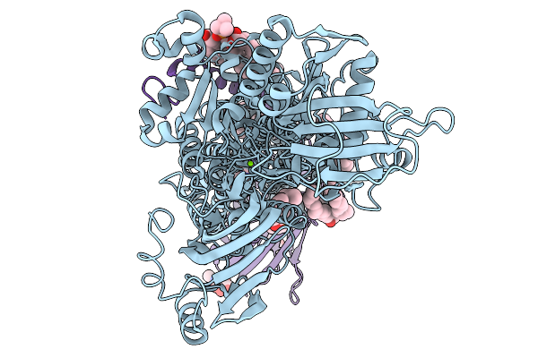 |
Cryo-Em Structure Of Gastric Proton Pump Bound To Yk01
Organism: Sus scrofa
Method: ELECTRON MICROSCOPY Release Date: 2026-01-14 Classification: MEMBRANE PROTEIN Ligands: PCW, MG, A1MA7, NAG, CLR |
Organism: Sus scrofa
Method: ELECTRON MICROSCOPY
Release Date: 2026-01-14
Ligands: PCW, MG, A1MA7, NAG, CLR
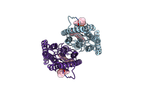 |
Channel Rhodospin From Klebsormidium Nitens (Knchr)
Organism: Klebsormidium nitens
Method: ELECTRON MICROSCOPY Release Date: 2025-07-02 Classification: ELECTRON TRANSPORT Ligands: PC1, RET |
Organism: Klebsormidium nitens
Method: ELECTRON MICROSCOPY
Release Date: 2025-07-02
Ligands: PC1, RET
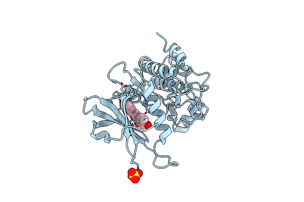 |
Crystal Structure Of Dapk1 In Complex With Isoliquiritigenin
Organism: Homo sapiens
Method: X-RAY DIFFRACTION Resolution:1.61 Å Release Date: 2024-10-09 Classification: TRANSFERASE Ligands: HCC, SO4 |
Organism: Homo sapiens
Method: X-RAY DIFFRACTION
Release Date: 2024-10-09
Ligands: HCC, SO4
 |
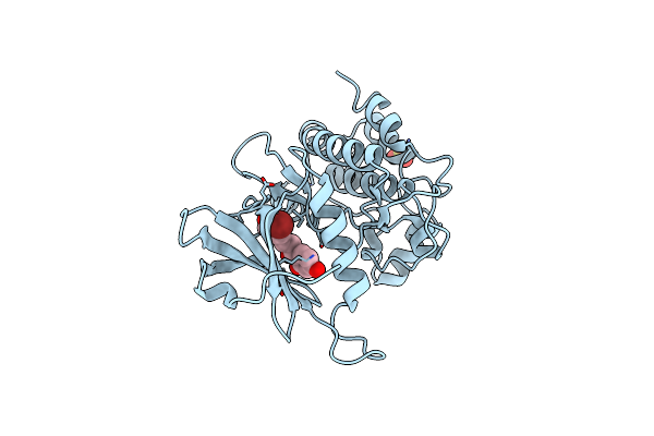
Crystal Structure Of Dapk1 In Complex With Compound 9
Organism: Homo sapiens
Method: X-RAY DIFFRACTION Resolution:1.52 Å Release Date: 2024-10-09 Classification: TRANSFERASE Ligands: A1L2V, SO4 |
Organism: Homo sapiens
Method: X-RAY DIFFRACTION
Release Date: 2024-10-09
Ligands: A1L2V, SO4
 |
Crystal Structure Of Dapk1 In Complex With Compound 10
Organism: Homo sapiens
Method: X-RAY DIFFRACTION Resolution:1.72 Å Release Date: 2024-10-09 Classification: TRANSFERASE Ligands: A1L2W, SO4 |
Organism: Homo sapiens
Method: X-RAY DIFFRACTION
Release Date: 2024-10-09
Ligands: A1L2W, SO4
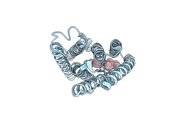 |
Cryo-Em Structure Of The Channelrhodopsin Gtccr2 Focused On The Monomer
Organism: Guillardia theta ccmp2712
Method: ELECTRON MICROSCOPY Resolution:2.86 Å Release Date: 2024-09-04 Classification: MEMBRANE PROTEIN Ligands: RET |
Organism: Guillardia theta ccmp2712
Method: ELECTRON MICROSCOPY
Release Date: 2024-09-04
Ligands: RET
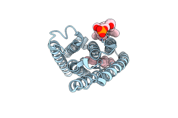 |
Cryo-Em Structure Of The Channelrhodopsin Gtccr2
Organism: Guillardia theta ccmp2712
Method: ELECTRON MICROSCOPY Resolution:2.73 Å Release Date: 2024-09-04 Classification: MEMBRANE PROTEIN Ligands: RET, PC1 |
Organism: Guillardia theta ccmp2712
Method: ELECTRON MICROSCOPY
Release Date: 2024-09-04
Ligands: RET, PC1
 |
Cryo-Em Structure Of The Channelrhodopsin Gtccr4
Organism: Guillardia theta
Method: ELECTRON MICROSCOPY Resolution:2.71 Å Release Date: 2024-09-04 Classification: MEMBRANE PROTEIN Ligands: RET, PC1 |
Organism: Guillardia theta
Method: ELECTRON MICROSCOPY
Release Date: 2024-09-04
Ligands: RET, PC1
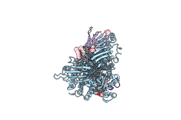 |
Cryo-Em Structure Of The Gastric Proton Pump With Bound Dq-02
Organism: Sus scrofa
Method: ELECTRON MICROSCOPY Release Date: 2023-08-30 Classification: MEMBRANE PROTEIN Ligands: PCW, MG, PWI, NAG, CLR |
Organism: Sus scrofa
Method: ELECTRON MICROSCOPY
Release Date: 2023-08-30
Ligands: PCW, MG, PWI, NAG, CLR
 |
Cryo-Em Structure Of The Gastric Proton Pump With Bound Dq-06
Organism: Sus scrofa
Method: ELECTRON MICROSCOPY Release Date: 2023-08-30 Classification: MEMBRANE PROTEIN Ligands: PCW, MG, PXR, NAG, CLR |
Organism: Sus scrofa
Method: ELECTRON MICROSCOPY
Release Date: 2023-08-30
Ligands: PCW, MG, PXR, NAG, CLR
 |
Cryo-Em Structure Of The Gastric Proton Pump With Bound Dq-18
Organism: Sus scrofa
Method: ELECTRON MICROSCOPY Release Date: 2023-08-30 Classification: MEMBRANE PROTEIN Ligands: PCW, MG, PZ0, NAG, CLR |
Organism: Sus scrofa
Method: ELECTRON MICROSCOPY
Release Date: 2023-08-30
Ligands: PCW, MG, PZ0, NAG, CLR
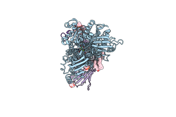 |
Cryo-Em Structure Of The Gastric Proton Pump With Bound Dq-21
Organism: Sus scrofa
Method: ELECTRON MICROSCOPY Release Date: 2023-08-30 Classification: MEMBRANE PROTEIN Ligands: PCW, MG, UOU, NAG, CLR |
Organism: Sus scrofa
Method: ELECTRON MICROSCOPY
Release Date: 2023-08-30
Ligands: PCW, MG, UOU, NAG, CLR
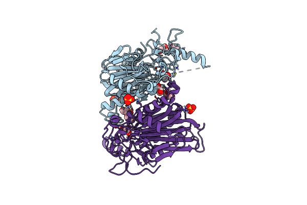 |
Structure Of 6-Aminohexanoate-Oligomer Hydrolase Nylc, D122G/H130Y/T267C Mutant, Hydroxylamine-Treated
Organism: Arthrobacter
Method: X-RAY DIFFRACTION Resolution:1.21 Å Release Date: 2023-03-29 Classification: HYDROLASE Ligands: GOL, SO4 |
Organism: Arthrobacter
Method: X-RAY DIFFRACTION
Release Date: 2023-03-29
Ligands: GOL, SO4
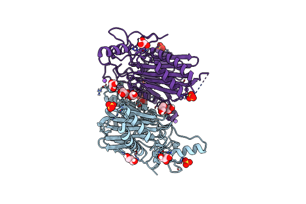 |
Structure Of 6-Aminohexanoate-Oligomer Hydrolase Nylc Precursor, H130Y/N266A/T267A Mutant
Organism: Arthrobacter
Method: X-RAY DIFFRACTION Resolution:1.35 Å Release Date: 2023-03-01 Classification: HYDROLASE Ligands: GOL, SO4, NA |
Organism: Arthrobacter
Method: X-RAY DIFFRACTION
Release Date: 2023-03-01
Ligands: GOL, SO4, NA
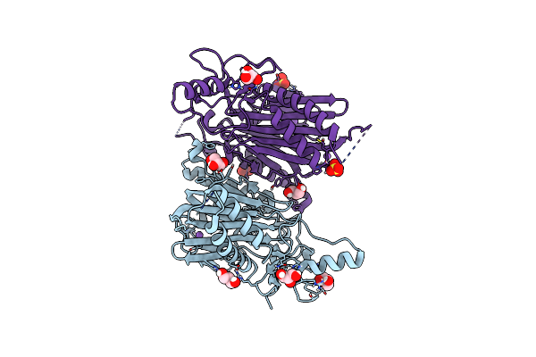 |
Structure Of 6-Aminohexanoate-Oligomer Hydrolase Nylc Precursor, D122G/H130Y/T267C Mutant
Organism: Arthrobacter
Method: X-RAY DIFFRACTION Resolution:1.13 Å Release Date: 2023-03-01 Classification: HYDROLASE Ligands: GOL, NA, SO4 |
Organism: Arthrobacter
Method: X-RAY DIFFRACTION
Release Date: 2023-03-01
Ligands: GOL, NA, SO4
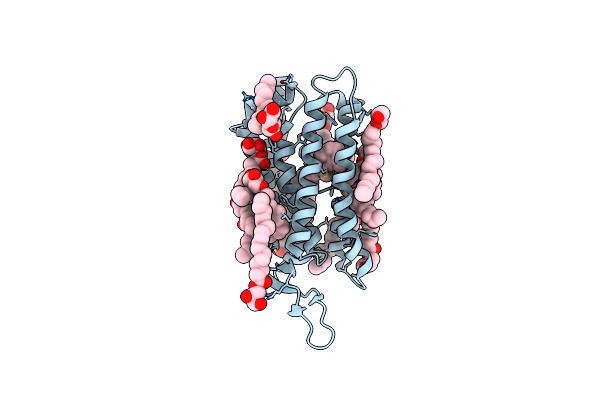 |
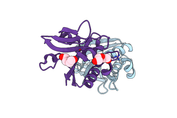
Crystal Structure Of Thermoplasmatales Archaeon Heliorhodopsin
Organism: Thermoplasmatales archaeon sg8-52-1
Method: X-RAY DIFFRACTION Resolution:2.40 Å Release Date: 2019-09-25 Classification: MEMBRANE PROTEIN Ligands: OLC, RET |
Organism: Thermoplasmatales archaeon sg8-52-1
Method: X-RAY DIFFRACTION
Release Date: 2019-09-25
Ligands: OLC, RET
 |
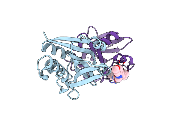
Crystal Structure Of The Peptidase Domain Of Streptococcus Mutans Coma
Organism: Streptococcus mutans serotype c
Method: X-RAY DIFFRACTION Resolution:3.10 Å Release Date: 2017-06-28 Classification: HYDROLASE/HYDROLASE INHIBITOR Ligands: PEG |
Organism: Streptococcus mutans serotype c
Method: X-RAY DIFFRACTION
Release Date: 2017-06-28
Ligands: PEG
 |
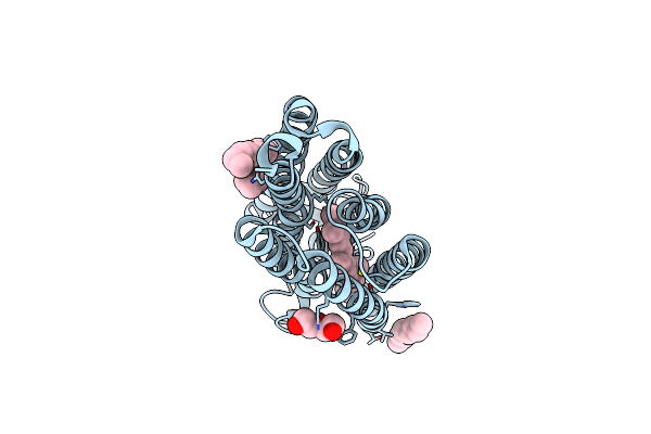
Crystal Structure Of The Complex Of The Peptidase Domain Of Streptococcus Mutans Coma With A Small Molecule Inhibitor.
Organism: Streptococcus mutans serotype c
Method: X-RAY DIFFRACTION Resolution:3.10 Å Release Date: 2017-06-28 Classification: HYDROLASE/HYDROLASE INHIBITOR Ligands: 6CH |
Organism: Streptococcus mutans serotype c
Method: X-RAY DIFFRACTION
Release Date: 2017-06-28
Ligands: 6CH
 |
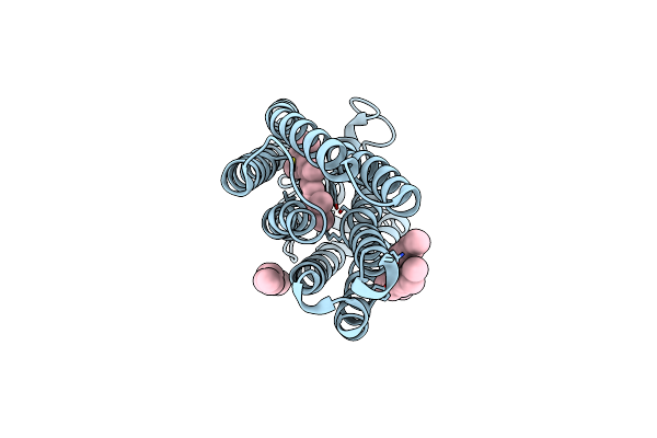
Crystal Structure Of The Light-Driven Sodium Pump Kr2 In Acidic State
Organism: Dokdonia eikasta
Method: X-RAY DIFFRACTION Resolution:2.30 Å Release Date: 2015-04-08 Classification: MEMBRANE PROTEIN Ligands: RET, OLA, PEG |
Organism: Dokdonia eikasta
Method: X-RAY DIFFRACTION
Release Date: 2015-04-08
Ligands: RET, OLA, PEG
 |
Crystal Structure Of The Light-Driven Sodium Pump Kr2 In Neutral State
Organism: Dokdonia eikasta
Method: X-RAY DIFFRACTION Resolution:2.30 Å Release Date: 2015-04-08 Classification: MEMBRANE PROTEIN Ligands: RET, OLA |
Organism: Dokdonia eikasta
Method: X-RAY DIFFRACTION
Release Date: 2015-04-08
Ligands: RET, OLA