

Search Count: 51
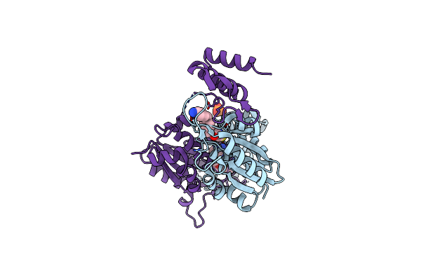 |
Crystal Structure Of Trmd From Pseudomonas Aeruginosa In Complex With Active-Site Inhibitor
Organism: Pseudomonas aeruginosa ucbpp-pa14
Method: X-RAY DIFFRACTION Resolution:2.21 Å Release Date: 2019-09-04 Classification: TRANSFERASE Ligands: BWR, PO4, SAM |
Organism: Pseudomonas aeruginosa ucbpp-pa14
Method: X-RAY DIFFRACTION
Release Date: 2019-09-04
Ligands: BWR, PO4, SAM
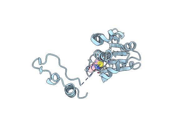 |
Crystal Structure Of Trmd From Mycobacterium Tuberculosis In Complex With Active-Site Inhibitor
Organism: Mycobacterium tuberculosis h37rv
Method: X-RAY DIFFRACTION Resolution:2.20 Å Release Date: 2019-09-04 Classification: TRANSFERASE/INHIBITOR Ligands: BWR |
Organism: Mycobacterium tuberculosis h37rv
Method: X-RAY DIFFRACTION
Release Date: 2019-09-04
Ligands: BWR
 |
Crystal Structure Of Trmd From Pseudomonas Aeruginosa In Complex With Active-Site Inhibitor
Organism: Pseudomonas aeruginosa ucbpp-pa14
Method: X-RAY DIFFRACTION Resolution:2.65 Å Release Date: 2019-03-27 Classification: TRANSFERASE Ligands: 9D0 |
Organism: Pseudomonas aeruginosa ucbpp-pa14
Method: X-RAY DIFFRACTION
Release Date: 2019-03-27
Ligands: 9D0
 |
Apo Crystal Structure Of Trmd From Mycobacterium Tuberculosis
Organism: Mycobacterium tuberculosis (strain cdc 1551 / oshkosh)
Method: X-RAY DIFFRACTION Resolution:2.20 Å Release Date: 2019-03-06 Classification: TRANSFERASE |
Organism: Mycobacterium tuberculosis (strain cdc 1551 / oshkosh)
Method: X-RAY DIFFRACTION
Release Date: 2019-03-06
 |
Crystal Structure Of Trmd From Mycobacterium Tuberculosis In Complex With S-Adenosyl Homocysteine (Sah)
Organism: Mycobacterium tuberculosis h37rv
Method: X-RAY DIFFRACTION Resolution:1.75 Å Release Date: 2019-03-06 Classification: TRANSFERASE Ligands: SAH, GOL |
Organism: Mycobacterium tuberculosis h37rv
Method: X-RAY DIFFRACTION
Release Date: 2019-03-06
Ligands: SAH, GOL
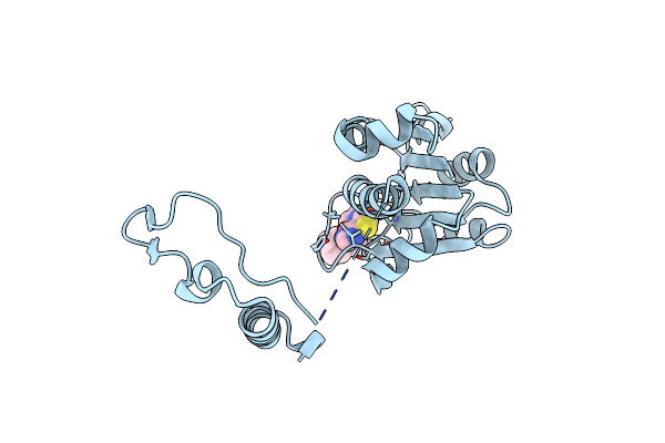 |
Crystal Structure Of Trmd From Mycobacterium Tuberculosis In Complex With Active-Site Inhibitor
Organism: Mycobacterium tuberculosis h37rv
Method: X-RAY DIFFRACTION Resolution:2.30 Å Release Date: 2019-03-06 Classification: TRANSFERASE Ligands: 9D6 |
Organism: Mycobacterium tuberculosis h37rv
Method: X-RAY DIFFRACTION
Release Date: 2019-03-06
Ligands: 9D6
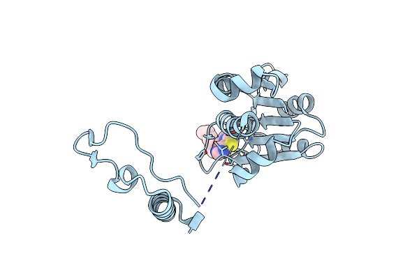 |
Crystal Structure Of Trmd From Mycobacterium Tuberculosis In Complex With Active-Site Inhibitor
Organism: Mycobacterium tuberculosis cdc 1551
Method: X-RAY DIFFRACTION Resolution:2.25 Å Release Date: 2019-03-06 Classification: TRANSFERASE Ligands: 9D0 |
Organism: Mycobacterium tuberculosis cdc 1551
Method: X-RAY DIFFRACTION
Release Date: 2019-03-06
Ligands: 9D0
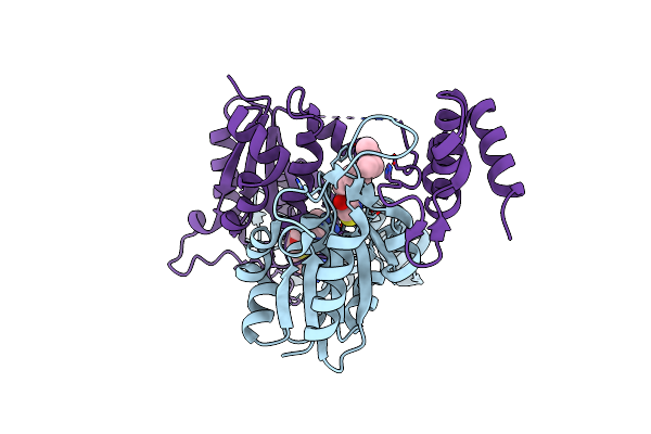 |
Crystal Structure Of Trmd From Pseudomonas Aeruginosa In Complex With Active-Site Inhibitor
Organism: Pseudomonas aeruginosa
Method: X-RAY DIFFRACTION Resolution:2.76 Å Release Date: 2019-03-06 Classification: TRANSFERASE Ligands: 9D3, SAM |
Organism: Pseudomonas aeruginosa
Method: X-RAY DIFFRACTION
Release Date: 2019-03-06
Ligands: 9D3, SAM
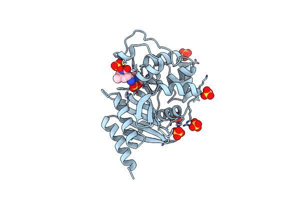 |
Diazabicyclooctenone Etx2514 Bound To Class D Beta Lactamase Oxa-24 From A. Baumannii
Organism: Acinetobacter baumannii
Method: X-RAY DIFFRACTION Resolution:1.93 Å Release Date: 2017-06-07 Classification: HYDROLASE/HYDROLASE INHIBITOR Ligands: SO4, 9CP, 9CM |
Organism: Acinetobacter baumannii
Method: X-RAY DIFFRACTION
Release Date: 2017-06-07
Ligands: SO4, 9CP, 9CM
 |
Tmk From S.Aureus In Complex With The Piperidinyl Thymine Class Inhibitor With A C5 Ethyl-Amine
Organism: Staphylococcus aureus (strain mu3 / atcc 700698)
Method: X-RAY DIFFRACTION Resolution:1.89 Å Release Date: 2015-02-11 Classification: TRANSFERASE Ligands: 43N |
Organism: Staphylococcus aureus (strain mu3 / atcc 700698)
Method: X-RAY DIFFRACTION
Release Date: 2015-02-11
Ligands: 43N
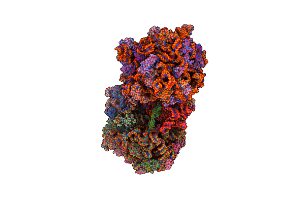 |
Crystal Structure Of The E. Coli Ribosome Bound To Negamycin.
Organism: Escherichia coli str. k-12 substr. mg1655, Escherichia coli, Thermus thermophilus
Method: X-RAY DIFFRACTION Resolution:3.09 Å Release Date: 2014-11-05 Classification: Ribosome/Antibiotic Ligands: MG, ZN, NEG |
Organism: Escherichia coli str. k-12 substr. mg1655, Escherichia coli, Thermus thermophilus
Method: X-RAY DIFFRACTION
Release Date: 2014-11-05
Ligands: MG, ZN, NEG
 |
Lpxc From A.Aaeolicus In Complex With The Mmp Inhibitor 4-[[4-(4-Chlorophenoxy)Phenyl]Sulfanylmethyl]Tetrahydropyran-4-Carbohydroxamic Acid - Compound 2
Organism: Aquifex aeolicus
Method: X-RAY DIFFRACTION Resolution:1.34 Å Release Date: 2014-10-01 Classification: HYDROLASE Ligands: IMD, ZN, CL, 3BW |
Organism: Aquifex aeolicus
Method: X-RAY DIFFRACTION
Release Date: 2014-10-01
Ligands: IMD, ZN, CL, 3BW
 |
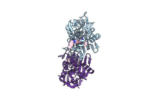
Lpxc From A.Aaeolicus In Complex With 4-[[4-[2-[4-(Morpholinomethyl)Phenyl]Ethynyl]Phenoxy]Methyl]Tetrahydropyran-4-Carbohydroxamic Acid (Compound 9)
Organism: Aquifex aeolicus
Method: X-RAY DIFFRACTION Resolution:1.25 Å Release Date: 2014-10-01 Classification: HYDROLASE Ligands: IMD, ZN, CL, 3BX |
Organism: Aquifex aeolicus
Method: X-RAY DIFFRACTION
Release Date: 2014-10-01
Ligands: IMD, ZN, CL, 3BX
 |
Lpxc From P.Aeruginosa With The Inhibitor 6-(Benzimidazol-1-Yl)-5-[4-[2-[6-[(4-Methylpiperazin-1-Yl)Methyl]-3-Pyridyl]Ethynyl]Phenyl]Pyridine-3-Carbohydroxamic Acid
Organism: Pseudomonas aeruginosa
Method: X-RAY DIFFRACTION Resolution:2.06 Å Release Date: 2014-09-24 Classification: HYDROLASE/HYDROLASE INHIBITOR Ligands: ZN, 2SZ |
Organism: Pseudomonas aeruginosa
Method: X-RAY DIFFRACTION
Release Date: 2014-09-24
Ligands: ZN, 2SZ
 |
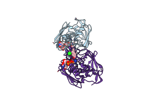
Avibactam And Class C Beta-Lactamases: Mechanism Of Inhibition, Conservation Of Binding Pocket And Implications For Resistance
Organism: Pseudomonas aeruginosa
Method: X-RAY DIFFRACTION Resolution:1.10 Å Release Date: 2014-08-20 Classification: hydrolase/hydrolase inhibitor Ligands: NXL |
Organism: Pseudomonas aeruginosa
Method: X-RAY DIFFRACTION
Release Date: 2014-08-20
Ligands: NXL
 |
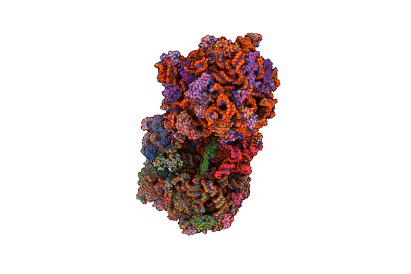
A.Aolicus Lpxc In Complex With Native Product
Organism: Aquifex aeolicus
Method: X-RAY DIFFRACTION Resolution:1.61 Å Release Date: 2014-08-13 Classification: HYDROLASE Ligands: ZN, CL, 24G |
Organism: Aquifex aeolicus
Method: X-RAY DIFFRACTION
Release Date: 2014-08-13
Ligands: ZN, CL, 24G
 |
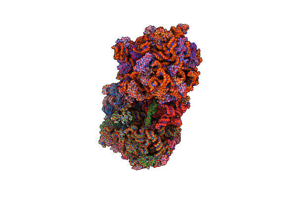
Crystal Structure Of The E. Coli Ribosome Bound To Quinupristin.
Organism: Synthetic construct, Escherichia coli str. k-12 substr. mds42, Escherichia coli, Thermus thermophilus
Method: X-RAY DIFFRACTION Resolution:2.95 Å Release Date: 2014-07-30 Classification: RIBOSOME Ligands: MG, ZN |
Organism: Synthetic construct, Escherichia coli str. k-12 substr. mds42, Escherichia coli, Thermus thermophilus
Method: X-RAY DIFFRACTION
Release Date: 2014-07-30
Ligands: MG, ZN
 |
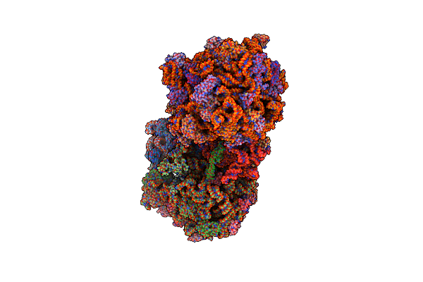
Crystal Structure Of The E. Coli Ribosome Bound To Linopristin.
Organism: Synthetic construct, Escherichia coli str. k-12 substr. mds42, Escherichia coli, Thermus thermophilus
Method: X-RAY DIFFRACTION Resolution:3.00 Å Release Date: 2014-07-30 Classification: RIBOSOME Ligands: MG, ZN |
Organism: Synthetic construct, Escherichia coli str. k-12 substr. mds42, Escherichia coli, Thermus thermophilus
Method: X-RAY DIFFRACTION
Release Date: 2014-07-30
Ligands: MG, ZN
 |
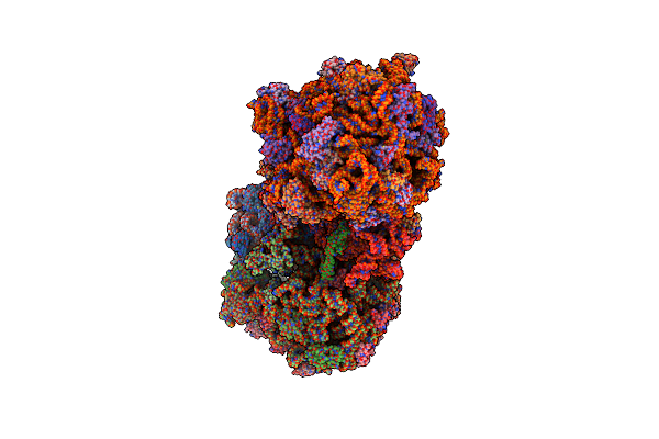
Crystal Structure Of The E. Coli Ribosome Bound To Flopristin.
Organism: Escherichia coli str. k-12 substr. mds42, Escherichia coli, Thermus thermophilus
Method: X-RAY DIFFRACTION Resolution:2.90 Å Release Date: 2014-07-30 Classification: RIBOSOME Ligands: MG, VIF, ZN |
Organism: Escherichia coli str. k-12 substr. mds42, Escherichia coli, Thermus thermophilus
Method: X-RAY DIFFRACTION
Release Date: 2014-07-30
Ligands: MG, VIF, ZN
 |
Crystal Structure Of The E. Coli Ribosome Bound To Dalfopristin.
Organism: Escherichia coli str. k-12 substr. mds42, Escherichia coli, Thermus thermophilus
Method: X-RAY DIFFRACTION Resolution:2.90 Å Release Date: 2014-07-30 Classification: RIBOSOME Ligands: MG, DOL, ZN |
Organism: Escherichia coli str. k-12 substr. mds42, Escherichia coli, Thermus thermophilus
Method: X-RAY DIFFRACTION
Release Date: 2014-07-30
Ligands: MG, DOL, ZN