

Search Count: 47
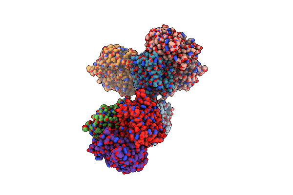 |
Crystal Structure Of A Dodecameric Multicopper Oxidase From M. Hydrothermalis In A Cubic Lattice
Organism: Marinithermus hydrothermalis
Method: X-RAY DIFFRACTION Resolution:1.92 Å Release Date: 2021-10-20 Classification: OXIDOREDUCTASE Ligands: CU, MG |
Organism: Marinithermus hydrothermalis
Method: X-RAY DIFFRACTION
Release Date: 2021-10-20
Ligands: CU, MG
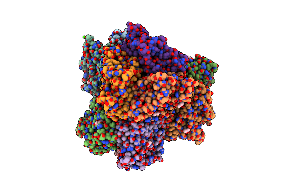 |
Crystal Structure Of A Dodecameric Multicopper Oxidase From M. Hydrothermalis In An Orthorhombic Lattice
Organism: Marinithermus hydrothermalis
Method: X-RAY DIFFRACTION Resolution:2.36 Å Release Date: 2021-10-20 Classification: OXIDOREDUCTASE Ligands: MPD, CU, CA |
Organism: Marinithermus hydrothermalis
Method: X-RAY DIFFRACTION
Release Date: 2021-10-20
Ligands: MPD, CU, CA
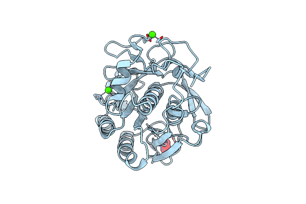 |
High-Viscosity Injector-Based Pink Beam Serial Crystallography Of Micro-Crystals At A Synchrotron Radiation Source.
Organism: Parengyodontium album
Method: X-RAY DIFFRACTION Resolution:1.80 Å Release Date: 2019-04-24 Classification: HYDROLASE Ligands: CA, NO3 |
Organism: Parengyodontium album
Method: X-RAY DIFFRACTION
Release Date: 2019-04-24
Ligands: CA, NO3
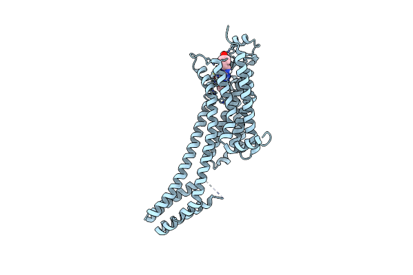 |
High-Viscosity Injector-Based Pink Beam Serial Crystallography Of Micro-Crystals At A Synchrotron Radiation Source
Organism: Homo sapiens, Escherichia coli
Method: X-RAY DIFFRACTION Resolution:4.20 Å Release Date: 2019-04-24 Classification: MEMBRANE PROTEIN Ligands: ZMA |
Organism: Homo sapiens, Escherichia coli
Method: X-RAY DIFFRACTION
Release Date: 2019-04-24
Ligands: ZMA
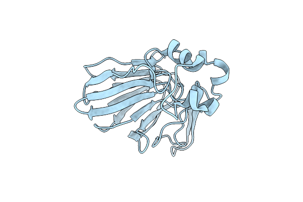 |
Crystal Structure Of Thaumatin From Microcrystals
Organism: Thaumatococcus daniellii
Method: X-RAY DIFFRACTION Resolution:2.50 Å Release Date: 2018-05-30 Classification: PLANT PROTEIN |
Organism: Thaumatococcus daniellii
Method: X-RAY DIFFRACTION
Release Date: 2018-05-30
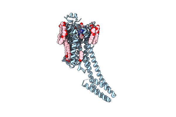 |
2.35-Angstrom In Situ Mylar Structure Of Human A2A Adenosine Receptor At 100 K
Organism: Homo sapiens, Escherichia coli
Method: X-RAY DIFFRACTION Resolution:2.35 Å Release Date: 2017-12-13 Classification: SIGNALING PROTEIN Ligands: ZMA, NA, CLR, OLC, OLA, OLB |
Organism: Homo sapiens, Escherichia coli
Method: X-RAY DIFFRACTION
Release Date: 2017-12-13
Ligands: ZMA, NA, CLR, OLC, OLA, OLB
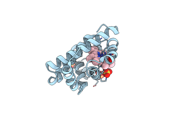 |
2.0-Angstrom In Situ Mylar Structure Of Sperm Whale Myoglobin (Swmb) At 293 K
Organism: Physeter catodon
Method: X-RAY DIFFRACTION Resolution:2.00 Å Release Date: 2017-12-13 Classification: OXYGEN TRANSPORT Ligands: HEM, CL, SO4 |
Organism: Physeter catodon
Method: X-RAY DIFFRACTION
Release Date: 2017-12-13
Ligands: HEM, CL, SO4
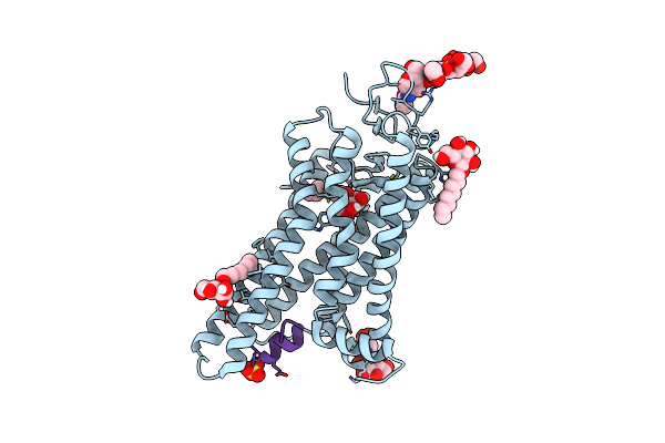 |
3.2-Angstrom In Situ Mylar Structure Of Bovine Opsin At 100 K
Organism: Bos taurus
Method: X-RAY DIFFRACTION Resolution:3.20 Å Release Date: 2017-12-13 Classification: SIGNALING PROTEIN Ligands: BOG, SO4 |
Organism: Bos taurus
Method: X-RAY DIFFRACTION
Release Date: 2017-12-13
Ligands: BOG, SO4
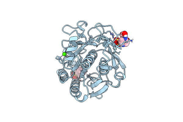 |
Single-Shot Pink Beam Serial Crystallography: Proteinase K
Organism: Engyodontium album
Method: X-RAY DIFFRACTION Resolution:2.21 Å Release Date: 2017-11-15 Classification: HYDROLASE Ligands: CA, CL, NHE, EPE |
Organism: Engyodontium album
Method: X-RAY DIFFRACTION
Release Date: 2017-11-15
Ligands: CA, CL, NHE, EPE
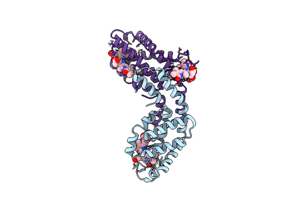 |
Single-Shot Pink Beam Serial Crystallography: Phycocyanin (Five Chips Merged)
Organism: Thermosynechococcus elongatus (strain bp-1), Synechococcus elongatus
Method: X-RAY DIFFRACTION Resolution:2.30 Å Release Date: 2017-11-15 Classification: PHOTOSYNTHESIS Ligands: CYC |
Organism: Thermosynechococcus elongatus (strain bp-1), Synechococcus elongatus
Method: X-RAY DIFFRACTION
Release Date: 2017-11-15
Ligands: CYC
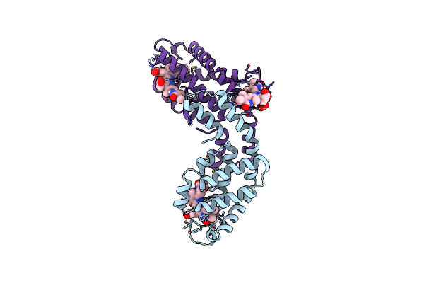 |
Multi-Bunch Pink Beam Serial Crystallography: Phycocyanin (One Chip)
Organism: Thermosynechococcus elongatus (strain bp-1)
Method: X-RAY DIFFRACTION Resolution:2.11 Å Release Date: 2017-11-15 Classification: PHOTOSYNTHESIS Ligands: CYC |
Organism: Thermosynechococcus elongatus (strain bp-1)
Method: X-RAY DIFFRACTION
Release Date: 2017-11-15
Ligands: CYC
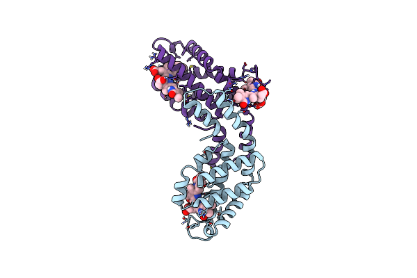 |
Single-Shot Pink Beam Serial Crystallography: Phycocyanin (One Chip)
Organism: Thermosynechococcus elongatus (strain bp-1)
Method: X-RAY DIFFRACTION Resolution:2.70 Å Release Date: 2017-11-15 Classification: PHOTOSYNTHESIS Ligands: CYC |
Organism: Thermosynechococcus elongatus (strain bp-1)
Method: X-RAY DIFFRACTION
Release Date: 2017-11-15
Ligands: CYC
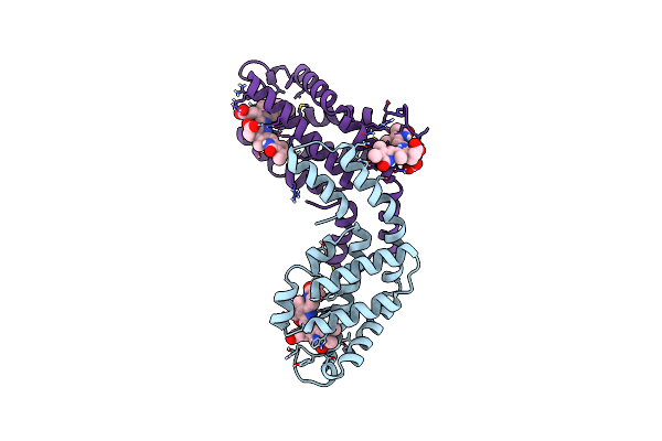 |
Single-Shot Pink Beam Serial Crystallography: Phycocyanin (One Chip, Chip_1)
Organism: Thermosynechococcus elongatus (strain bp-1)
Method: X-RAY DIFFRACTION Resolution:2.46 Å Release Date: 2017-11-15 Classification: PHOTOSYNTHESIS Ligands: CYC |
Organism: Thermosynechococcus elongatus (strain bp-1)
Method: X-RAY DIFFRACTION
Release Date: 2017-11-15
Ligands: CYC
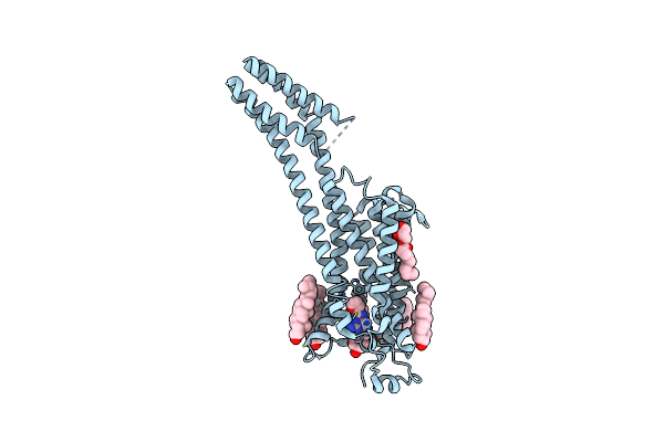 |
Serial Millisecond Crystallography Of Membrane And Soluble Protein Micro-Crystals Using Synchrotron Radiation
Organism: Homo sapiens, Escherichia coli
Method: X-RAY DIFFRACTION Resolution:3.20 Å Release Date: 2017-05-24 Classification: MEMBRANE PROTEIN Ligands: ZMA, CLR, OLC, OLA |
Organism: Homo sapiens, Escherichia coli
Method: X-RAY DIFFRACTION
Release Date: 2017-05-24
Ligands: ZMA, CLR, OLC, OLA
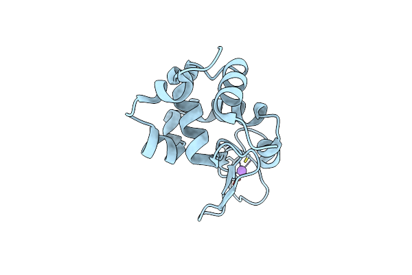 |
Serial Millisecond Crystallography Of Membrane And Soluble Protein Micro-Crystals Using Synchrotron Radiation
Organism: Gallus gallus
Method: X-RAY DIFFRACTION Resolution:2.05 Å Release Date: 2017-05-24 Classification: HYDROLASE Ligands: CL, NA |
Organism: Gallus gallus
Method: X-RAY DIFFRACTION
Release Date: 2017-05-24
Ligands: CL, NA
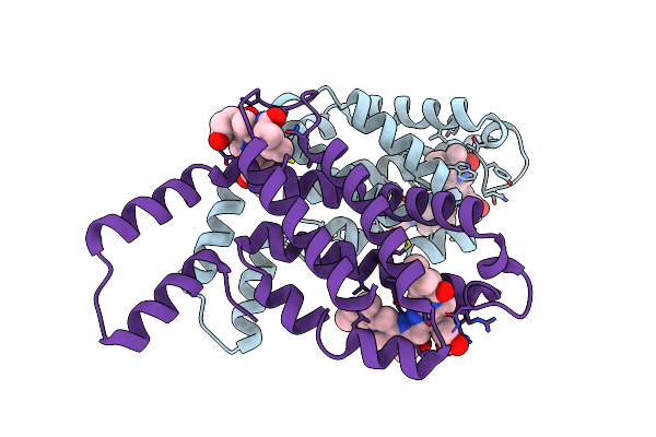 |
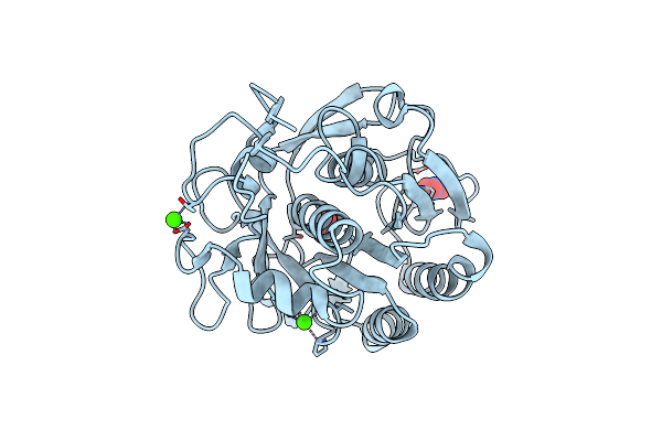
Serial Millisecond Crystallography Of Membrane And Soluble Protein Micro-Crystals Using Synchrotron Radiation
Organism: Thermosynechococcus elongatus (strain bp-1)
Method: X-RAY DIFFRACTION Resolution:3.10 Å Release Date: 2017-05-24 Classification: ELECTRON TRANSPORT Ligands: CYC |
Organism: Thermosynechococcus elongatus (strain bp-1)
Method: X-RAY DIFFRACTION
Release Date: 2017-05-24
Ligands: CYC
 |
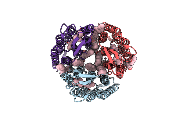
Serial Millisecond Crystallography Of Membrane And Soluble Protein Micro-Crystals Using Synchrotron Radiation
Organism: Parengyodontium album
Method: X-RAY DIFFRACTION Resolution:2.65 Å Release Date: 2017-05-24 Classification: HYDROLASE Ligands: NO3, CA |
Organism: Parengyodontium album
Method: X-RAY DIFFRACTION
Release Date: 2017-05-24
Ligands: NO3, CA
 |
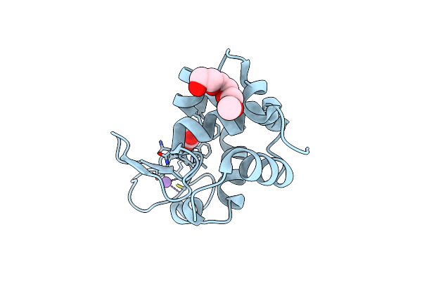
2.1-Angstrom In Situ Mylar Structure Of Bacteriorhodopsin From Haloquadratum Walsbyi (Hwbr) At 100 K
Organism: Haloquadratum walsbyi (strain dsm 16790 / hbsq001)
Method: X-RAY DIFFRACTION Resolution:2.13 Å Release Date: 2017-02-15 Classification: MEMBRANE PROTEIN Ligands: OLC, OLB, RET |
Organism: Haloquadratum walsbyi (strain dsm 16790 / hbsq001)
Method: X-RAY DIFFRACTION
Release Date: 2017-02-15
Ligands: OLC, OLB, RET
 |
1.7-Angstrom In Situ Mylar Structure Of Hen Egg-White Lysozyme (Hewl) At 100 K
Organism: Gallus gallus
Method: X-RAY DIFFRACTION Resolution:1.70 Å Release Date: 2017-02-15 Classification: HYDROLASE |
Organism: Gallus gallus
Method: X-RAY DIFFRACTION
Release Date: 2017-02-15
 |
2.0-Angstrom In Situ Mylar Structure Of Hen Egg-White Lysozyme (Hewl) At 293 K
Organism: Gallus gallus
Method: X-RAY DIFFRACTION Resolution:2.00 Å Release Date: 2017-02-15 Classification: HYDROLASE |
Organism: Gallus gallus
Method: X-RAY DIFFRACTION
Release Date: 2017-02-15