

Search Count: 21
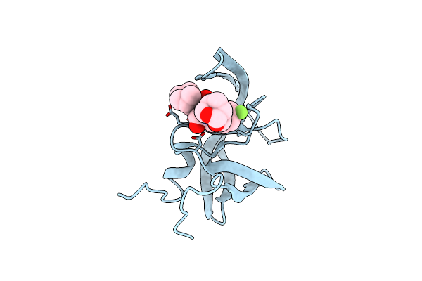 |
X-Ray Crystal Structure Of Wild Type Hiv-1 Protease In Complex With Grl-004
Organism: Human immunodeficiency virus 1
Method: X-RAY DIFFRACTION Resolution:1.46 Å Release Date: 2020-05-20 Classification: VIRAL PROTEIN/INHIBITOR Ligands: NKA |
Organism: Human immunodeficiency virus 1
Method: X-RAY DIFFRACTION
Release Date: 2020-05-20
Ligands: NKA
 |
X-Ray Crystal Structure Of Wild Type Hiv-1 Protease In Complex With Grl-002
Organism: Human immunodeficiency virus 1
Method: X-RAY DIFFRACTION Resolution:1.54 Å Release Date: 2020-05-20 Classification: VIRAL PROTEIN/INHIBITOR Ligands: NJY |
Organism: Human immunodeficiency virus 1
Method: X-RAY DIFFRACTION
Release Date: 2020-05-20
Ligands: NJY
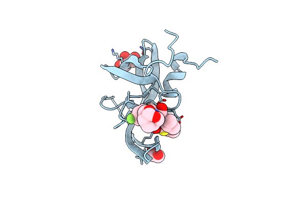 |
X-Ray Crystal Structure Of Darunavir-Resistant Hiv-1 Protease (P51) In Complex With Grl-003
Organism: Human immunodeficiency virus 1
Method: X-RAY DIFFRACTION Resolution:1.21 Å Release Date: 2020-04-08 Classification: VIRAL PROTEIN/INHIBITOR Ligands: GOL, EDO, JDV |
Organism: Human immunodeficiency virus 1
Method: X-RAY DIFFRACTION
Release Date: 2020-04-08
Ligands: GOL, EDO, JDV
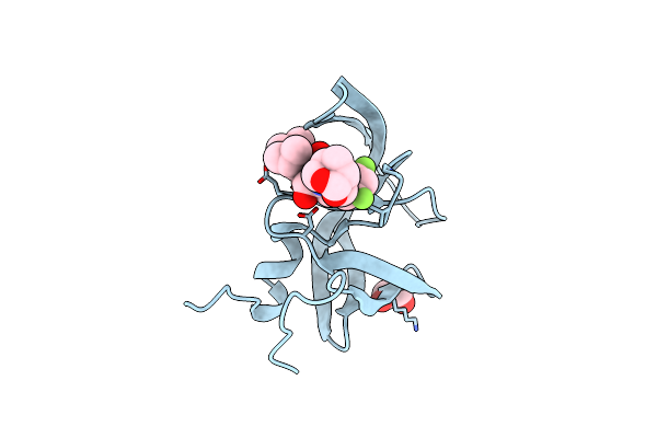 |
X-Ray Crystal Structure Of Wild Type Hiv-1 Protease In Complex With Grl-063
Organism: Human immunodeficiency virus 1
Method: X-RAY DIFFRACTION Resolution:1.53 Å Release Date: 2020-04-08 Classification: VIRAL PROTEIN/INHIBITOR Ligands: EDO, MJD |
Organism: Human immunodeficiency virus 1
Method: X-RAY DIFFRACTION
Release Date: 2020-04-08
Ligands: EDO, MJD
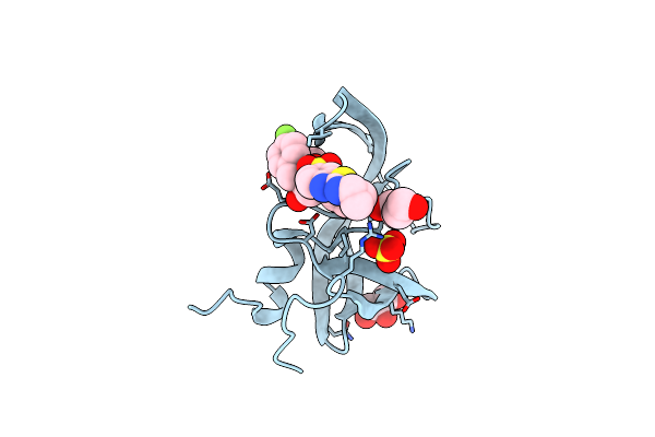 |
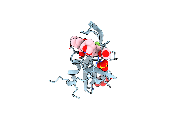
X-Ray Crystal Structure Of Darunavir-Resistant Hiv-1 Protease (P30) In Complex With Grl-003
Organism: Human immunodeficiency virus 1
Method: X-RAY DIFFRACTION Resolution:1.41 Å Release Date: 2020-04-08 Classification: VIRAL PROTEIN/INHIBITOR Ligands: GOL, PEG, JDV, SO4 |
Organism: Human immunodeficiency virus 1
Method: X-RAY DIFFRACTION
Release Date: 2020-04-08
Ligands: GOL, PEG, JDV, SO4
 |
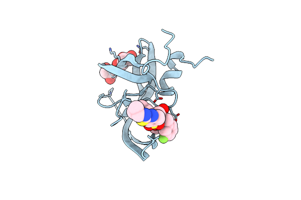
X-Ray Crystal Structure Of Darunavir-Resistant Hiv-1 Protease (P30) In Complex With Grl-001
Organism: Human immunodeficiency virus 1
Method: X-RAY DIFFRACTION Resolution:1.27 Å Release Date: 2020-04-08 Classification: VIRAL PROTEIN/INHIBITOR Ligands: PEG, GOL, JDY, SO4 |
Organism: Human immunodeficiency virus 1
Method: X-RAY DIFFRACTION
Release Date: 2020-04-08
Ligands: PEG, GOL, JDY, SO4
 |
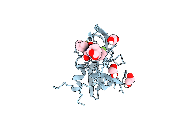
X-Ray Crystal Structure Of Darunavir-Resistant Hiv-1 Protease (P51) In Complex With Grl-001
Organism: Human immunodeficiency virus 1
Method: X-RAY DIFFRACTION Resolution:1.21 Å Release Date: 2020-04-08 Classification: VIRAL PROTEIN/INHIBITOR Ligands: GOL, JDY, EDO |
Organism: Human immunodeficiency virus 1
Method: X-RAY DIFFRACTION
Release Date: 2020-04-08
Ligands: GOL, JDY, EDO
 |
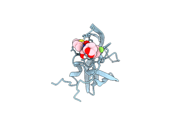
X-Ray Crystal Structure Of Wild Type Hiv-1 Protease In Complex With Grl-001
Organism: Human immunodeficiency virus 1
Method: X-RAY DIFFRACTION Resolution:1.48 Å Release Date: 2019-04-24 Classification: VIRAL PROTEIN Ligands: EDO, JDY |
Organism: Human immunodeficiency virus 1
Method: X-RAY DIFFRACTION
Release Date: 2019-04-24
Ligands: EDO, JDY
 |
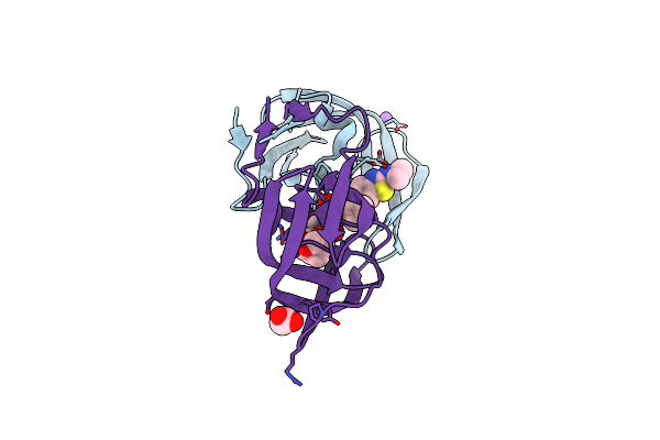
X-Ray Crystal Structure Of Wild Type Hiv-1 Protease In Complex With Grl-003
Organism: Human immunodeficiency virus 1
Method: X-RAY DIFFRACTION Resolution:1.52 Å Release Date: 2019-04-24 Classification: VIRAL PROTEIN Ligands: JDV |
Organism: Human immunodeficiency virus 1
Method: X-RAY DIFFRACTION
Release Date: 2019-04-24
Ligands: JDV
 |
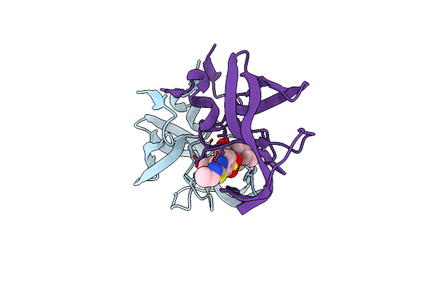
Crystal Structure Of Wild-Type Hiv-1 Protease With A Novel Hiv-1 Inhibitor Grl-14213A Of 6-5-5-Ring Fused Crown-Like Tetrahydropyranofuran As The P2-Ligand, A Cyclopropylaminobenzothiazole As The P2'-Ligand And 3,5-Difluorophenylmethyl As The P1-Ligand
Organism: Human immunodeficiency virus 1
Method: X-RAY DIFFRACTION Resolution:1.67 Å Release Date: 2018-02-28 Classification: HYDROLASE/HYDROLASE inhibitor |
Organism: Human immunodeficiency virus 1
Method: X-RAY DIFFRACTION
Release Date: 2018-02-28
 |
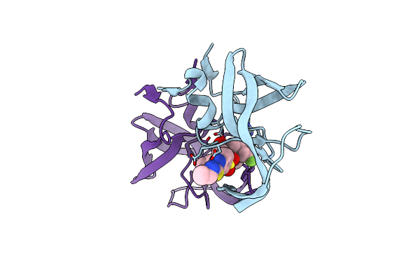
X-Ray Crystal Structure Of Wild Type Hiv-1 Protease In Complex With Grl-121
Organism: Human immunodeficiency virus 1
Method: X-RAY DIFFRACTION Resolution:1.80 Å Release Date: 2017-10-18 Classification: HYDROLASE/HYDROLASE INHIBITOR Ligands: 7O7 |
Organism: Human immunodeficiency virus 1
Method: X-RAY DIFFRACTION
Release Date: 2017-10-18
Ligands: 7O7
 |
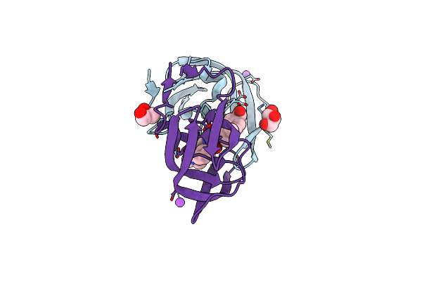
X-Ray Crystal Structure Of Wild Type Hiv-1 Protease In Complex With Grl-142
Organism: Human immunodeficiency virus 1
Method: X-RAY DIFFRACTION Resolution:2.01 Å Release Date: 2017-10-18 Classification: HYDROLASE/HYDROLASE INHIBITOR Ligands: 7OA |
Organism: Human immunodeficiency virus 1
Method: X-RAY DIFFRACTION
Release Date: 2017-10-18
Ligands: 7OA
 |
Hiv-1 Wild Type Protease With Grl-1118A , An Isophthalamide-Derived P2-P3 Ligand With The Sulfonamide Isostere As The P2' Group
Organism: Human immunodeficiency virus 1
Method: X-RAY DIFFRACTION Resolution:1.33 Å Release Date: 2017-05-10 Classification: hydrolase/hydrolase inhibitor Ligands: 8FP, NA, CL, GOL, ACT |
Organism: Human immunodeficiency virus 1
Method: X-RAY DIFFRACTION
Release Date: 2017-05-10
Ligands: 8FP, NA, CL, GOL, ACT
 |
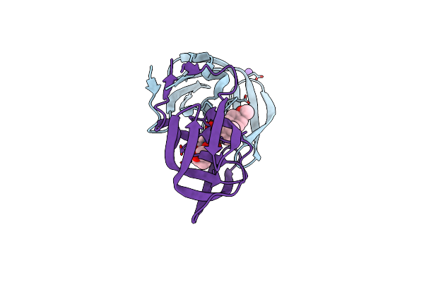
Hiv-1 Wild Type Protease With Grl-0518A , An Isophthalamide-Derived P2-P3 Ligand With The Para-Hydoxymethyl Sulfonamide Isostere As The P2' Group
Organism: Human immunodeficiency virus 1
Method: X-RAY DIFFRACTION Resolution:1.27 Å Release Date: 2017-05-10 Classification: hydrolase/hydrolase inhibitor Ligands: 8HD, NA, CL, GOL |
Organism: Human immunodeficiency virus 1
Method: X-RAY DIFFRACTION
Release Date: 2017-05-10
Ligands: 8HD, NA, CL, GOL
 |
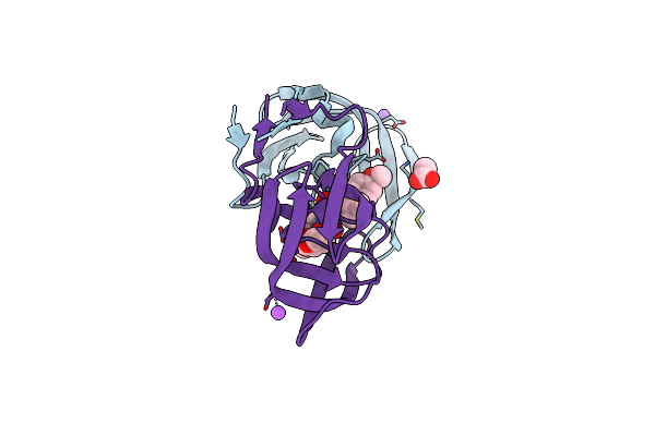
Hiv-1 Wild Type Protease With Grl-100-13A (A Crown-Like Oxotricyclic Core As The P2-Ligand With The Sulfonamide Isostere As The P2' Group)
Organism: Human immunodeficiency virus 1
Method: X-RAY DIFFRACTION Resolution:1.53 Å Release Date: 2017-05-03 Classification: hydrolase/hydrolase inhibitor |
Organism: Human immunodeficiency virus 1
Method: X-RAY DIFFRACTION
Release Date: 2017-05-03
 |
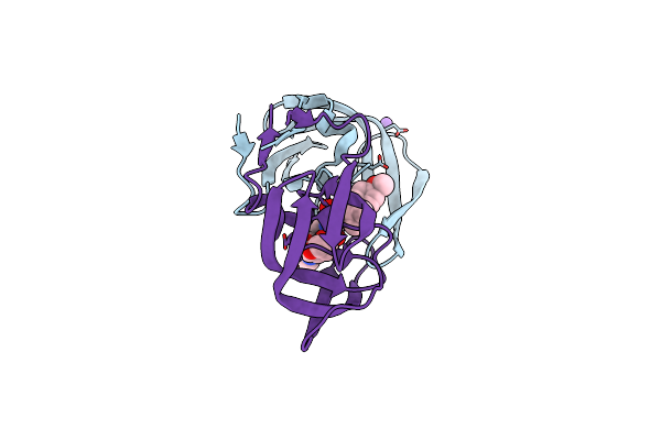
Crystal Structure Of Hiv-1 Protease Inhibitors Containing Substituted Fused-Tetrahydropyranyl Tetrahydrofuran As P2-Ligand Grl-004-11A
Organism: Human immunodeficiency virus 1
Method: X-RAY DIFFRACTION Resolution:1.22 Å Release Date: 2015-10-28 Classification: HYDROLASE Ligands: 5B7, NA, CL, ACT |
Organism: Human immunodeficiency virus 1
Method: X-RAY DIFFRACTION
Release Date: 2015-10-28
Ligands: 5B7, NA, CL, ACT
 |
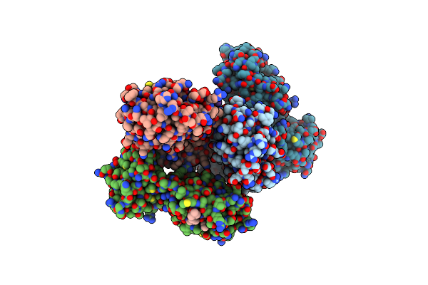
Crystal Structure Of Hiv-1 Protease Inhibitor Grl-105-11A Containing Substituted Fused-Tetrahydropyranyl Tetrahydrofuran As P2-Ligand
Organism: Human immunodeficiency virus 1
Method: X-RAY DIFFRACTION Resolution:1.62 Å Release Date: 2015-10-28 Classification: HYDROLASE Ligands: 5B5, NA, CL |
Organism: Human immunodeficiency virus 1
Method: X-RAY DIFFRACTION
Release Date: 2015-10-28
Ligands: 5B5, NA, CL
 |
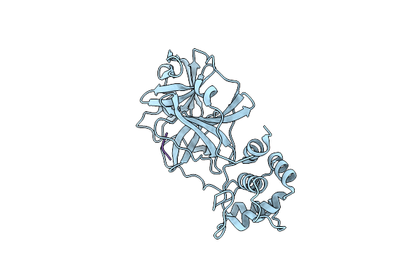
2.1 A X-Ray Structure Of Fipv-3Clpro Bound To Covalent Inhibitor
Organism: Feline coronavirus (strain fipv wsu-79/1146), Synthetic construct
Method: X-RAY DIFFRACTION Resolution:2.06 Å Release Date: 2015-10-14 Classification: Hydrolase/Hydrolase Inhibitor Ligands: DMS |
Organism: Feline coronavirus (strain fipv wsu-79/1146), Synthetic construct
Method: X-RAY DIFFRACTION
Release Date: 2015-10-14
Ligands: DMS
 |
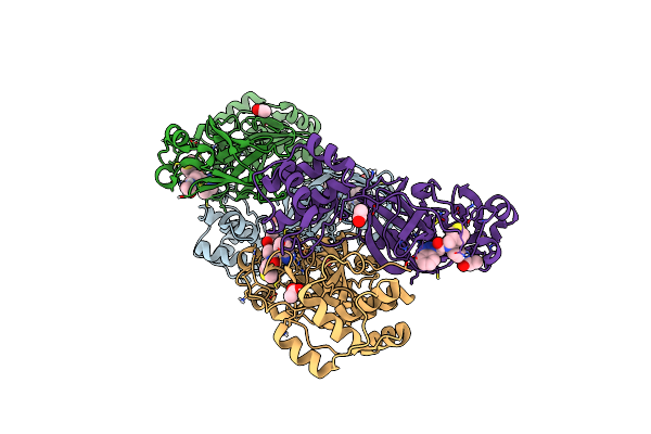
X-Ray Structure Of Mers-Cov Nsp5 Protease Bound With A Designed Inhibitor
Organism: Middle east respiratory syndrome coronavirus
Method: X-RAY DIFFRACTION Resolution:1.62 Å Release Date: 2015-06-17 Classification: Hydrolase/hydrolase Inhibitor |
Organism: Middle east respiratory syndrome coronavirus
Method: X-RAY DIFFRACTION
Release Date: 2015-06-17
 |
X-Ray Structure Of Mers-Cov Nsp5 Protease Bound With A Non-Covalent Inhibitor
Organism: Middle east respiratory syndrome coronavirus
Method: X-RAY DIFFRACTION Resolution:2.10 Å Release Date: 2015-06-17 Classification: HYDROLASE/HYDROLASE INHIBITOR Ligands: R30, ACT |
Organism: Middle east respiratory syndrome coronavirus
Method: X-RAY DIFFRACTION
Release Date: 2015-06-17
Ligands: R30, ACT