

Search Count: 31
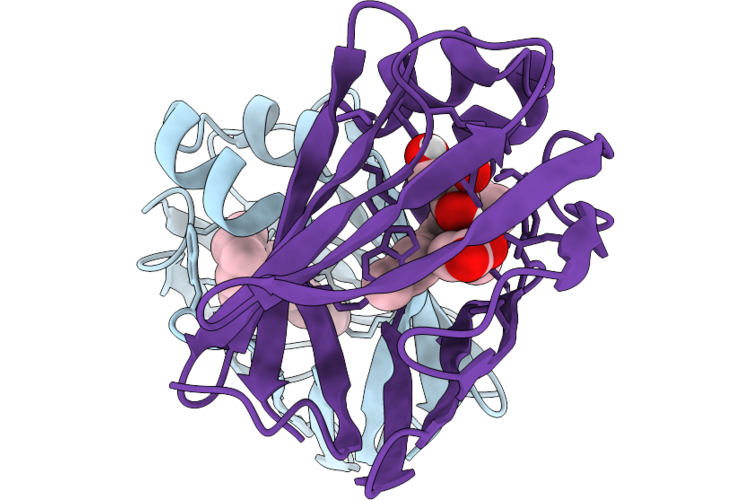 |
Crystal Structure Of Q108K:K40L:T51V:T53C:R58W:T29L:Y19W:Q4A Mutant Of Cellular Retinol Binding Protein Ii Complex With 15-Cis-Retinal
Organism: Homo sapiens
Method: X-RAY DIFFRACTION Resolution:1.26 Å Release Date: 2026-06-10 Classification: TRANSPORT PROTEIN Ligands: ACT, RET, GOL |
Organism: Homo sapiens
Method: X-RAY DIFFRACTION
Release Date: 2026-06-10
Ligands: ACT, RET, GOL
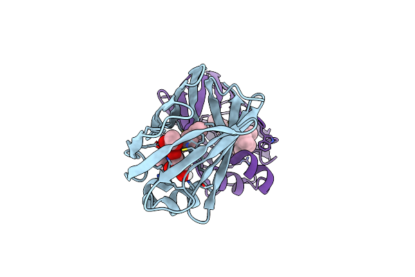 |
Crystal Structure Of Q108K:K40L:T51V:T53C:R58W:T29L:Y19W:Q4A:Q38L Mutant Of Cellular Retinol Binding Protein Ii Complex With All-Trans-Retinal
Organism: Homo sapiens
Method: X-RAY DIFFRACTION Resolution:1.50 Å Release Date: 2021-04-14 Classification: CYTOSOLIC PROTEIN Ligands: ACT, RET, GOL |
Organism: Homo sapiens
Method: X-RAY DIFFRACTION
Release Date: 2021-04-14
Ligands: ACT, RET, GOL
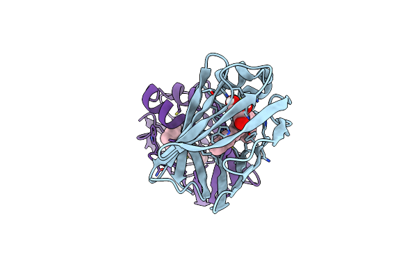 |
Crystal Structure Of Q108K:K40L:T51V:T53C:R58W:T29L:Y19W:Q4A Mutant Of Cellular Retinol Binding Protein Ii Complex With All-Trans-Retinal After Exposure To Visible Light
Organism: Homo sapiens
Method: X-RAY DIFFRACTION Resolution:2.11 Å Release Date: 2021-04-14 Classification: CYTOSOLIC PROTEIN Ligands: GOL, RET |
Organism: Homo sapiens
Method: X-RAY DIFFRACTION
Release Date: 2021-04-14
Ligands: GOL, RET
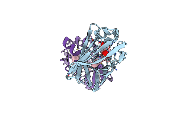 |
Crystal Structure Of Q108K:K40L:T51V:T53C:R58W:T29L:Y19W:Q4A Mutant Of Cellular Retinol Binding Protein Ii Complex With All-Trans-Retinal In The Dark
Organism: Homo sapiens
Method: X-RAY DIFFRACTION Resolution:1.40 Å Release Date: 2021-04-14 Classification: TRANSPORT PROTEIN Ligands: GOL, RET |
Organism: Homo sapiens
Method: X-RAY DIFFRACTION
Release Date: 2021-04-14
Ligands: GOL, RET
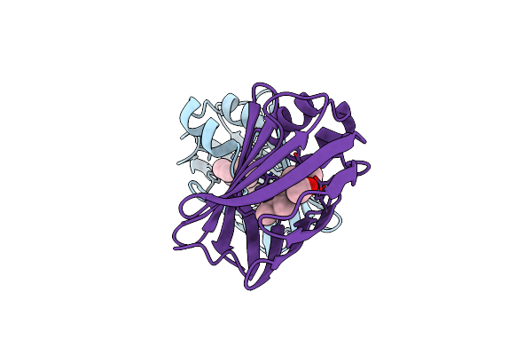 |
Crystal Structure Of The Q108K:K40L:T51V:R58F Mutant Of Human Cellular Retinol Binding Protein Ii In Complex With Synthetic Ligand Julolidine
Organism: Homo sapiens
Method: X-RAY DIFFRACTION Resolution:1.42 Å Release Date: 2018-04-25 Classification: TRANSPORT PROTEIN Ligands: ACT, JLO |
Organism: Homo sapiens
Method: X-RAY DIFFRACTION
Release Date: 2018-04-25
Ligands: ACT, JLO
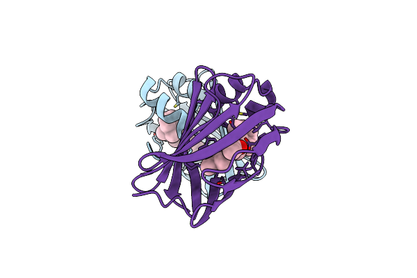 |
Crystal Structure Of The Q108K:K40L:T53C Mutant Of Human Cellular Retinol Binding Protein Ii In Complex With All-Trans-Retinal After 24 Hours Of Incubation At 1.55 Angstrom Resolution
Organism: Homo sapiens
Method: X-RAY DIFFRACTION Resolution:1.55 Å Release Date: 2016-12-21 Classification: TRANSPORT PROTEIN Ligands: RET, ACT |
Organism: Homo sapiens
Method: X-RAY DIFFRACTION
Release Date: 2016-12-21
Ligands: RET, ACT
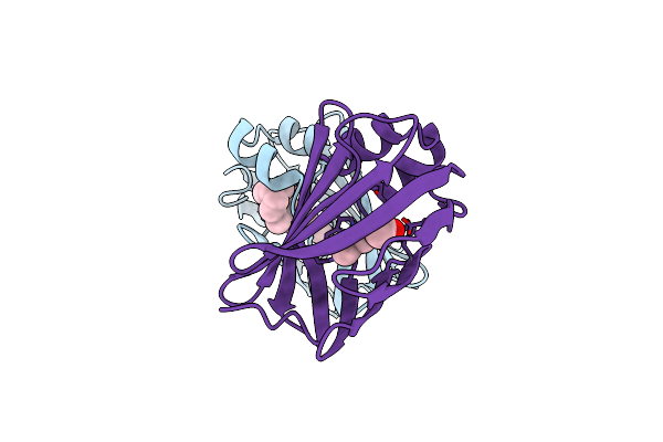 |
Crystal Structure Of The Q108K:K40L:T51V:R58W:Y19W Mutant Of Human Cellular Retinol Binding Protein Ii In Complex With All-Trans-Retinal At 1.68 Angstrom Resolution
Organism: Homo sapiens
Method: X-RAY DIFFRACTION Resolution:1.68 Å Release Date: 2016-12-21 Classification: TRANSPORT PROTEIN Ligands: ACT, RET |
Organism: Homo sapiens
Method: X-RAY DIFFRACTION
Release Date: 2016-12-21
Ligands: ACT, RET
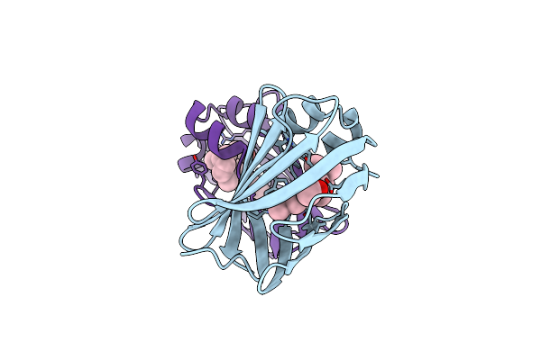 |
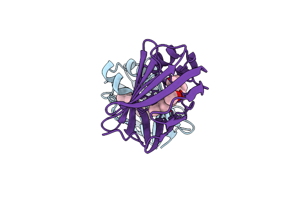
Crystal Structure Of The Q108K:K40L:T51V:R58Y:Y19W:Q38L Mutant Of Human Cellular Retinol Binding Protein Ii In Complex With All-Trans-Retinal At 1.48 Angstrom Resolution
Organism: Homo sapiens
Method: X-RAY DIFFRACTION Resolution:1.48 Å Release Date: 2016-12-14 Classification: TRANSPORT PROTEIN Ligands: RET, ACT |
Organism: Homo sapiens
Method: X-RAY DIFFRACTION
Release Date: 2016-12-14
Ligands: RET, ACT
 |
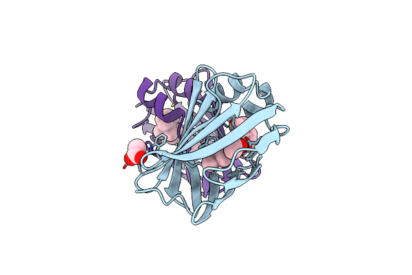
Crystal Structure Of The Q108K:K40L:T51V Mutant Of Human Cellular Retinol Binding Protein Ii In Complex With All-Trans-Retinal After 24 Hours Of Incubation At 1.4 Angstrom Resolution
Organism: Homo sapiens
Method: X-RAY DIFFRACTION Resolution:1.40 Å Release Date: 2016-12-14 Classification: TRANSPORT PROTEIN Ligands: ACT, RET |
Organism: Homo sapiens
Method: X-RAY DIFFRACTION
Release Date: 2016-12-14
Ligands: ACT, RET
 |
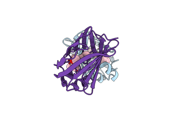
Crystal Structure Of The Q108K:K40L:T51V:R58F Mutant Of Human Cellular Retinol Binding Protein Ii In Complex With All-Trans-Retinal After 24 Hours Of Incubation At 1.54 Angstrom Resolution
Organism: Homo sapiens
Method: X-RAY DIFFRACTION Resolution:1.54 Å Release Date: 2016-12-07 Classification: TRANSPORT PROTEIN Ligands: RET, ACT |
Organism: Homo sapiens
Method: X-RAY DIFFRACTION
Release Date: 2016-12-07
Ligands: RET, ACT
 |
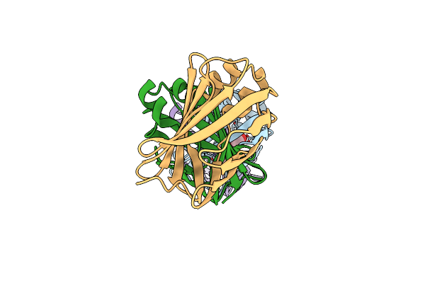
Crystal Structure Of The Q108K:K40L:T51V:R58Y:Y19W Mutant Of Human Cellular Retinol Binding Protein Ii In Complex With All-Trans-Retinal At 1.3 Angstrom Resolution
Organism: Homo sapiens
Method: X-RAY DIFFRACTION Resolution:1.31 Å Release Date: 2016-12-07 Classification: TRANSPORT PROTEIN Ligands: RET, ACT |
Organism: Homo sapiens
Method: X-RAY DIFFRACTION
Release Date: 2016-12-07
Ligands: RET, ACT
 |
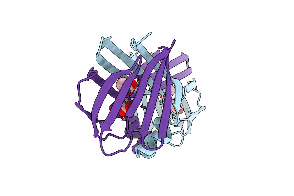
Crystal Structure Of Monomer Human Cellular Retinol Binding Protein Ii-Y60L
Organism: Homo sapiens
Method: X-RAY DIFFRACTION Resolution:1.50 Å Release Date: 2016-09-21 Classification: TRANSPORT PROTEIN Ligands: ACT |
Organism: Homo sapiens
Method: X-RAY DIFFRACTION
Release Date: 2016-09-21
Ligands: ACT
 |
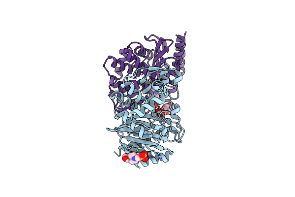
Crystal Structure Of E72A Mutant Of Domain Swapped Dimer Human Cellular Retinol Binding Protein
Organism: Homo sapiens
Method: X-RAY DIFFRACTION Resolution:1.78 Å Release Date: 2016-09-14 Classification: Retinol Binding Protein Ligands: ACT |
Organism: Homo sapiens
Method: X-RAY DIFFRACTION
Release Date: 2016-09-14
Ligands: ACT
 |
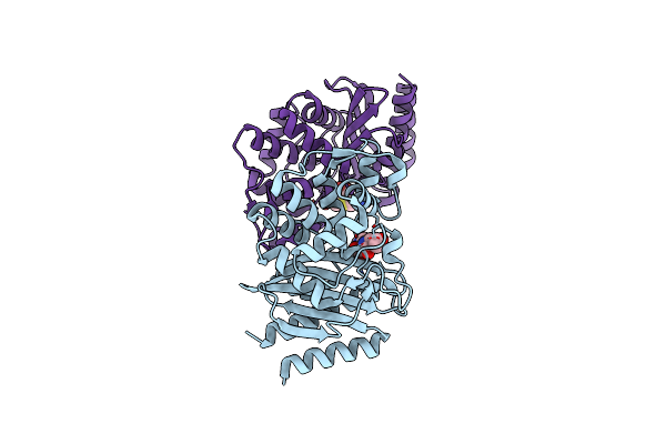
Apo Ges-5 C69G Mutant
Organism: Pseudomonas aeruginosa
Method: X-RAY DIFFRACTION Resolution:0.96 Å Release Date: 2016-09-07 Classification: HYDROLASE Ligands: EPE |
Organism: Pseudomonas aeruginosa
Method: X-RAY DIFFRACTION
Release Date: 2016-09-07
Ligands: EPE
 |
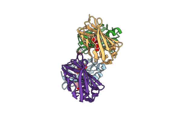
Imipenem Complex Of The Ges-5 C69G Mutant
Organism: Pseudomonas aeruginosa
Method: X-RAY DIFFRACTION Resolution:1.38 Å Release Date: 2016-09-07 Classification: HYDROLASE/ANTIBIOTIC Ligands: IM2 |
Organism: Pseudomonas aeruginosa
Method: X-RAY DIFFRACTION
Release Date: 2016-09-07
Ligands: IM2
 |
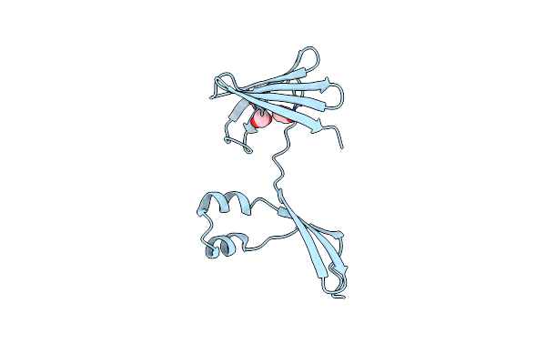
Crystal Structure Of Monomer Y60W Hcrbpii
Organism: Homo sapiens
Method: X-RAY DIFFRACTION Resolution:1.49 Å Release Date: 2016-06-08 Classification: TRANSPORT PROTEIN Ligands: ACT |
Organism: Homo sapiens
Method: X-RAY DIFFRACTION
Release Date: 2016-06-08
Ligands: ACT
 |
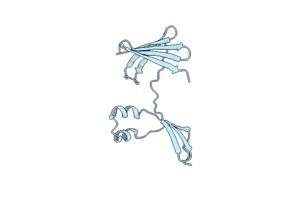
Crystal Structure Of The Domain-Swapped Dimer Y60L Mutant Of Human Cellular Retinol Binding Protein Ii
Organism: Homo sapiens
Method: X-RAY DIFFRACTION Resolution:1.55 Å Release Date: 2016-06-08 Classification: TRANSPORT PROTEIN Ligands: ACT |
Organism: Homo sapiens
Method: X-RAY DIFFRACTION
Release Date: 2016-06-08
Ligands: ACT
 |
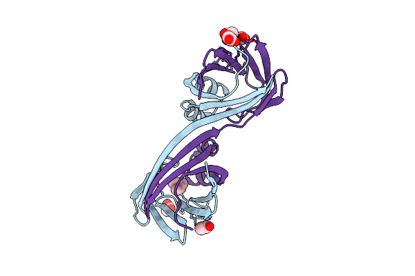
Crystal Structure Of The Domain-Swapped Dimer Wild-Type Of Human Cellular Retinol Binding Protein Ii
Organism: Homo sapiens
Method: X-RAY DIFFRACTION Resolution:2.66 Å Release Date: 2016-06-08 Classification: TRANSPORT PROTEIN |
Organism: Homo sapiens
Method: X-RAY DIFFRACTION
Release Date: 2016-06-08
 |
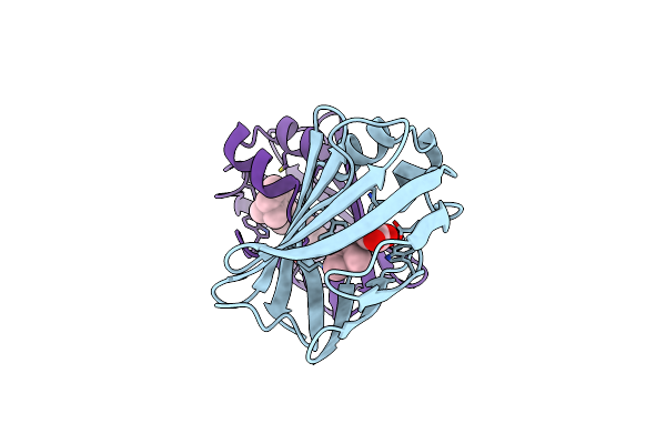
Crystal Structure Of The Domain-Swapped Dimer K40L:Q108K:Y60W Mutant Of Human Cellular Retinol Binding Protein Ii
Organism: Homo sapiens
Method: X-RAY DIFFRACTION Resolution:1.80 Å Release Date: 2016-06-08 Classification: LIPID BINDING PROTEIN Ligands: RET, ACT |
Organism: Homo sapiens
Method: X-RAY DIFFRACTION
Release Date: 2016-06-08
Ligands: RET, ACT
 |
The Crystal Structure Of Monomer Q108K:K40L:Y60W Crbpii Bound To All-Trans-Retinal
Organism: Homo sapiens
Method: X-RAY DIFFRACTION Resolution:1.50 Å Release Date: 2016-05-11 Classification: TRANSPORT PROTEIN Ligands: ACT, RET |
Organism: Homo sapiens
Method: X-RAY DIFFRACTION
Release Date: 2016-05-11
Ligands: ACT, RET