

Search Count: 27
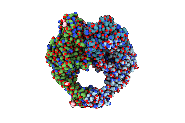 |
A 2.58A Crystal Structure Of S. Aureus Dna Gyrase And Dna With Metals Identified Through Anomalous Scattering
Organism: Staphylococcus aureus, Dna molecule
Method: X-RAY DIFFRACTION Resolution:2.58 Å Release Date: 2024-10-23 Classification: ISOMERASE Ligands: GOL, MN, BTB |
Organism: Staphylococcus aureus, Dna molecule
Method: X-RAY DIFFRACTION
Release Date: 2024-10-23
Ligands: GOL, MN, BTB
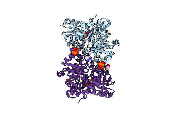 |
Human Serine Racemase Bound To Atp And Malonate.
Organism: Homo sapiens
Method: X-RAY DIFFRACTION Resolution:1.60 Å Release Date: 2021-03-03 Classification: ISOMERASE Ligands: IMD, MG, MLI, EDO, GOL, ATP |
Organism: Homo sapiens
Method: X-RAY DIFFRACTION
Release Date: 2021-03-03
Ligands: IMD, MG, MLI, EDO, GOL, ATP
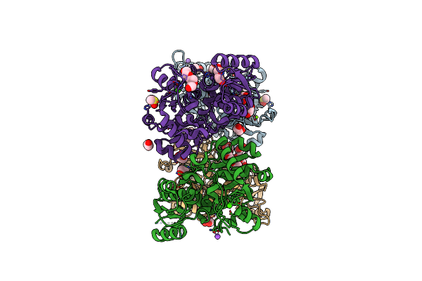 |
Human Serine Racemase Holoenzyme From 20% Dmso Soak (Xchem Crystallographic Fragment Screen).
Organism: Homo sapiens
Method: X-RAY DIFFRACTION Resolution:1.80 Å Release Date: 2021-03-03 Classification: ISOMERASE Ligands: PGE, DMS, MG, CA, NA, EDO, GOL |
Organism: Homo sapiens
Method: X-RAY DIFFRACTION
Release Date: 2021-03-03
Ligands: PGE, DMS, MG, CA, NA, EDO, GOL
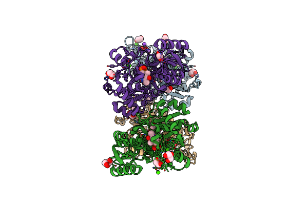 |
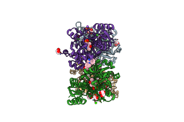
Crystal Structure Of Human Serine Racemase In Complex With Dsip Fragment Z2856434779, Xchem Fragment Screen.
Organism: Homo sapiens
Method: X-RAY DIFFRACTION Resolution:1.71 Å Release Date: 2021-03-03 Classification: ISOMERASE Ligands: PLP, GOL, CA, MG, EDO, NA, PG0, BP4 |
Organism: Homo sapiens
Method: X-RAY DIFFRACTION
Release Date: 2021-03-03
Ligands: PLP, GOL, CA, MG, EDO, NA, PG0, BP4
 |
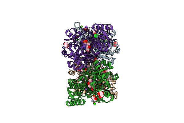
Crystal Structure Of Human Serine Racemase In Complex With Dsip Fragment Z235449082, Xchem Fragment Screen.
Organism: Homo sapiens
Method: X-RAY DIFFRACTION Resolution:1.87 Å Release Date: 2021-03-03 Classification: ISOMERASE Ligands: PLP, GOL, CA, MG, JFS, EDO, PGE, NA |
Organism: Homo sapiens
Method: X-RAY DIFFRACTION
Release Date: 2021-03-03
Ligands: PLP, GOL, CA, MG, JFS, EDO, PGE, NA
 |
Crystal Structure Of Human Serine Racemase In Complex With Dsip Fragment Z126932614, Xchem Fragment Screen.
Organism: Homo sapiens
Method: X-RAY DIFFRACTION Resolution:1.60 Å Release Date: 2021-03-03 Classification: ISOMERASE Ligands: PLP, GOL, CA, MG, DMS, T6J, NA, EDO, PGE, CL |
Organism: Homo sapiens
Method: X-RAY DIFFRACTION
Release Date: 2021-03-03
Ligands: PLP, GOL, CA, MG, DMS, T6J, NA, EDO, PGE, CL
 |
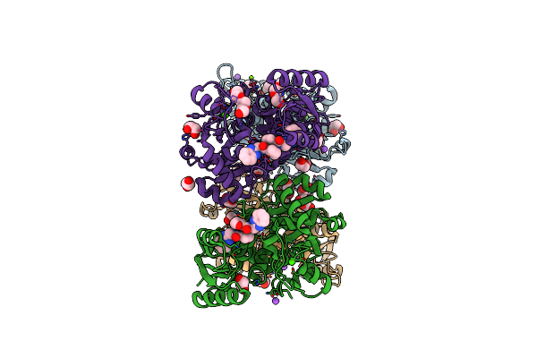
Crystal Structure Of Human Serine Racemase In Complex With Dsip Fragment Z52314092, Xchem Fragment Screen.
Organism: Homo sapiens
Method: X-RAY DIFFRACTION Resolution:1.53 Å Release Date: 2021-03-03 Classification: ISOMERASE Ligands: PLP, EDO, NA, CA, GOL, U78, MG, PG0, PGE, OXE |
Organism: Homo sapiens
Method: X-RAY DIFFRACTION
Release Date: 2021-03-03
Ligands: PLP, EDO, NA, CA, GOL, U78, MG, PG0, PGE, OXE
 |
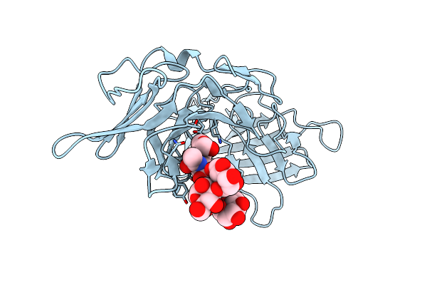
Crystal Structure Of Human Serine Racemase In Complex With Dsip Fragment Z26781964, Xchem Fragment Screen.
Organism: Homo sapiens
Method: X-RAY DIFFRACTION Resolution:1.77 Å Release Date: 2021-03-03 Classification: ISOMERASE Ligands: EDO, NA, CA, GOL, W0D, MG, CL, PGE |
Organism: Homo sapiens
Method: X-RAY DIFFRACTION
Release Date: 2021-03-03
Ligands: EDO, NA, CA, GOL, W0D, MG, CL, PGE
 |
Human Cation-Independent Mannose 6-Phosphate/ Igf2 Receptor Domains 9-10
Organism: Homo sapiens
Method: X-RAY DIFFRACTION Resolution:1.50 Å Release Date: 2020-08-19 Classification: SUGAR BINDING PROTEIN |
Organism: Homo sapiens
Method: X-RAY DIFFRACTION
Release Date: 2020-08-19
 |
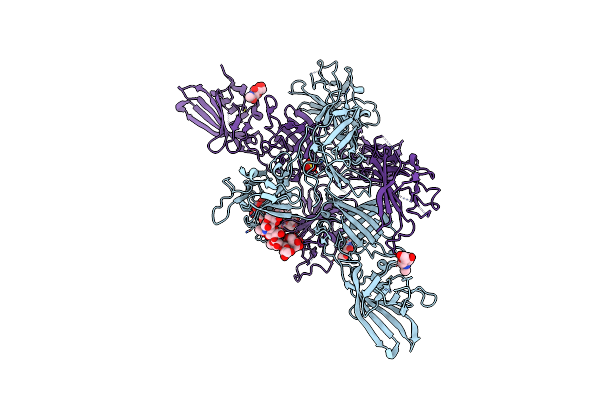
Human Cation-Independent Mannose 6-Phosphate/ Igf2 Receptor Domain 8
Organism: Homo sapiens
Method: X-RAY DIFFRACTION Resolution:2.56 Å Release Date: 2020-08-19 Classification: SUGAR BINDING PROTEIN Ligands: CL, NA |
Organism: Homo sapiens
Method: X-RAY DIFFRACTION
Release Date: 2020-08-19
Ligands: CL, NA
 |
Human Cation-Independent Mannose 6-Phosphate/Igf2 Receptor Domains 7-11
Organism: Homo sapiens
Method: X-RAY DIFFRACTION Resolution:3.47 Å Release Date: 2020-08-19 Classification: SUGAR BINDING PROTEIN Ligands: SO4, NAG |
Organism: Homo sapiens
Method: X-RAY DIFFRACTION
Release Date: 2020-08-19
Ligands: SO4, NAG
 |
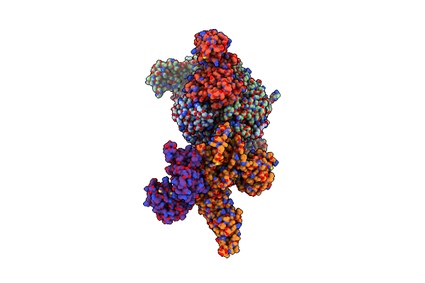
X-Ray Crystallographic Structure Of A High Affinity Igf2 Antagonist (Domain11 Ab5 Rhh) Based On Human Igf2R Domain 11
Organism: Homo sapiens
Method: X-RAY DIFFRACTION Resolution:2.80 Å Release Date: 2016-05-18 Classification: TRANSPORT PROTEIN Ligands: GOL, EDO, SO4 |
Organism: Homo sapiens
Method: X-RAY DIFFRACTION
Release Date: 2016-05-18
Ligands: GOL, EDO, SO4
 |
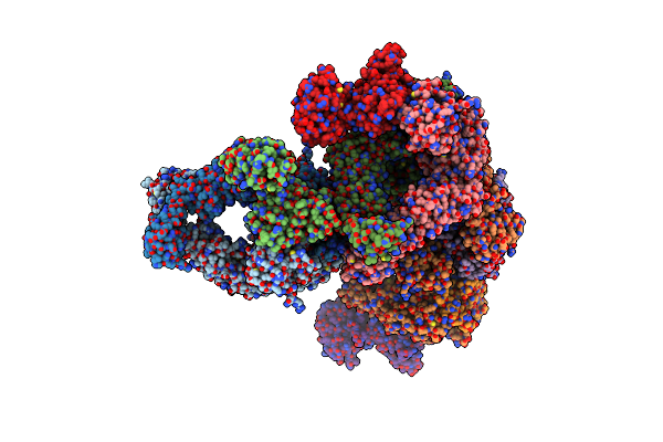
Muts In Complex With The N-Terminal Domain Of Mutl - Crystal Form 1
Organism: Escherichia coli k-12
Method: X-RAY DIFFRACTION Resolution:4.71 Å Release Date: 2015-07-22 Classification: DNA BINDING PROTEIN Ligands: ANP |
Organism: Escherichia coli k-12
Method: X-RAY DIFFRACTION
Release Date: 2015-07-22
Ligands: ANP
 |
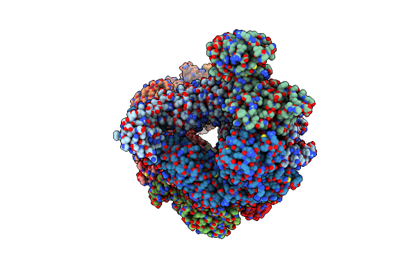
Muts In Complex With The N-Terminal Domain Of Mutl - Crystal Form 2
Organism: Escherichia coli k-12
Method: X-RAY DIFFRACTION Resolution:6.60 Å Release Date: 2015-07-22 Classification: HYDROLASE Ligands: ANP |
Organism: Escherichia coli k-12
Method: X-RAY DIFFRACTION
Release Date: 2015-07-22
Ligands: ANP
 |
Muts In Complex With The N-Terminal Domain Of Mutl - Crystal Form 3
Organism: Escherichia coli
Method: X-RAY DIFFRACTION Resolution:7.60 Å Release Date: 2015-07-22 Classification: DNA BINDING PROTEIN Ligands: ANP |
Organism: Escherichia coli
Method: X-RAY DIFFRACTION
Release Date: 2015-07-22
Ligands: ANP
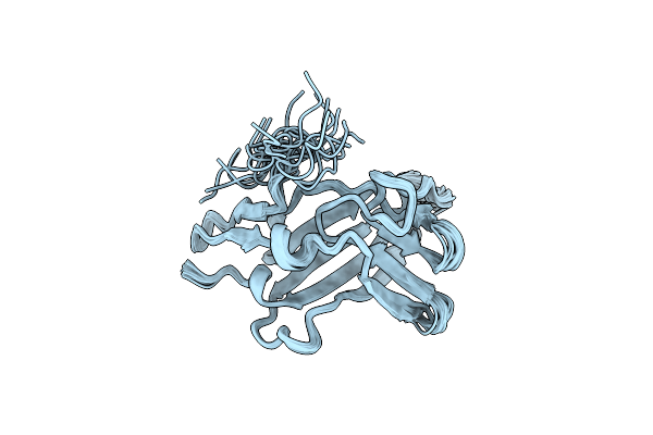 |
Nmr Solution Structure Ensemble Of 3-4D Mutant Domain 11 Igf2R In Complex With Igf2 (Domain 11 Structure Only)
Organism: Homo sapiens
Method: SOLUTION NMR Release Date: 2014-10-15 Classification: ANTITUMOR PROTEIN |
Organism: Homo sapiens
Method: SOLUTION NMR
Release Date: 2014-10-15
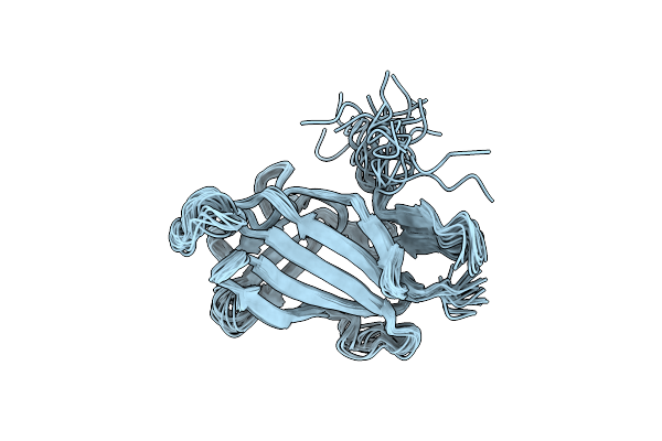 |
Nmr Solution Structure Ensemble Of 3-4D Mutant Domain 11 Igf2R
Organism: Homo sapiens
Method: SOLUTION NMR Release Date: 2014-10-15 Classification: ANTITUMOR PROTEIN |
Organism: Homo sapiens
Method: SOLUTION NMR
Release Date: 2014-10-15
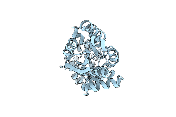 |
The Putative Substrate Binding Domain Of Abc-Type Transporter From Agrobacterium Tumefaciens In Open Conformation
Organism: Agrobacterium fabrum
Method: X-RAY DIFFRACTION Resolution:2.30 Å Release Date: 2013-12-11 Classification: TRANSPORT PROTEIN |
Organism: Agrobacterium fabrum
Method: X-RAY DIFFRACTION
Release Date: 2013-12-11
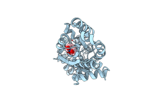 |
Crystal Structure Of Abc Transporter Substrate Binding Protein Prox From Agrobacterium Tumefaciens Cocrystalized With Btb
Organism: Agrobacterium fabrum
Method: X-RAY DIFFRACTION Resolution:1.73 Å Release Date: 2013-11-27 Classification: TRANSPORT PROTEIN Ligands: BTB, CL |
Organism: Agrobacterium fabrum
Method: X-RAY DIFFRACTION
Release Date: 2013-11-27
Ligands: BTB, CL
 |
Crystal Structure Of Dethiobiotin Synthetase (Biod) From Helicobacter Pylori Complexed With Atp
Organism: Helicobacter pylori
Method: X-RAY DIFFRACTION Resolution:1.34 Å Release Date: 2011-03-30 Classification: LIGASE Ligands: ATP, MG, NO3, EDO, GOL, PEG |
Organism: Helicobacter pylori
Method: X-RAY DIFFRACTION
Release Date: 2011-03-30
Ligands: ATP, MG, NO3, EDO, GOL, PEG