

Search Count: 44
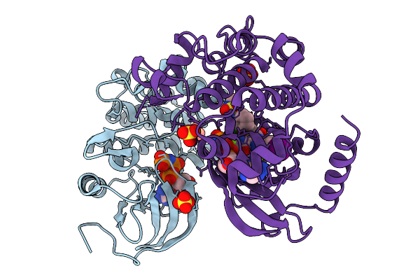 |
Crystal Structure Of The Inactive Braf/Mek1 Complex Bound To Ik-595
Organism: Homo sapiens
Method: X-RAY DIFFRACTION Resolution:3.46 Å Release Date: 2026-01-14 Classification: ONCOPROTEIN/INHIBITOR Ligands: ANP, MG, SO4, A1BIS |
Organism: Homo sapiens
Method: X-RAY DIFFRACTION
Release Date: 2026-01-14
Ligands: ANP, MG, SO4, A1BIS
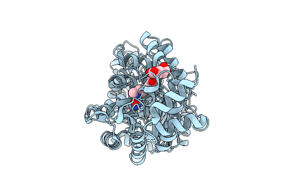 |
Human Methionine Synthase With Methyltetrahydrofolate, N-Half From Full-Length
Organism: Homo sapiens
Method: ELECTRON MICROSCOPY Release Date: 2025-10-15 Classification: TRANSFERASE Ligands: THH |
Organism: Homo sapiens
Method: ELECTRON MICROSCOPY
Release Date: 2025-10-15
Ligands: THH
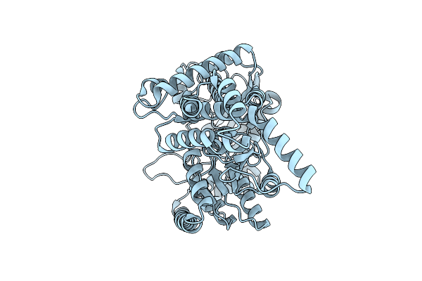 |
Human Methionine Synthase With Methyltetrahydrofolate, C-Half From Full-Length
Organism: Homo sapiens
Method: ELECTRON MICROSCOPY Release Date: 2025-10-15 Classification: TRANSFERASE |
Organism: Homo sapiens
Method: ELECTRON MICROSCOPY
Release Date: 2025-10-15
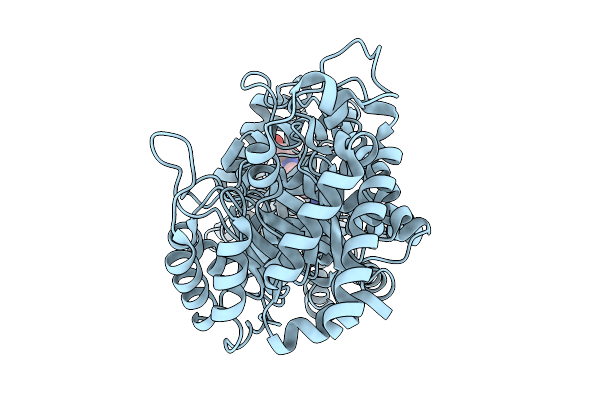 |
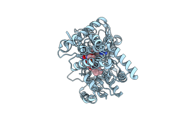
Human Methionine Synthase With Methyltetrahydrofolate, Hydroxocobalamin, And Sam, N-Half From Full-Length
Organism: Homo sapiens
Method: ELECTRON MICROSCOPY Release Date: 2025-10-15 Classification: TRANSFERASE Ligands: THH |
Organism: Homo sapiens
Method: ELECTRON MICROSCOPY
Release Date: 2025-10-15
Ligands: THH
 |
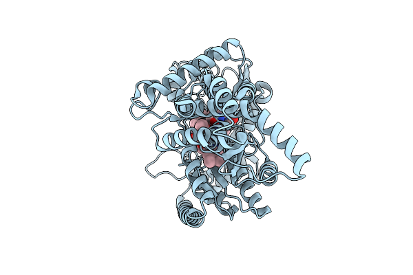
Human Methionine Synthase With Methyltetrahydrofolate, Hydroxocobalamin, And Sam, C-Half His-On From Full-Length
Organism: Homo sapiens
Method: ELECTRON MICROSCOPY Release Date: 2025-10-15 Classification: TRANSFERASE Ligands: B12 |
Organism: Homo sapiens
Method: ELECTRON MICROSCOPY
Release Date: 2025-10-15
Ligands: B12
 |
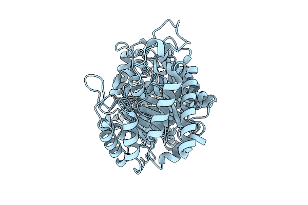
Human Methionine Synthase With Methyltetrahydrofolate, Hydroxocobalamin, And Sam, C-Half His-Off From Full-Length
Organism: Homo sapiens
Method: ELECTRON MICROSCOPY Release Date: 2025-10-15 Classification: TRANSFERASE Ligands: B12 |
Organism: Homo sapiens
Method: ELECTRON MICROSCOPY
Release Date: 2025-10-15
Ligands: B12
 |
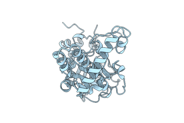
Human Methionine Synthase With Methylcobalamin, N-Half From Full-Length
Organism: Homo sapiens
Method: ELECTRON MICROSCOPY Release Date: 2025-10-15 Classification: TRANSFERASE |
Organism: Homo sapiens
Method: ELECTRON MICROSCOPY
Release Date: 2025-10-15
 |
Human Methionine Synthase With Methylcobalamin, Activation Domain From Full-Length
Organism: Homo sapiens
Method: ELECTRON MICROSCOPY Release Date: 2025-10-15 Classification: TRANSFERASE |
Organism: Homo sapiens
Method: ELECTRON MICROSCOPY
Release Date: 2025-10-15
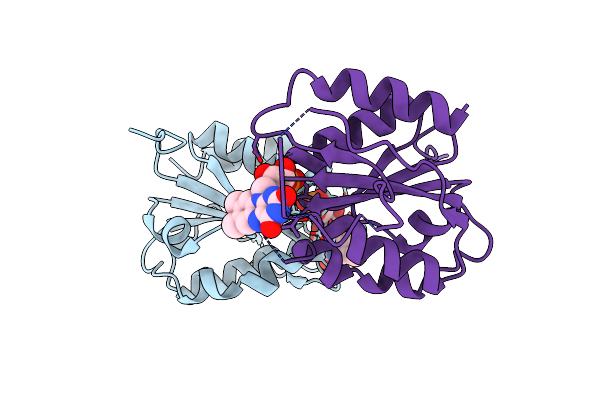 |
X-Ray Crystal Structure Of Fmn-Bound Long-Chain Flavodoxin From Rhodopseudomonas Palustris
Organism: Rhodopseudomonas palustris
Method: X-RAY DIFFRACTION Resolution:2.17 Å Release Date: 2024-03-13 Classification: FLAVOPROTEIN Ligands: FMN |
Organism: Rhodopseudomonas palustris
Method: X-RAY DIFFRACTION
Release Date: 2024-03-13
Ligands: FMN
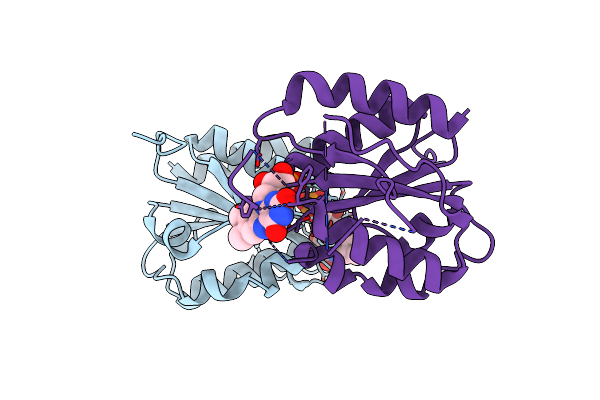 |
Room Temperature X-Ray Crystal Structure Of Fmn-Bound Long-Chain Flavodoxin From Rhodopseudomonas Palustris
Organism: Rhodopseudomonas palustris
Method: X-RAY DIFFRACTION Resolution:2.86 Å Release Date: 2024-03-13 Classification: FLAVOPROTEIN Ligands: FMN |
Organism: Rhodopseudomonas palustris
Method: X-RAY DIFFRACTION
Release Date: 2024-03-13
Ligands: FMN
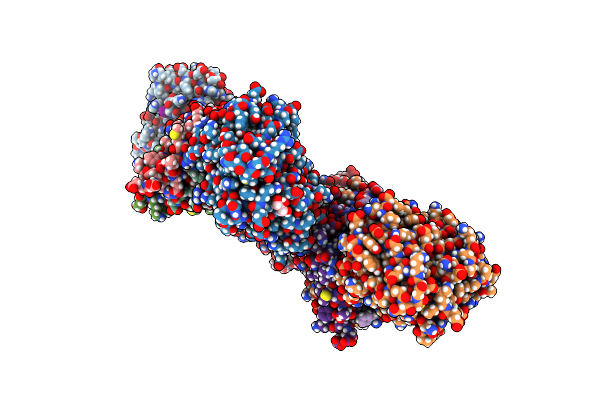 |
Bacillus Phage Vmy22 P56 In Complex With Bacillus Weidmannii Ung
Organism: Bacillus wiedmannii, Bacillus phage vmy22
Method: X-RAY DIFFRACTION Resolution:2.45 Å Release Date: 2023-06-21 Classification: PROTEIN BINDING Ligands: GOL, IOD |
Organism: Bacillus wiedmannii, Bacillus phage vmy22
Method: X-RAY DIFFRACTION
Release Date: 2023-06-21
Ligands: GOL, IOD
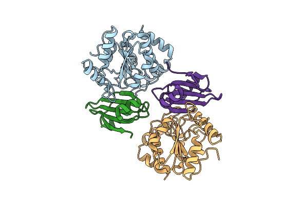 |
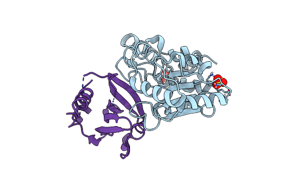
Ugi-2 Saung Complex
Organism: Bacillus phage vb_bpum-bpsp, Staphylococcus aureus
Method: X-RAY DIFFRACTION Resolution:2.60 Å Release Date: 2023-06-21 Classification: PROTEIN BINDING |
Organism: Bacillus phage vb_bpum-bpsp, Staphylococcus aureus
Method: X-RAY DIFFRACTION
Release Date: 2023-06-21
 |
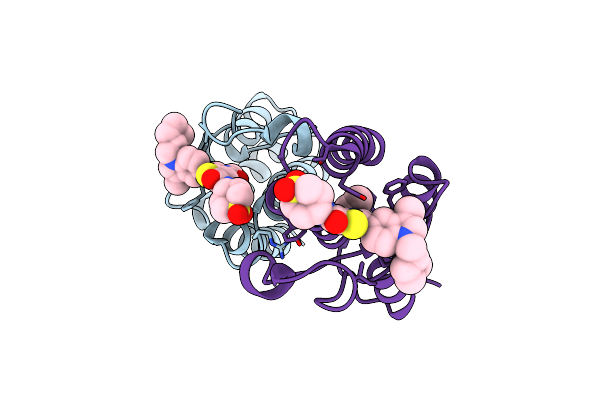
Mcugi Saung Complex
Organism: Staphylococcus aureus, Macrococcus caseolyticus
Method: X-RAY DIFFRACTION Resolution:2.70 Å Release Date: 2023-06-21 Classification: PROTEIN BINDING Ligands: EDO, SO4 |
Organism: Staphylococcus aureus, Macrococcus caseolyticus
Method: X-RAY DIFFRACTION
Release Date: 2023-06-21
Ligands: EDO, SO4
 |
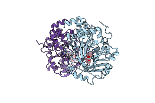
Crystal Structure Of Brd9 Bromodomain In Complex With Compound Ea-89
Organism: Homo sapiens
Method: X-RAY DIFFRACTION Resolution:1.76 Å Release Date: 2022-08-10 Classification: TRANSCRIPTION Ligands: L8L |
Organism: Homo sapiens
Method: X-RAY DIFFRACTION
Release Date: 2022-08-10
Ligands: L8L
 |
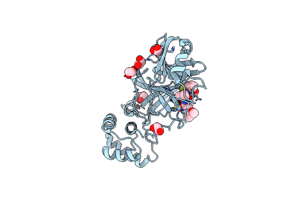
Crystal Structure Of Kdm4B In Complex With Qc6352
Organism: Homo sapiens
Method: X-RAY DIFFRACTION Resolution:2.70 Å Release Date: 2022-02-02 Classification: OXIDOREDUCTASE/OXIDOREDUCTASE inhibitor Ligands: NI, 9DJ, ZN |
Organism: Homo sapiens
Method: X-RAY DIFFRACTION
Release Date: 2022-02-02
Ligands: NI, 9DJ, ZN
 |
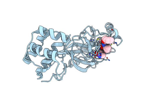
Crystal Structure Of Sar-Cov-2 3Cl Protease Complex With Inhibitor Yh-53
Organism: Severe acute respiratory syndrome coronavirus 2
Method: X-RAY DIFFRACTION Resolution:1.65 Å Release Date: 2021-06-23 Classification: HYDROLASE Ligands: HUR, EDO |
Organism: Severe acute respiratory syndrome coronavirus 2
Method: X-RAY DIFFRACTION
Release Date: 2021-06-23
Ligands: HUR, EDO
 |
Crystal Structure Of Sar-Cov-2 3Cl Protease Complex With Inhibitor Sh-5
Organism: Severe acute respiratory syndrome coronavirus 2
Method: X-RAY DIFFRACTION Resolution:2.15 Å Release Date: 2021-06-23 Classification: HYDROLASE Ligands: HUO |
Organism: Severe acute respiratory syndrome coronavirus 2
Method: X-RAY DIFFRACTION
Release Date: 2021-06-23
Ligands: HUO
 |
Structure Of The Pscd-Region Of The Cell Wall Protein Pleuralin-1
Organism: Cylindrotheca fusiformis
Method: SOLUTION NMR Release Date: 2016-12-21 Classification: STRUCTURAL PROTEIN |
Organism: Cylindrotheca fusiformis
Method: SOLUTION NMR
Release Date: 2016-12-21
 |
Structure Of E. Coli Beta-Glucuronidase Bound With A Novel, Potent Inhibitor 1-((6,8-Dimethyl-2-Oxo-1,2-Dihydroquinolin-3-Yl)Methyl)-1-(2-Hydroxyethyl)-3-(4-Hydroxyphenyl)Thiourea
Organism: Escherichia coli
Method: X-RAY DIFFRACTION Resolution:2.39 Å Release Date: 2015-10-14 Classification: Hydrolase/Hydrolase Inhibitor Ligands: 57Z |
Organism: Escherichia coli
Method: X-RAY DIFFRACTION
Release Date: 2015-10-14
Ligands: 57Z
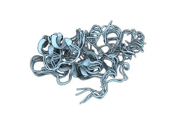 |
Structure Of The Pscd4-Domain Of The Cell Wall Protein Pleuralin-1 From The Diatom Cylindrotheca Fusiformis
Organism: Cylindrotheca fusiformis
Method: SOLUTION NMR Release Date: 2015-02-25 Classification: STRUCTURAL PROTEIN |
Organism: Cylindrotheca fusiformis
Method: SOLUTION NMR
Release Date: 2015-02-25