

Search Count: 47
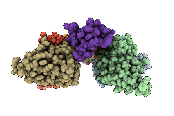 |
D11 Mabs Bound To Alpha-Bungarotoxin At Ph 7.5
Organism: Homo sapiens, Bungarus multicinctus
Method: X-RAY DIFFRACTION Resolution:1.33 Å Release Date: 2025-11-19 Classification: TOXIN Ligands: EDO |
Organism: Homo sapiens, Bungarus multicinctus
Method: X-RAY DIFFRACTION
Release Date: 2025-11-19
Ligands: EDO
 |
A01 Mabs Bound To Cobratoxin At Ph 5.5
Organism: Homo sapiens, Naja kaouthia
Method: X-RAY DIFFRACTION Resolution:1.60 Å Release Date: 2025-11-19 Classification: TOXIN Ligands: GOL, CL, K, NA, SO4 |
Organism: Homo sapiens, Naja kaouthia
Method: X-RAY DIFFRACTION
Release Date: 2025-11-19
Ligands: GOL, CL, K, NA, SO4
 |
A01 Mabs Bound To Cobratoxin At Ph 6.0
Organism: Homo sapiens, Naja kaouthia
Method: X-RAY DIFFRACTION Resolution:1.49 Å Release Date: 2025-11-19 Classification: TOXIN Ligands: CL, GOL, TRS, SO4, NA |
Organism: Homo sapiens, Naja kaouthia
Method: X-RAY DIFFRACTION
Release Date: 2025-11-19
Ligands: CL, GOL, TRS, SO4, NA
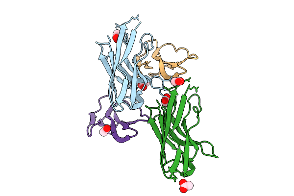 |
Co-Crystal Of Broadly Neutralizing Biparatopic Vhh In Complex With Cardiotoxin (P01468) Naja Pallida
Organism: Vicugna pacos, Naja pallida
Method: X-RAY DIFFRACTION Resolution:1.60 Å Release Date: 2025-11-05 Classification: TOXIN Ligands: ACT |
Organism: Vicugna pacos, Naja pallida
Method: X-RAY DIFFRACTION
Release Date: 2025-11-05
Ligands: ACT
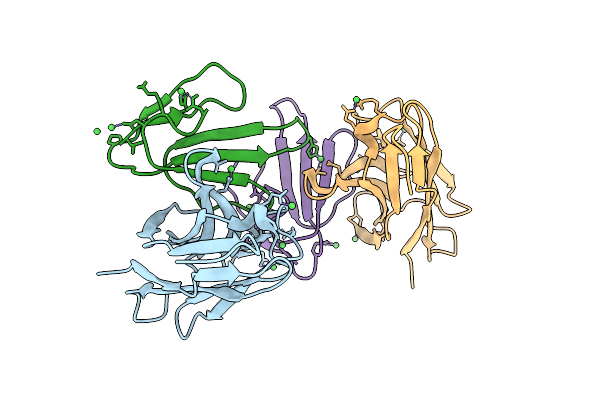 |
Co-Crystal Of Broadly Neutralizing Vhh In Complex With Short Neurotoxin 1 (P01426) Naja Pallida
Organism: Vicugna pacos, Naja pallida
Method: X-RAY DIFFRACTION Resolution:2.50 Å Release Date: 2025-11-05 Classification: TOXIN Ligands: NI |
Organism: Vicugna pacos, Naja pallida
Method: X-RAY DIFFRACTION
Release Date: 2025-11-05
Ligands: NI
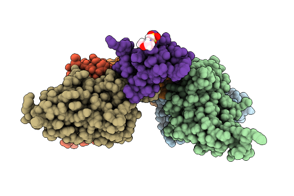 |
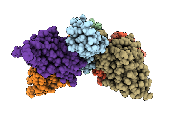
D11 Mabs Bound To Alpha-Bungarotoxin
Organism: Homo sapiens, Bungarus multicinctus
Method: X-RAY DIFFRACTION Resolution:1.32 Å Release Date: 2025-07-16 Classification: TOXIN Ligands: MES |
Organism: Homo sapiens, Bungarus multicinctus
Method: X-RAY DIFFRACTION
Release Date: 2025-07-16
Ligands: MES
 |
Mabs In Complex With Cobratoxin At Ph 4.5
Organism: Naja kaouthia, Homo sapiens
Method: X-RAY DIFFRACTION Resolution:1.55 Å Release Date: 2025-07-16 Classification: TOXIN Ligands: CL, GOL, SO4, NA |
Organism: Naja kaouthia, Homo sapiens
Method: X-RAY DIFFRACTION
Release Date: 2025-07-16
Ligands: CL, GOL, SO4, NA
 |
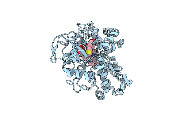
The Crystal Structure Of T252E Cyp199A4 Bound To 4-(Pyridin-2-Yl)Benzoic Acid
Organism: Rhodopseudomonas palustris haa2
Method: X-RAY DIFFRACTION Resolution:1.73 Å Release Date: 2023-10-25 Classification: OXIDOREDUCTASE Ligands: CL, HEM, PQS |
Organism: Rhodopseudomonas palustris haa2
Method: X-RAY DIFFRACTION
Release Date: 2023-10-25
Ligands: CL, HEM, PQS
 |
The Crystal Structure Of T252E Cyp199A4 Bound To 4-(Thiophen-3-Yl)Benzoic Acid
Organism: Rhodopseudomonas palustris haa2
Method: X-RAY DIFFRACTION Resolution:2.02 Å Release Date: 2023-10-25 Classification: OXIDOREDUCTASE Ligands: EJD, HEM, CL |
Organism: Rhodopseudomonas palustris haa2
Method: X-RAY DIFFRACTION
Release Date: 2023-10-25
Ligands: EJD, HEM, CL
 |
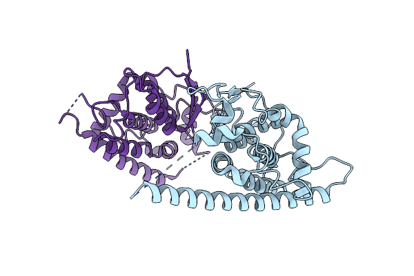
The Crystal Structure Of The T252E Mutant Of Cyp199A4 Bound To 4-Benzylbenzoic Acid
Organism: Rhodopseudomonas palustris haa2
Method: X-RAY DIFFRACTION Resolution:1.79 Å Release Date: 2023-10-25 Classification: OXIDOREDUCTASE Ligands: IRJ, HEM, CL |
Organism: Rhodopseudomonas palustris haa2
Method: X-RAY DIFFRACTION
Release Date: 2023-10-25
Ligands: IRJ, HEM, CL
 |
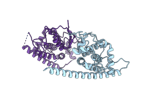
Human Gdap1, A247V Mutant
Organism: Homo sapiens
Method: X-RAY DIFFRACTION Resolution:2.68 Å Release Date: 2023-04-26 Classification: MEMBRANE PROTEIN |
Organism: Homo sapiens
Method: X-RAY DIFFRACTION
Release Date: 2023-04-26
 |
Human Gdap1, R282H Mutant
Organism: Homo sapiens
Method: X-RAY DIFFRACTION Resolution:1.95 Å Release Date: 2023-04-26 Classification: MEMBRANE PROTEIN |
Organism: Homo sapiens
Method: X-RAY DIFFRACTION
Release Date: 2023-04-26
 |
Crystal Structure Of The Human Gdap1 Cmt2 Mutant-H123R
Organism: Homo sapiens
Method: X-RAY DIFFRACTION Resolution:2.20 Å Release Date: 2022-06-08 Classification: SIGNALING PROTEIN Ligands: PEG |
Organism: Homo sapiens
Method: X-RAY DIFFRACTION
Release Date: 2022-06-08
Ligands: PEG
 |
Crystal Structure Of The Human Gdap1 Cmt2 Mutant-R120W
Organism: Homo sapiens
Method: X-RAY DIFFRACTION Resolution:3.41 Å Release Date: 2022-06-08 Classification: SIGNALING PROTEIN |
Organism: Homo sapiens
Method: X-RAY DIFFRACTION
Release Date: 2022-06-08
 |
Human Gdap1 Core Domain, Trigonal Crystal Form
Organism: Homo sapiens
Method: X-RAY DIFFRACTION Resolution:3.20 Å Release Date: 2022-06-08 Classification: MEMBRANE PROTEIN |
Organism: Homo sapiens
Method: X-RAY DIFFRACTION
Release Date: 2022-06-08
 |
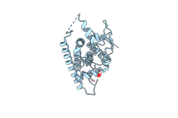
Crystal Structure Of T252E Cyp199A4 Bound To 4-Methoxybenzoic Acid
Organism: Rhodopseudomonas palustris (strain haa2)
Method: X-RAY DIFFRACTION Resolution:1.55 Å Release Date: 2022-02-02 Classification: OXIDOREDUCTASE Ligands: ANN, CL, MG, ACT, HEM |
Organism: Rhodopseudomonas palustris (strain haa2)
Method: X-RAY DIFFRACTION
Release Date: 2022-02-02
Ligands: ANN, CL, MG, ACT, HEM
 |
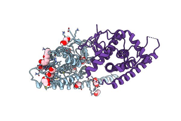
Crystal Structure Of R120Q Gdap1 Mutant
Organism: Homo sapiens
Method: X-RAY DIFFRACTION Resolution:3.00 Å Release Date: 2021-12-08 Classification: SIGNALING PROTEIN Ligands: GOL |
Organism: Homo sapiens
Method: X-RAY DIFFRACTION
Release Date: 2021-12-08
Ligands: GOL
 |
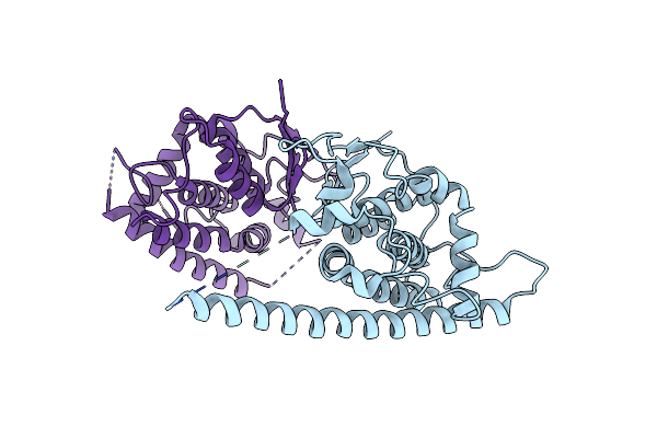
Complex Of Human Gdap1 With Hexadecanedioic Acid
Organism: Homo sapiens
Method: X-RAY DIFFRACTION Resolution:2.20 Å Release Date: 2021-02-24 Classification: SIGNALING PROTEIN Ligands: GOL, EOH, RF8 |
Organism: Homo sapiens
Method: X-RAY DIFFRACTION
Release Date: 2021-02-24
Ligands: GOL, EOH, RF8
 |
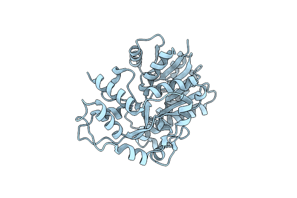
Crystal Structure Of Human Gdap1 At 2.8 Angstrom Resolution.
Organism: Homo sapiens
Method: X-RAY DIFFRACTION Resolution:2.80 Å Release Date: 2021-02-24 Classification: SIGNALING PROTEIN |
Organism: Homo sapiens
Method: X-RAY DIFFRACTION
Release Date: 2021-02-24
 |
Crystal Structure Of Gre2 From Candida Albicans
Organism: Candida albicans sc5314
Method: X-RAY DIFFRACTION Resolution:2.80 Å Release Date: 2020-09-09 Classification: OXIDOREDUCTASE |
Organism: Candida albicans sc5314
Method: X-RAY DIFFRACTION
Release Date: 2020-09-09