

Search Count: 25
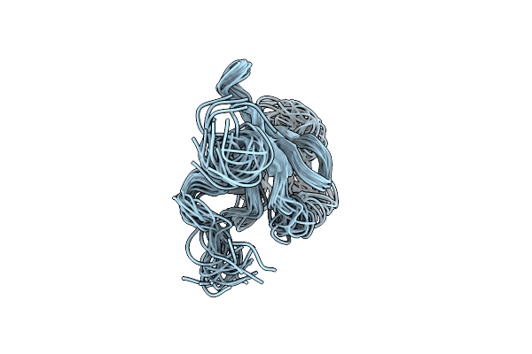 |
Sipa Solution Structure
Organism: Synechocystis
Method: SOLUTION NMR Release Date: 2024-03-06 Classification: UNKNOWN FUNCTION |
Organism: Synechocystis
Method: SOLUTION NMR
Release Date: 2024-03-06
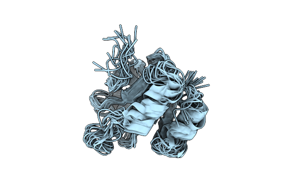 |
Structure Of Thioredoxin M From Pea
|
Organism: Pisum sativum
Method: SOLUTION NMR
Release Date: 2024-02-14
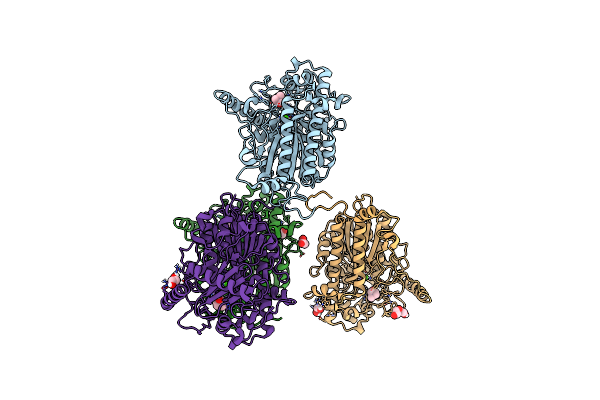 |
C54S Mutant Of Choline-Sulfatase From E. Meliloti Cect4857 Bound To Choline
Organism: Rhizobium meliloti
Method: X-RAY DIFFRACTION Resolution:1.85 Å Release Date: 2022-08-03 Classification: HYDROLASE Ligands: CHT, CA, ACT, EDO, PEG |
Organism: Rhizobium meliloti
Method: X-RAY DIFFRACTION
Release Date: 2022-08-03
Ligands: CHT, CA, ACT, EDO, PEG
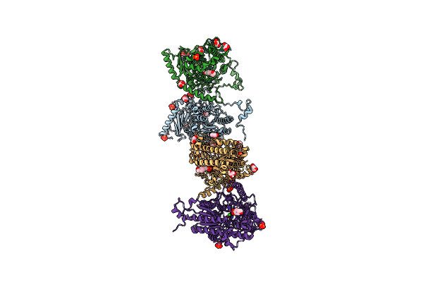 |
C54S Mutant Of Choline-Sulfatase From E. Meliloti Cect4857 Bound To Hepes
Organism: Rhizobium meliloti
Method: X-RAY DIFFRACTION Resolution:2.10 Å Release Date: 2022-08-03 Classification: HYDROLASE Ligands: EPE, SO4, GOL, CA |
Organism: Rhizobium meliloti
Method: X-RAY DIFFRACTION
Release Date: 2022-08-03
Ligands: EPE, SO4, GOL, CA
 |
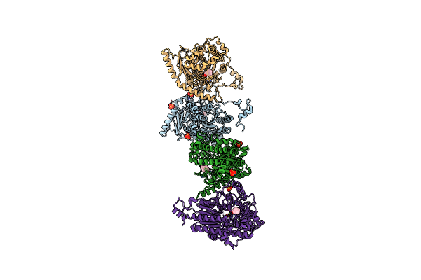
Choline Sulfatase From Ensifer (Sinorhizobium) Meliloti
Organism: Sinorhizobium meliloti cect 4857
Method: X-RAY DIFFRACTION Resolution:1.98 Å Release Date: 2019-04-10 Classification: HYDROLASE Ligands: MG, SO4 |
Organism: Sinorhizobium meliloti cect 4857
Method: X-RAY DIFFRACTION
Release Date: 2019-04-10
Ligands: MG, SO4
 |
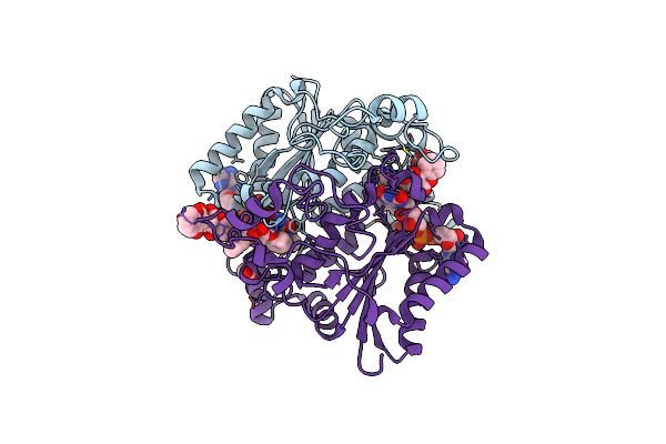
Choline Sulfatase From Ensifer (Sinorhizobium) Meliloti Cocrystalized With Choline
Organism: Rhizobium meliloti (strain 1021)
Method: X-RAY DIFFRACTION Resolution:1.84 Å Release Date: 2019-04-10 Classification: HYDROLASE Ligands: CHT, MG, SO4 |
Organism: Rhizobium meliloti (strain 1021)
Method: X-RAY DIFFRACTION
Release Date: 2019-04-10
Ligands: CHT, MG, SO4
 |
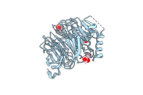
Crystal Structure Of The H80R Variant Of Nqo1 Bound To Dicoumarol
Organism: Homo sapiens
Method: X-RAY DIFFRACTION Resolution:2.04 Å Release Date: 2017-02-22 Classification: OXIDOREDUCTASE Ligands: FAD, DTC, ACT |
Organism: Homo sapiens
Method: X-RAY DIFFRACTION
Release Date: 2017-02-22
Ligands: FAD, DTC, ACT
 |
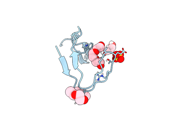
The Carboxy-Terminal Domain Of Erb1 Is A Seven-Bladed Beta-Propeller That Binds Rna.
Organism: Saccharomyces cerevisiae
Method: X-RAY DIFFRACTION Resolution:1.60 Å Release Date: 2015-04-29 Classification: PROTEIN BINDING Ligands: EDO, GOL, EOH |
Organism: Saccharomyces cerevisiae
Method: X-RAY DIFFRACTION
Release Date: 2015-04-29
Ligands: EDO, GOL, EOH
 |
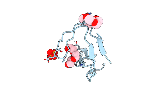
Crystal Structure Of The Intertwined Dimer Of The C-Src Tyrosine Kinase Sh3 Domain Mutant N113S
Organism: Gallus gallus
Method: X-RAY DIFFRACTION Resolution:1.90 Å Release Date: 2015-01-28 Classification: TRANSFERASE Ligands: PGE, PEG, GOL, SO4 |
Organism: Gallus gallus
Method: X-RAY DIFFRACTION
Release Date: 2015-01-28
Ligands: PGE, PEG, GOL, SO4
 |
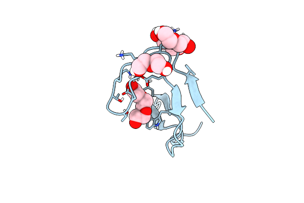
Crystal Structure Of The Intertwined Dimer Of The C-Src Tyrosine Kinase Sh3 Domain Mutant S94A
Organism: Gallus gallus
Method: X-RAY DIFFRACTION Resolution:2.00 Å Release Date: 2015-01-28 Classification: TRANSFERASE Ligands: PGE, PEG, SO4 |
Organism: Gallus gallus
Method: X-RAY DIFFRACTION
Release Date: 2015-01-28
Ligands: PGE, PEG, SO4
 |
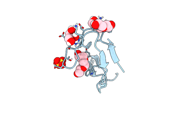
Crystal Structure Of The Intertwined Dimer Of The C-Src Tyrosine Kinase Sh3 Domain Mutant Q128R
Organism: Gallus gallus
Method: X-RAY DIFFRACTION Resolution:1.60 Å Release Date: 2014-12-10 Classification: TRANSFERASE Ligands: PGE, PEG |
Organism: Gallus gallus
Method: X-RAY DIFFRACTION
Release Date: 2014-12-10
Ligands: PGE, PEG
 |
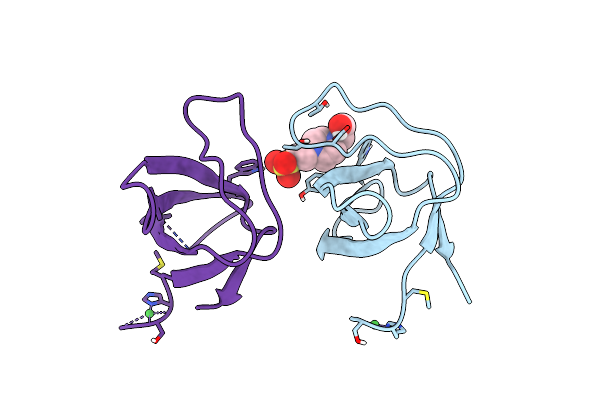
Crystal Structure Of The Intertwined Dimer Of The C-Src Tyrosine Kinase Sh3 Domain Mutant Q128E
Organism: Gallus gallus
Method: X-RAY DIFFRACTION Resolution:1.50 Å Release Date: 2014-12-10 Classification: TRANSFERASE Ligands: PGE, PEG, SO4, GOL |
Organism: Gallus gallus
Method: X-RAY DIFFRACTION
Release Date: 2014-12-10
Ligands: PGE, PEG, SO4, GOL
 |
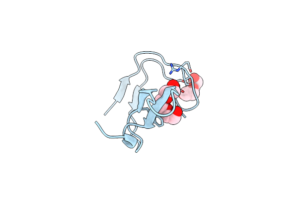
Crystal Structure Of The C-Src Tyrosine Kinase Sh3 Domain Mutant Q128E
Organism: Gallus gallus
Method: X-RAY DIFFRACTION Resolution:1.04 Å Release Date: 2014-12-10 Classification: TRANSFERASE Ligands: NI, EPE |
Organism: Gallus gallus
Method: X-RAY DIFFRACTION
Release Date: 2014-12-10
Ligands: NI, EPE
 |
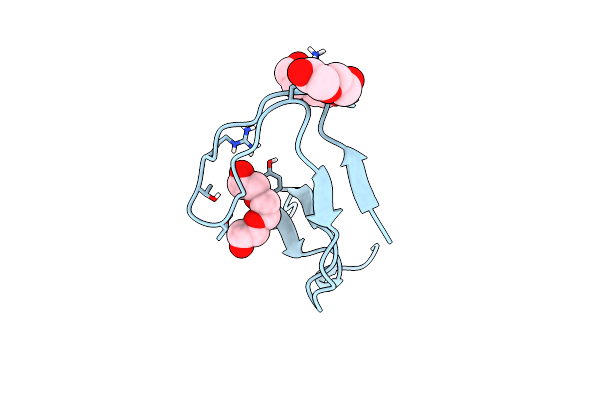
Crystal Structure Of The Intertwined Dimer Of The C-Src Tyrosine Kinase Sh3 Domain Mutant Q128K
Organism: Gallus gallus
Method: X-RAY DIFFRACTION Resolution:2.00 Å Release Date: 2014-12-10 Classification: TRANSFERASE Ligands: PGE, PEG |
Organism: Gallus gallus
Method: X-RAY DIFFRACTION
Release Date: 2014-12-10
Ligands: PGE, PEG
 |
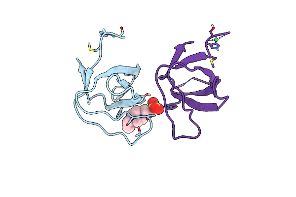
Crystal Structure Of The Chicken C-Src-Sh3 Domain Intertwined Dimer
Organism: Gallus gallus
Method: X-RAY DIFFRACTION Resolution:1.85 Å Release Date: 2014-04-23 Classification: SIGNALING PROTEIN Ligands: PGE, PEG |
Organism: Gallus gallus
Method: X-RAY DIFFRACTION
Release Date: 2014-04-23
Ligands: PGE, PEG
 |
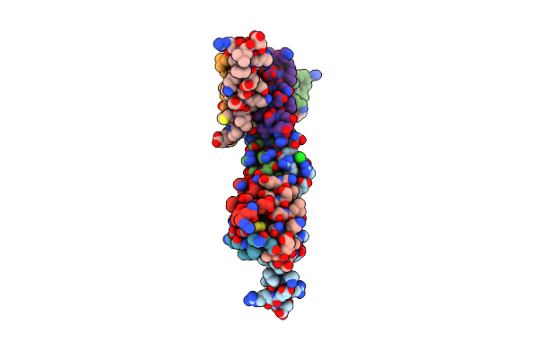
Crystal Structure Of Chicken C-Src-Sh3 Domain: Monomeric Form
Organism: Gallus gallus
Method: X-RAY DIFFRACTION Resolution:1.56 Å Release Date: 2014-04-23 Classification: SIGNALING PROTEIN Ligands: NI, EPE |
Organism: Gallus gallus
Method: X-RAY DIFFRACTION
Release Date: 2014-04-23
Ligands: NI, EPE
 |
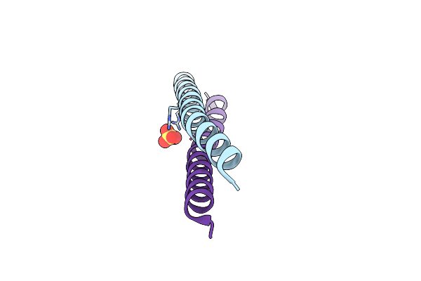
Crystal Structure Of The Brms1 N-Terminal Region
Organism: Homo sapiens
Method: X-RAY DIFFRACTION Resolution:2.00 Å Release Date: 2013-03-20 Classification: APOPTOSIS Ligands: CL, SO4, ACY |
Organism: Homo sapiens
Method: X-RAY DIFFRACTION
Release Date: 2013-03-20
Ligands: CL, SO4, ACY
 |
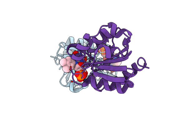
Crystal Structure Of The Brms1 N-Terminal Region
Organism: Homo sapiens
Method: X-RAY DIFFRACTION Resolution:1.91 Å Release Date: 2011-07-27 Classification: PROTEIN BINDING Ligands: CL, SO4 |
Organism: Homo sapiens
Method: X-RAY DIFFRACTION
Release Date: 2011-07-27
Ligands: CL, SO4
 |
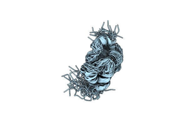
Flavodoxin From Helicobacter Pylori In Complex With The C3 Inhibitor
Organism: Helicobacter pylori
Method: X-RAY DIFFRACTION Resolution:2.62 Å Release Date: 2009-12-22 Classification: ELECTRON TRANSPORT Ligands: FMN, IC3 |
Organism: Helicobacter pylori
Method: X-RAY DIFFRACTION
Release Date: 2009-12-22
Ligands: FMN, IC3
 |
The Solution Structure Of The Monomeric Species Of The C Terminal Domain Of The Ca Protein Of Hiv-1
Organism: Human immunodeficiency virus 1
Method: SOLUTION NMR Release Date: 2007-07-31 Classification: VIRAL PROTEIN |
Organism: Human immunodeficiency virus 1
Method: SOLUTION NMR
Release Date: 2007-07-31