

Search Count: 28
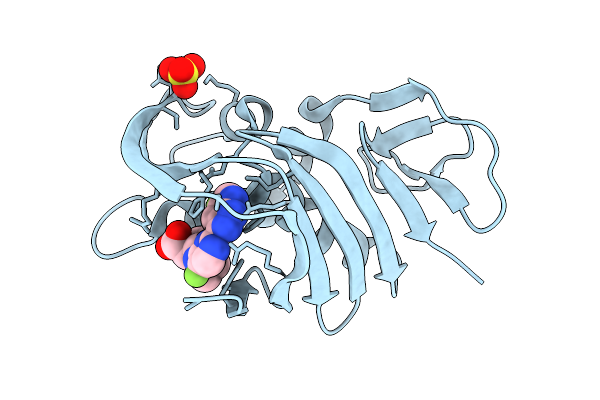 |
Crystal Structure Of H5N1 Influenza Polymerase Pb2 Domain In Complex With Compound 3
Organism: Influenza a virus (a/duck/shantou/4610/2003(h5n1))
Method: X-RAY DIFFRACTION Resolution:0.91 Å Release Date: 2026-01-28 Classification: TRANSFERASE Ligands: A1JV9, SO4 |
Organism: Influenza a virus (a/duck/shantou/4610/2003(h5n1))
Method: X-RAY DIFFRACTION
Release Date: 2026-01-28
Ligands: A1JV9, SO4
 |
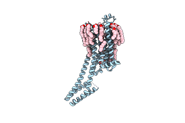
Dark Structure Of The Human Adenosine A2A Receptor Bound To Synthetic Photoswitch "Stilswitch3" Determined By Serial Synchrotron Crystallography
Organism: Homo sapiens
Method: X-RAY DIFFRACTION Resolution:2.65 Å Release Date: 2025-01-08 Classification: MEMBRANE PROTEIN Ligands: OLA, A1H3H, CLR, NA |
Organism: Homo sapiens
Method: X-RAY DIFFRACTION
Release Date: 2025-01-08
Ligands: OLA, A1H3H, CLR, NA
 |
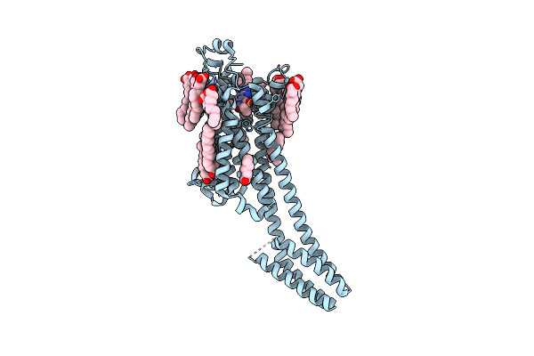
Crystal Structure Of The Adenosine A2A Receptor In Complex With Istradefylline
Organism: Homo sapiens
Method: X-RAY DIFFRACTION Resolution:1.94 Å Release Date: 2025-01-08 Classification: MEMBRANE PROTEIN Ligands: JQ9, CLR, OLA, NA |
Organism: Homo sapiens
Method: X-RAY DIFFRACTION
Release Date: 2025-01-08
Ligands: JQ9, CLR, OLA, NA
 |
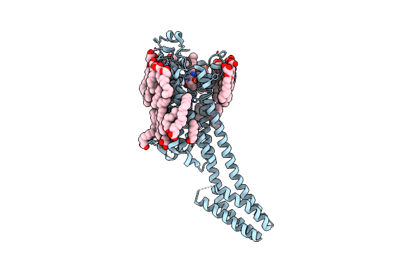
Crystal Structure Of The Adenosine A2A Receptor In Complex With The Synthetic Photoswitch 'Azoswitch2
Organism: Homo sapiens
Method: X-RAY DIFFRACTION Resolution:2.20 Å Release Date: 2025-01-08 Classification: MEMBRANE PROTEIN Ligands: A1H3L, CLR, OLA, NA |
Organism: Homo sapiens
Method: X-RAY DIFFRACTION
Release Date: 2025-01-08
Ligands: A1H3L, CLR, OLA, NA
 |
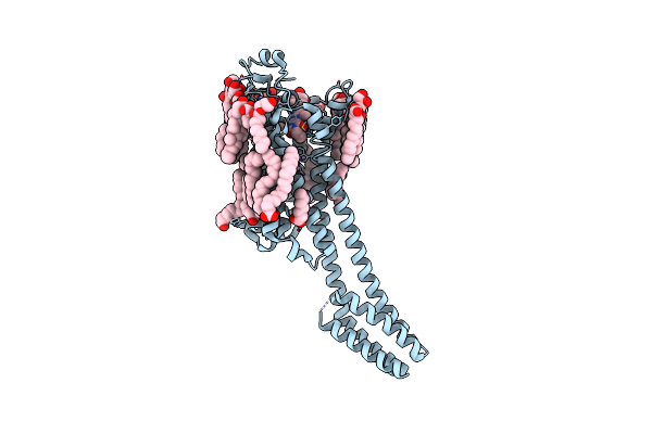
Crystal Structure Of The Adenosine A2A Receptor In Complex With The Synthetic Photoswitch 'Stilswitch1
Organism: Homo sapiens
Method: X-RAY DIFFRACTION Resolution:2.25 Å Release Date: 2025-01-08 Classification: MEMBRANE PROTEIN Ligands: A1H3J, CLR, OLA, OLC, OLB, NA |
Organism: Homo sapiens
Method: X-RAY DIFFRACTION
Release Date: 2025-01-08
Ligands: A1H3J, CLR, OLA, OLC, OLB, NA
 |
Crystal Structure Of The Adenosine A2A Receptor In Complex With The Synthetic Photoswitch 'Stilswitch2
Organism: Homo sapiens
Method: X-RAY DIFFRACTION Resolution:2.31 Å Release Date: 2025-01-08 Classification: MEMBRANE PROTEIN Ligands: A1H3I, CLR, OLC, OLA, NA |
Organism: Homo sapiens
Method: X-RAY DIFFRACTION
Release Date: 2025-01-08
Ligands: A1H3I, CLR, OLC, OLA, NA
 |
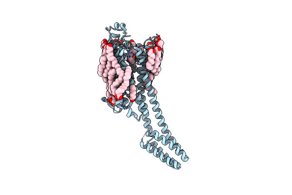
Crystal Structure Of The Adenosine A2A Receptor In Complex With The Synthetic Photoswitch 'Stilswitch3
Organism: Homo sapiens
Method: X-RAY DIFFRACTION Resolution:2.05 Å Release Date: 2025-01-08 Classification: MEMBRANE PROTEIN Ligands: A1H3H, CLR, OLA, NA |
Organism: Homo sapiens
Method: X-RAY DIFFRACTION
Release Date: 2025-01-08
Ligands: A1H3H, CLR, OLA, NA
 |
Crystal Structure Of The Adenosine A2A Receptor In Complex With The Synthetic Photoswitch 'Stilswitch4
Organism: Homo sapiens
Method: X-RAY DIFFRACTION Resolution:2.20 Å Release Date: 2025-01-08 Classification: MEMBRANE PROTEIN Ligands: A1H3K, CLR, OLA, OLC, NA |
Organism: Homo sapiens
Method: X-RAY DIFFRACTION
Release Date: 2025-01-08
Ligands: A1H3K, CLR, OLA, OLC, NA
 |
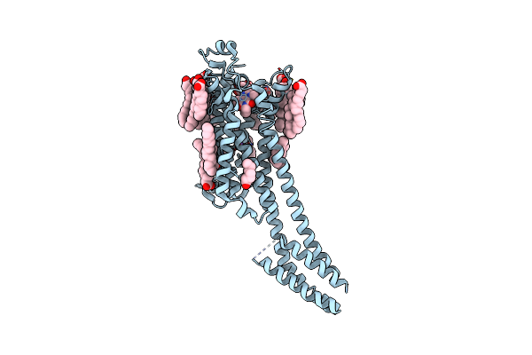
Dark Structure Of The Human Adenosine A2A Receptor Bound To Synthetic Photoswitch 'Stilswitch2' Determined By Serial Synchrotron Crystallography
Organism: Homo sapiens
Method: X-RAY DIFFRACTION Resolution:2.45 Å Release Date: 2025-01-08 Classification: MEMBRANE PROTEIN Ligands: A1H3I, CLR, OLA, OLB, OLC, NA |
Organism: Homo sapiens
Method: X-RAY DIFFRACTION
Release Date: 2025-01-08
Ligands: A1H3I, CLR, OLA, OLB, OLC, NA
 |
Steady State Structure Of The Human Adenosine A2A Receptor Bound To Synthetic Photoswitch 'Stilswitch2' Determined By Serial Synchrotron Crystallography
Organism: Homo sapiens
Method: X-RAY DIFFRACTION Resolution:2.80 Å Release Date: 2025-01-08 Classification: MEMBRANE PROTEIN Ligands: A1H3I, CLR, OLA, NA |
Organism: Homo sapiens
Method: X-RAY DIFFRACTION
Release Date: 2025-01-08
Ligands: A1H3I, CLR, OLA, NA
 |
Steady State Structure Of The Human Adenosine A2A Receptor Bound To Synthetic Photoswitch 'Stilswitch3' Determined By Serial Synchrotron Crystallography
Organism: Homo sapiens, Escherichia coli
Method: X-RAY DIFFRACTION Resolution:3.05 Å Release Date: 2025-01-08 Classification: MEMBRANE PROTEIN Ligands: A1H3H, OLA, NA |
Organism: Homo sapiens, Escherichia coli
Method: X-RAY DIFFRACTION
Release Date: 2025-01-08
Ligands: A1H3H, OLA, NA
 |
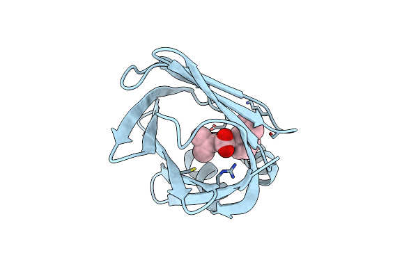
Fabp5 In Complex With 6-Chloro-4-Phenyl-2-Piperidin-1-Yl-3-(1H-Tetrazol-5-Yl)-Quinoline
Organism: Homo sapiens
Method: X-RAY DIFFRACTION Resolution:1.40 Å Release Date: 2017-01-25 Classification: LIPID BINDING PROTEIN Ligands: DMS, SO4, 65X |
Organism: Homo sapiens
Method: X-RAY DIFFRACTION
Release Date: 2017-01-25
Ligands: DMS, SO4, 65X
 |
Fabp4 In Complex With 6-Chloro-2-Isopropyl-4-(3-Isopropyl-Phenyl)-Quinoline-3-Carboxylic Acid
Organism: Homo sapiens
Method: X-RAY DIFFRACTION Resolution:1.14 Å Release Date: 2016-12-14 Classification: TRANSPORT PROTEIN Ligands: 65Y |
Organism: Homo sapiens
Method: X-RAY DIFFRACTION
Release Date: 2016-12-14
Ligands: 65Y
 |
Fabp4_3 In Complex With 6,8-Dichloro-4-Phenyl-2-Piperidin-1-Yl-Quinoline-3-Carboxylic Acid
Organism: Homo sapiens
Method: X-RAY DIFFRACTION Resolution:1.12 Å Release Date: 2016-12-14 Classification: TRANSPORT PROTEIN Ligands: 65Z, DMS |
Organism: Homo sapiens
Method: X-RAY DIFFRACTION
Release Date: 2016-12-14
Ligands: 65Z, DMS
 |
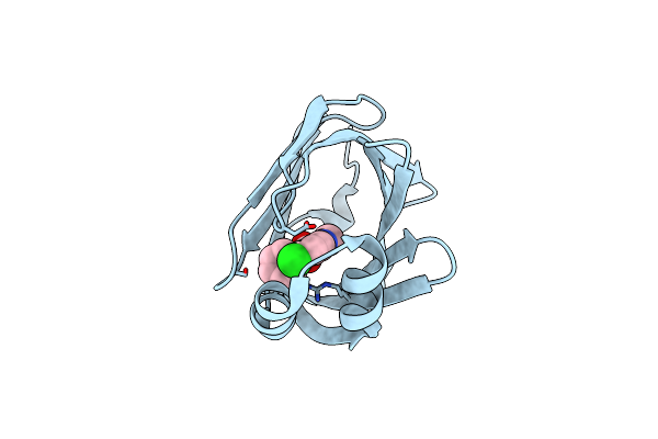
Human Fabp3 In Complex With 6-Chloro-2-Methyl-4-Phenyl-Quinoline-3-Carboxylic Acid
Organism: Homo sapiens
Method: X-RAY DIFFRACTION Resolution:2.30 Å Release Date: 2016-12-14 Classification: LIPID BINDING PROTEIN Ligands: SO4, 5M8, CL |
Organism: Homo sapiens
Method: X-RAY DIFFRACTION
Release Date: 2016-12-14
Ligands: SO4, 5M8, CL
 |
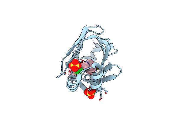
Human Fatty Acid Binding Protein 4 In Complex With 6-Chloro-2-Methyl-4-Phenyl-Quinoline-3-Carboxylic Acid At 1.18A
Organism: Homo sapiens
Method: X-RAY DIFFRACTION Resolution:1.18 Å Release Date: 2016-03-09 Classification: LIPID BINDING PROTEIN Ligands: 5M8 |
Organism: Homo sapiens
Method: X-RAY DIFFRACTION
Release Date: 2016-03-09
Ligands: 5M8
 |
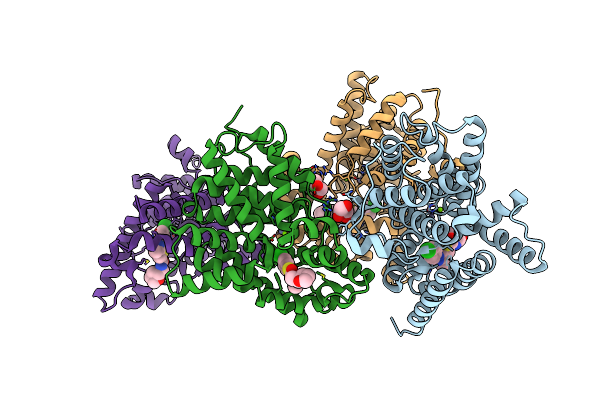
Human Fabp4 In Complex With 6-Chloro-4-Phenyl-2-Piperidin-1-Yl-Quinoline-3-Carboxylic Acid At 1.29A
Organism: Homo sapiens
Method: X-RAY DIFFRACTION Resolution:1.29 Å Release Date: 2016-03-09 Classification: LIPID BINDING PROTEIN Ligands: 5M7, SO4 |
Organism: Homo sapiens
Method: X-RAY DIFFRACTION
Release Date: 2016-03-09
Ligands: 5M7, SO4
 |
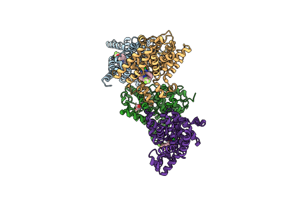
Crystal Structure Of Human Phosphodiesterase 10 In Complex With C13C(Cc(S1)C(Ncc2Occc2)=O)C(Nn3C4Ccc(Cc4)Cl)C, Micromolar Ic50=0.217
Organism: Homo sapiens
Method: X-RAY DIFFRACTION Resolution:2.20 Å Release Date: 2016-03-09 Classification: HYDROLASE Ligands: ZN, MG, GOL, 5M6 |
Organism: Homo sapiens
Method: X-RAY DIFFRACTION
Release Date: 2016-03-09
Ligands: ZN, MG, GOL, 5M6
 |
Crystal Structure Of Human Phosphodiesterase 10 In Complex With C1(C(Nc([Nh]1)Cl)C2Ccccc2)C4=Nn(C3Cccc(C3)Oc(F)(F)F)C=Cc4=O, Micromolar Ic50=0.029618
Organism: Homo sapiens
Method: X-RAY DIFFRACTION Resolution:2.30 Å Release Date: 2016-03-09 Classification: HYDROLASE Ligands: ZN, MG, 5MG |
Organism: Homo sapiens
Method: X-RAY DIFFRACTION
Release Date: 2016-03-09
Ligands: ZN, MG, 5MG
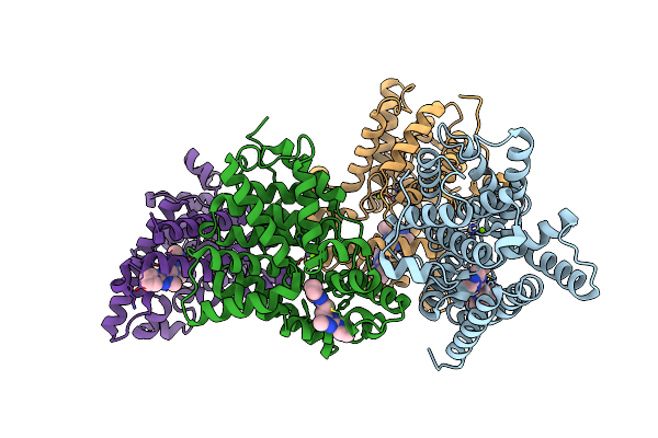 |
Crystal Structure Of Human Phosphodiesterase 10 In Complex With N4C(C)N1C(Nc(N1)Ccc2Nc(Nn2C)N3Cccc3)C(C4)Cc, Micromolar Ic50=0.0037753
Organism: Homo sapiens
Method: X-RAY DIFFRACTION Resolution:2.03 Å Release Date: 2016-03-09 Classification: HYDROLASE Ligands: ZN, MG, 5MF |
Organism: Homo sapiens
Method: X-RAY DIFFRACTION
Release Date: 2016-03-09
Ligands: ZN, MG, 5MF