

Search Count: 44
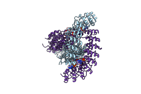 |
Crystal Structure Of Rnase L In Complex With Sunitinib
Organism: Sus scrofa
Method: X-RAY DIFFRACTION Resolution:2.46 Å Release Date: 2020-09-02 Classification: HYDROLASE Ligands: 25A, B49, PO4 |
Organism: Sus scrofa
Method: X-RAY DIFFRACTION
Release Date: 2020-09-02
Ligands: 25A, B49, PO4
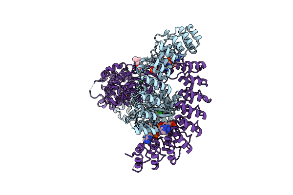 |
Crystal Structure Of Rnase L In Complex With Su11652
Organism: Sus scrofa
Method: X-RAY DIFFRACTION Resolution:2.40 Å Release Date: 2020-09-02 Classification: HYDROLASE Ligands: J60, 25L, PO4 |
Organism: Sus scrofa
Method: X-RAY DIFFRACTION
Release Date: 2020-09-02
Ligands: J60, 25L, PO4
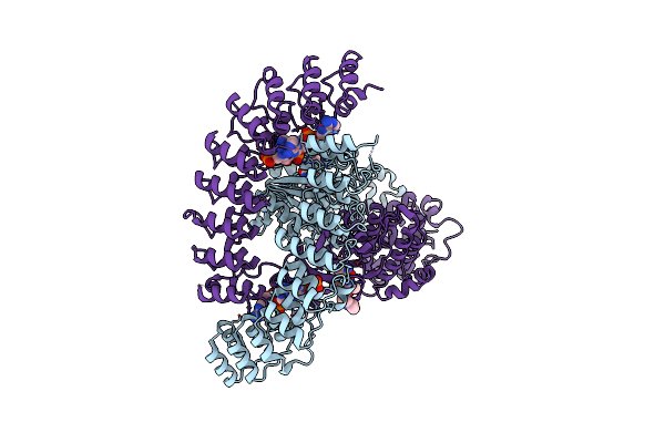 |
Crystal Structure Of Rnase L In Complex With Toceranib
Organism: Sus scrofa
Method: X-RAY DIFFRACTION Resolution:2.56 Å Release Date: 2020-09-02 Classification: HYDROLASE Ligands: BWC, 25L, PO4 |
Organism: Sus scrofa
Method: X-RAY DIFFRACTION
Release Date: 2020-09-02
Ligands: BWC, 25L, PO4
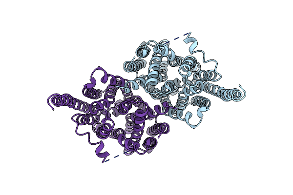 |
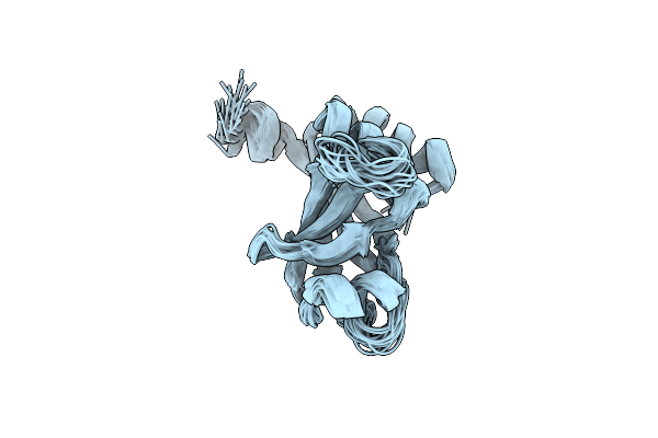
Cryo-Em Structure Of The Human Mct2
Organism: Homo sapiens
Method: ELECTRON MICROSCOPY Release Date: 2020-06-03 Classification: TRANSPORT PROTEIN |
Organism: Homo sapiens
Method: ELECTRON MICROSCOPY
Release Date: 2020-06-03
 |
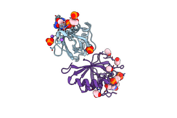
Nmr Structure Of The Hltf Hiran Domain
Organism: Homo sapiens
Method: SOLUTION NMR Release Date: 2016-06-08 Classification: DNA BINDING PROTEIN |
Organism: Homo sapiens
Method: SOLUTION NMR
Release Date: 2016-06-08
 |
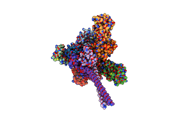
Crystal Structure Of The Hltf Hiran Domain With A Ssdna Fragment
Organism: Homo sapiens, Synthetic construct
Method: X-RAY DIFFRACTION Resolution:1.70 Å Release Date: 2016-05-25 Classification: DNA BINDING PROTEIN/DNA |
Organism: Homo sapiens, Synthetic construct
Method: X-RAY DIFFRACTION
Release Date: 2016-05-25
 |
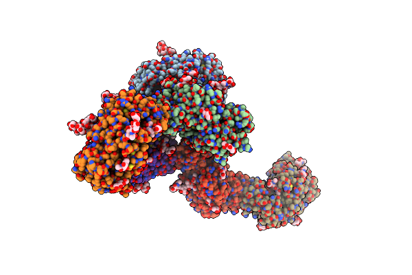
Crystal Structure Of Rnf8 Bound To The Ubc13/Mms2 Heterodimer
Organism: Homo sapiens
Method: X-RAY DIFFRACTION Resolution:4.80 Å Release Date: 2014-02-26 Classification: PROTEIN BINDING/LIGASE Ligands: ZN |
Organism: Homo sapiens
Method: X-RAY DIFFRACTION
Release Date: 2014-02-26
Ligands: ZN
 |
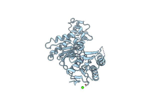
Structure Of The Lysosomal Domain Of Limp-2
Organism: Homo sapiens
Method: X-RAY DIFFRACTION Resolution:3.00 Å Release Date: 2013-10-09 Classification: CELL ADHESION Ligands: NAG, PO4, PEG |
Organism: Homo sapiens
Method: X-RAY DIFFRACTION
Release Date: 2013-10-09
Ligands: NAG, PO4, PEG
 |
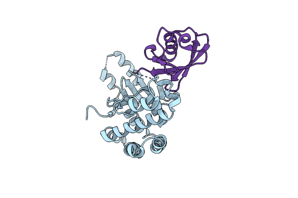
Crystal Structure Of Yeast Qri7 Homodimer
Organism: Saccharomyces cerevisiae
Method: X-RAY DIFFRACTION Resolution:2.88 Å Release Date: 2013-05-08 Classification: BIOSYNTHETIC PROTEIN Ligands: ZN, CA |
Organism: Saccharomyces cerevisiae
Method: X-RAY DIFFRACTION
Release Date: 2013-05-08
Ligands: ZN, CA
 |
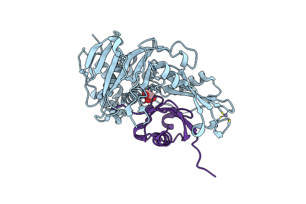
Crystal Structure Of Otub1 In Complex With Ubiquitin Variant
Organism: Homo sapiens
Method: X-RAY DIFFRACTION Resolution:2.49 Å Release Date: 2013-01-16 Classification: HYDROLASE |
Organism: Homo sapiens
Method: X-RAY DIFFRACTION
Release Date: 2013-01-16
 |
Crystal Structure Of Usp2 And A Mutant Form Of Ubiquitin
Organism: Homo sapiens
Method: X-RAY DIFFRACTION Resolution:2.10 Å Release Date: 2012-12-19 Classification: hydrolase/signaling protein Ligands: ZN, CL, GOL |
Organism: Homo sapiens
Method: X-RAY DIFFRACTION
Release Date: 2012-12-19
Ligands: ZN, CL, GOL
 |
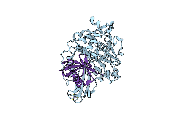
Human Cdc34 E2 In Complex With Cc0651 Inhibitor
Organism: Homo sapiens
Method: X-RAY DIFFRACTION Resolution:2.30 Å Release Date: 2011-07-06 Classification: LIGASE/LIGASE INHIBITOR Ligands: U94 |
Organism: Homo sapiens
Method: X-RAY DIFFRACTION
Release Date: 2011-07-06
Ligands: U94
 |
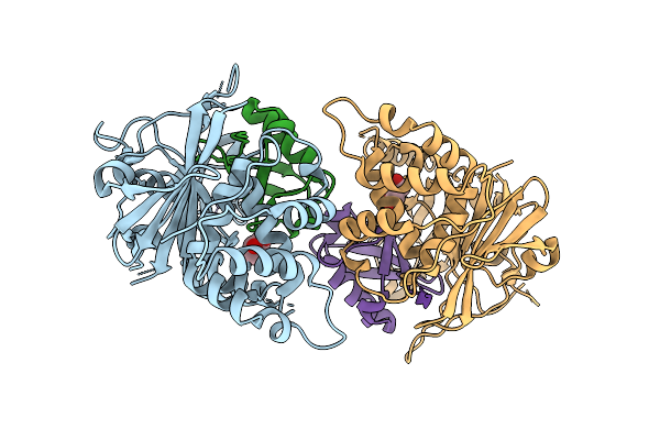
The Catalytic Domain Of Usp8 In Complex With A Usp8 Specific Inhibitor
Organism: Homo sapiens
Method: X-RAY DIFFRACTION Resolution:2.60 Å Release Date: 2010-06-23 Classification: HYDROLASE Ligands: ZN |
Organism: Homo sapiens
Method: X-RAY DIFFRACTION
Release Date: 2010-06-23
Ligands: ZN
 |
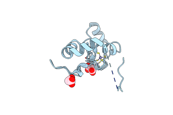
Usp21 In Complex With A Ubiquitin-Based, Usp21-Specific Inhibitor
Organism: Homo sapiens
Method: X-RAY DIFFRACTION Resolution:2.70 Å Release Date: 2010-06-09 Classification: HYDROLASE Ligands: ZN, CL, GOL |
Organism: Homo sapiens
Method: X-RAY DIFFRACTION
Release Date: 2010-06-09
Ligands: ZN, CL, GOL
 |
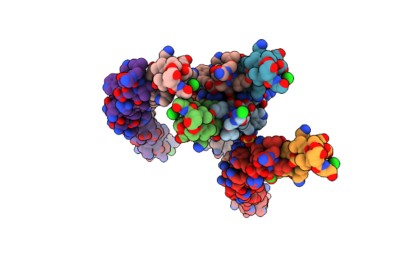
Crystal Structure Of The Ring Domain Of Rnf168
Organism: Homo sapiens
Method: X-RAY DIFFRACTION Resolution:2.12 Å Release Date: 2010-01-19 Classification: LIGASE Ligands: ZN, MLI |
Organism: Homo sapiens
Method: X-RAY DIFFRACTION
Release Date: 2010-01-19
Ligands: ZN, MLI
 |
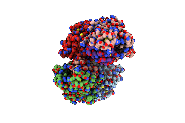
Coiled-Coil Domain Of Tumor Susceptibility Gene 101
Organism: Homo sapiens
Method: X-RAY DIFFRACTION Resolution:2.50 Å Release Date: 2009-11-03 Classification: HYDROLASE Ligands: SO4, CL |
Organism: Homo sapiens
Method: X-RAY DIFFRACTION
Release Date: 2009-11-03
Ligands: SO4, CL
 |
Crystal Structure Of Covalent Ubiquitin-Usp21 Complex
Organism: Homo sapiens
Method: X-RAY DIFFRACTION Resolution:2.59 Å Release Date: 2009-07-21 Classification: HYDROLASE Ligands: ZN, NEH |
Organism: Homo sapiens
Method: X-RAY DIFFRACTION
Release Date: 2009-07-21
Ligands: ZN, NEH
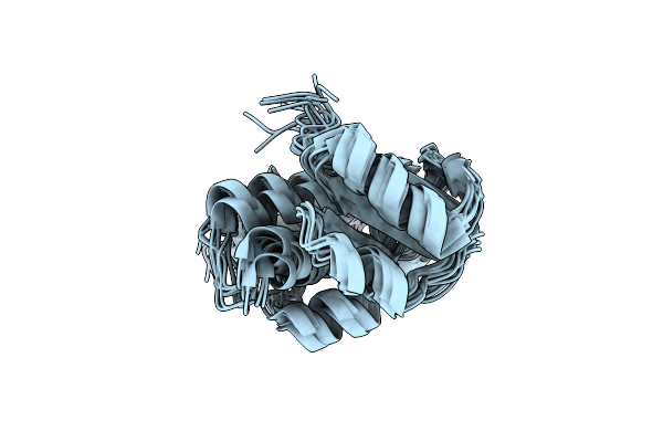 |
Solution Nmr Structure Of Cgi121 From Methanococcus Jannaschii. Northeast Structural Genomics Consortium Target Mj0187
Organism: Methanocaldococcus jannaschii
Method: SOLUTION NMR Release Date: 2008-11-04 Classification: structural genomics, unknown function |
Organism: Methanocaldococcus jannaschii
Method: SOLUTION NMR
Release Date: 2008-11-04
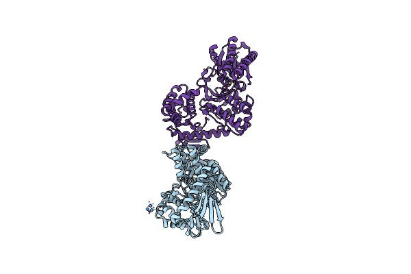 |
Structure Of The Methanococcus Jannaschii Kae1-Bud32 Fusion Protein
Organism: Methanocaldococcus jannaschii
Method: X-RAY DIFFRACTION Resolution:2.67 Å Release Date: 2008-11-04 Classification: HYDROLASE Ligands: MG, TBR |
Organism: Methanocaldococcus jannaschii
Method: X-RAY DIFFRACTION
Release Date: 2008-11-04
Ligands: MG, TBR
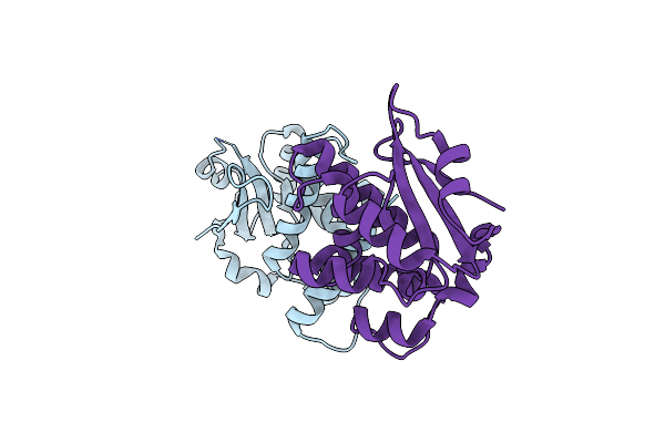 |
Crystal Structure Of Human Cgi121
Organism: Homo sapiens
Method: X-RAY DIFFRACTION Resolution:2.48 Å Release Date: 2008-11-04 Classification: HYDROLASE |
Organism: Homo sapiens
Method: X-RAY DIFFRACTION
Release Date: 2008-11-04