

Search Count: 20
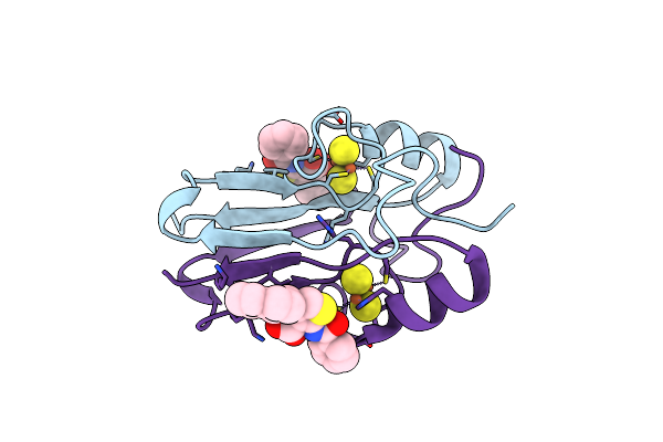 |
Mitoneet Bound To M1 Molecule
Organism: Homo sapiens
Method: X-RAY DIFFRACTION Resolution:1.65 Å Release Date: 2022-05-25 Classification: METAL BINDING PROTEIN Ligands: FES, 49I, 4OY |
Organism: Homo sapiens
Method: X-RAY DIFFRACTION
Release Date: 2022-05-25
Ligands: FES, 49I, 4OY
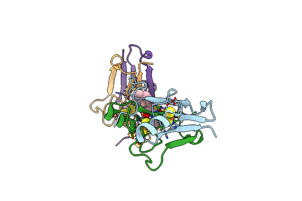 |
Naf-1 Bound To M1 Molecule
Organism: Homo sapiens
Method: X-RAY DIFFRACTION Resolution:1.74 Å Release Date: 2022-05-25 Classification: METAL BINDING PROTEIN Ligands: FES, 49I |
Organism: Homo sapiens
Method: X-RAY DIFFRACTION
Release Date: 2022-05-25
Ligands: FES, 49I
 |
Structure Of Low-Light Grown Chlorella Ohadii Photosystem I
|
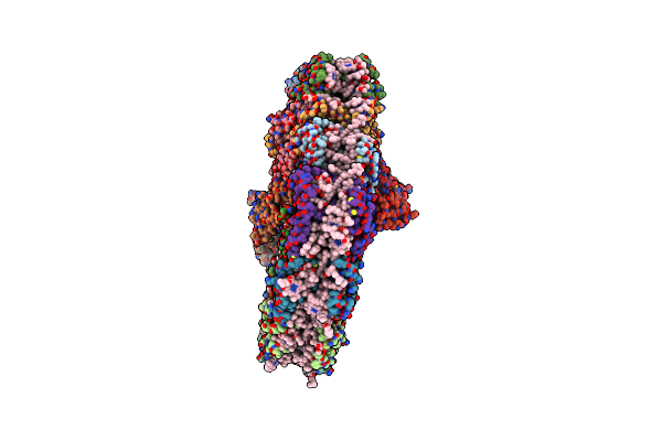 |
Structure Of High-Light Grown Chlorella Ohadii Photosystem I
|
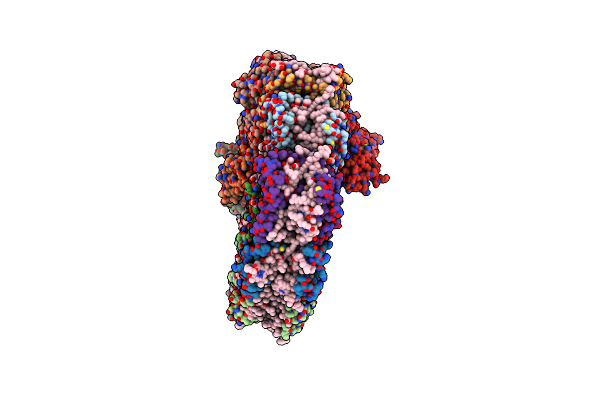 |
Structure Of Small High-Light Grown Chlorella Ohadii Photosystem I
|
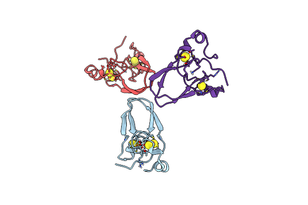 |
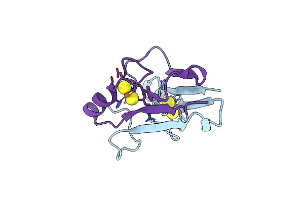
Crystal Structure Of Human Mitochondrial Inner Neet Protein (Mint)/Cisd3
Organism: Homo sapiens
Method: X-RAY DIFFRACTION Resolution:1.90 Å Release Date: 2017-12-20 Classification: METAL BINDING PROTEIN Ligands: FES |
Organism: Homo sapiens
Method: X-RAY DIFFRACTION
Release Date: 2017-12-20
Ligands: FES
 |
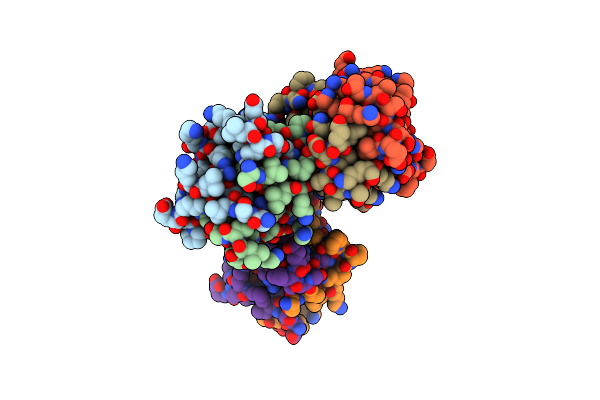
The 1.55A Crystal Structure Of Naf1 (Miner1): The Redox-Active 2Fe-2S Protein
Organism: Homo sapiens
Method: X-RAY DIFFRACTION Resolution:1.65 Å Release Date: 2014-07-02 Classification: METAL BINDING PROTEIN Ligands: FES |
Organism: Homo sapiens
Method: X-RAY DIFFRACTION
Release Date: 2014-07-02
Ligands: FES
 |
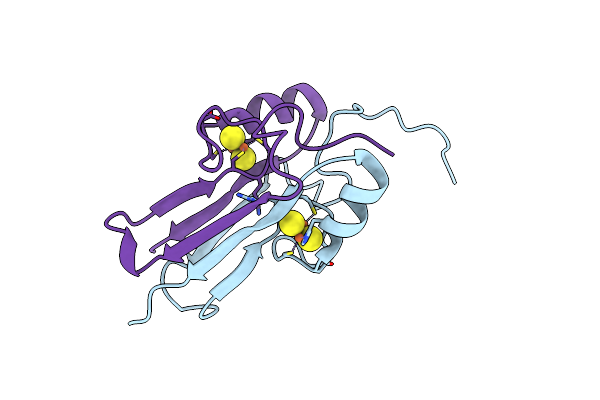
Crystal Structure Of Naf1 (Miner1): H114C The Redox-Active 2Fe-2S Protein
Organism: Homo sapiens
Method: X-RAY DIFFRACTION Resolution:1.58 Å Release Date: 2014-07-02 Classification: METAL BINDING PROTEIN Ligands: FES |
Organism: Homo sapiens
Method: X-RAY DIFFRACTION
Release Date: 2014-07-02
Ligands: FES
 |
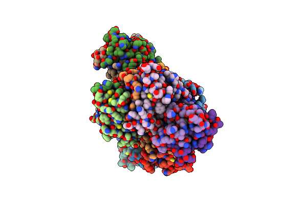
The Crystal Structure Of A Human Mitoneet Mutant With An Ala Inserted Between Asp 67 And Lys 68
Organism: Homo sapiens
Method: X-RAY DIFFRACTION Resolution:1.19 Å Release Date: 2012-12-26 Classification: METAL BINDING PROTEIN Ligands: FES |
Organism: Homo sapiens
Method: X-RAY DIFFRACTION
Release Date: 2012-12-26
Ligands: FES
 |
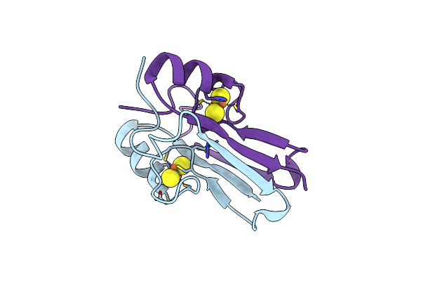
The Crystal Structure Of A Human Mitoneet Mutant With Asp 67 Replaced By A Gly
Organism: Homo sapiens
Method: X-RAY DIFFRACTION Resolution:2.40 Å Release Date: 2012-12-26 Classification: METAL BINDING PROTEIN Ligands: FES |
Organism: Homo sapiens
Method: X-RAY DIFFRACTION
Release Date: 2012-12-26
Ligands: FES
 |
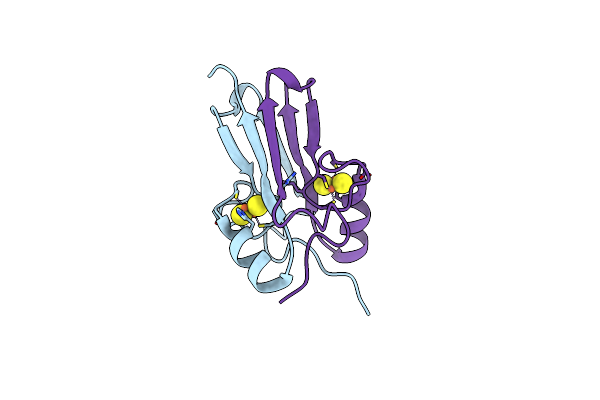
The Crystal Structure Of A Human Mitoneet Mutant With Met 62 Replaced By A Gly
Organism: Homo sapiens
Method: X-RAY DIFFRACTION Resolution:1.55 Å Release Date: 2012-12-26 Classification: METAL BINDING PROTEIN Ligands: FES |
Organism: Homo sapiens
Method: X-RAY DIFFRACTION
Release Date: 2012-12-26
Ligands: FES
 |
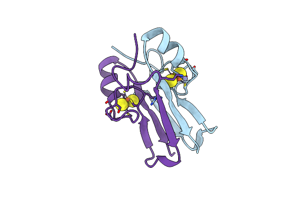
The Crystal Structure Of A Human Mitoneet Double Mutant In Which Gly 66 Are Asp 67 Are Both Replaced With Ala Residues
Organism: Homo sapiens
Method: X-RAY DIFFRACTION Resolution:1.35 Å Release Date: 2012-12-26 Classification: METAL BINDING PROTEIN Ligands: FES |
Organism: Homo sapiens
Method: X-RAY DIFFRACTION
Release Date: 2012-12-26
Ligands: FES
 |
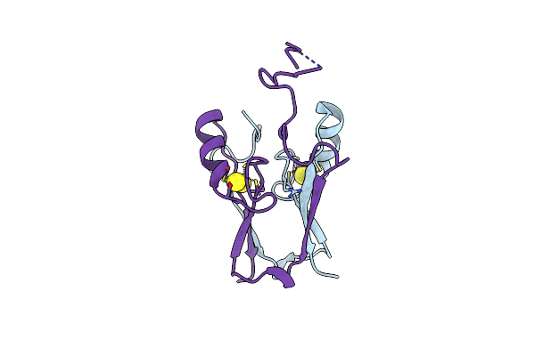
The Crystal Structure Of At5G51720 (At-Neet)
Organism: Arabidopsis thaliana
Method: X-RAY DIFFRACTION Resolution:1.75 Å Release Date: 2012-05-23 Classification: METAL BINDING PROTEIN Ligands: FES, ZN |
Organism: Arabidopsis thaliana
Method: X-RAY DIFFRACTION
Release Date: 2012-05-23
Ligands: FES, ZN
 |
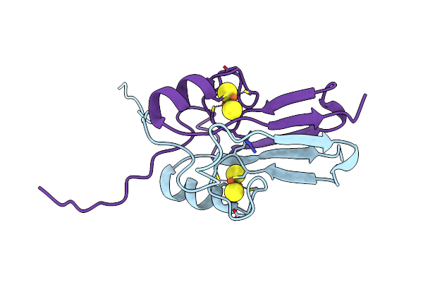
Atchloroneet (H87C Mutant)
Organism: Arabidopsis thaliana
Method: X-RAY DIFFRACTION Resolution:1.14 Å Release Date: 2012-05-23 Classification: METAL BINDING PROTEIN Ligands: FES |
Organism: Arabidopsis thaliana
Method: X-RAY DIFFRACTION
Release Date: 2012-05-23
Ligands: FES
 |
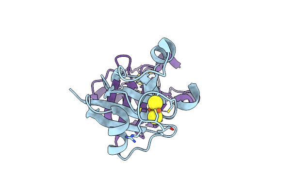
Human Mitoneet With 2Fe-2S Coordinating Ligand His 87 Replaced With Cys
Organism: Homo sapiens
Method: X-RAY DIFFRACTION Resolution:1.70 Å Release Date: 2011-06-01 Classification: METAL BINDING PROTEIN Ligands: FES |
Organism: Homo sapiens
Method: X-RAY DIFFRACTION
Release Date: 2011-06-01
Ligands: FES
 |
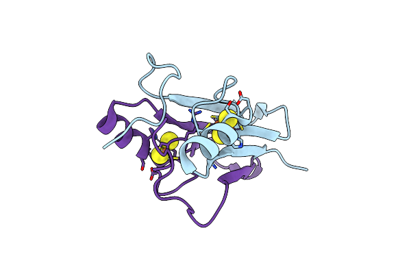
Structure Of M. Laminosus Ferredoxin With A Shorter L1,2 Loop
Organism: Mastigocladus laminosus
Method: X-RAY DIFFRACTION Resolution:2.30 Å Release Date: 2011-02-09 Classification: ELECTRON TRANSPORT Ligands: FES |
Organism: Mastigocladus laminosus
Method: X-RAY DIFFRACTION
Release Date: 2011-02-09
Ligands: FES
 |
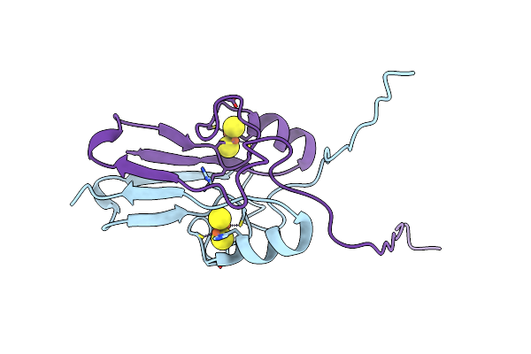
Crystal Structure Of Miner1: The Redox-Active 2Fe-2S Protein Causative In Wolfram Syndrome 2
Organism: Homo sapiens
Method: X-RAY DIFFRACTION Resolution:2.10 Å Release Date: 2009-08-18 Classification: METAL BINDING PROTEIN Ligands: FES |
Organism: Homo sapiens
Method: X-RAY DIFFRACTION
Release Date: 2009-08-18
Ligands: FES
 |
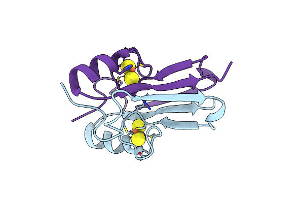
The Novel 2Fe-2S Outer Mitochondrial Protein Mitoneet Displays Conformational Flexibility In Its N-Terminal Cytoplasmic Tethering Domain
Organism: Homo sapiens
Method: X-RAY DIFFRACTION Resolution:1.40 Å Release Date: 2009-07-07 Classification: METAL BINDING PROTEIN Ligands: FES |
Organism: Homo sapiens
Method: X-RAY DIFFRACTION
Release Date: 2009-07-07
Ligands: FES
 |
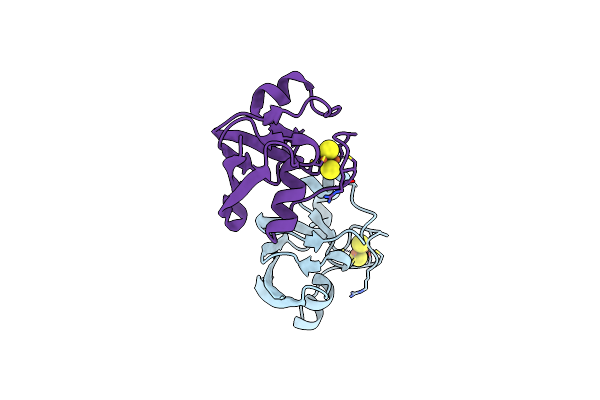
Mitoneet Is A Uniquely Folded 2Fe-2S Outer Mitochondrial Membrane Protein Stabilized By Pioglitazone
Organism: Homo sapiens
Method: X-RAY DIFFRACTION Resolution:1.50 Å Release Date: 2007-08-21 Classification: METAL BINDING PROTEIN Ligands: FES |
Organism: Homo sapiens
Method: X-RAY DIFFRACTION
Release Date: 2007-08-21
Ligands: FES
 |
Crystal Structure Of 2Fe2S Ferredoxin From Thermophilic Cyanobacterium Mastigocladus Laminosus
Organism: Mastigocladus laminosus
Method: X-RAY DIFFRACTION Resolution:1.25 Å Release Date: 2005-04-12 Classification: ELECTRON TRANSPORT Ligands: FES |
Organism: Mastigocladus laminosus
Method: X-RAY DIFFRACTION
Release Date: 2005-04-12
Ligands: FES