
Search Count: 142
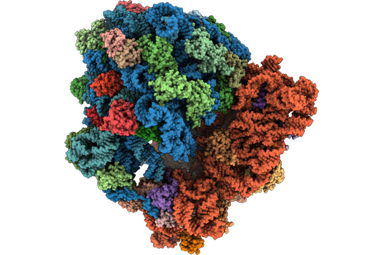 |
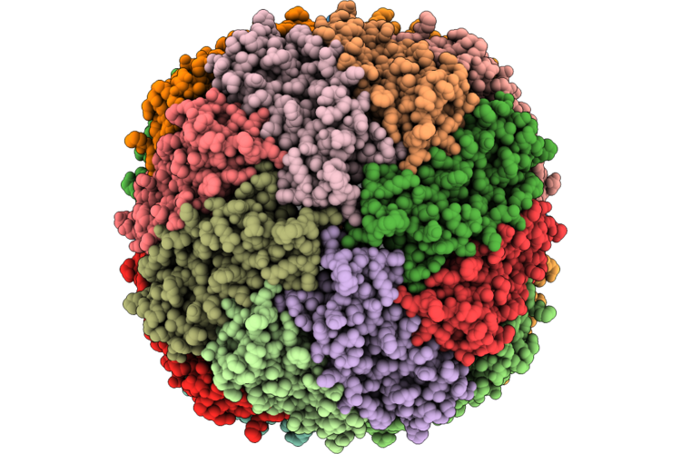
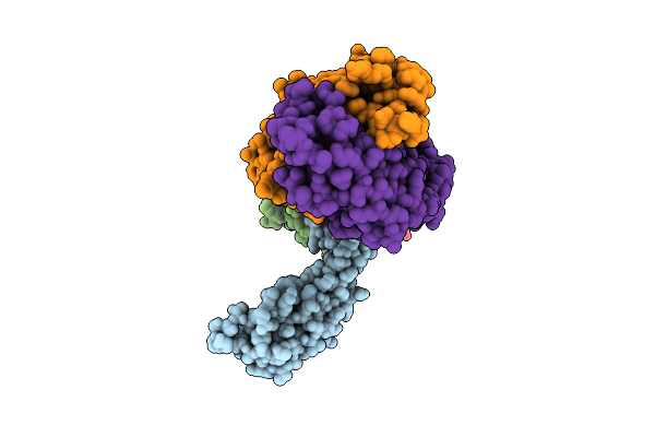
Subtomogram Average Of 70S Ribosome (11X11) Using Cryo Arm 300Ii
Organism: Escherichia coli
Method: ELECTRON MICROSCOPY Release Date: 2026-07-15 Classification: RECOMBINATION Ligands: MG, CL, NA, ZN |
Organism: Escherichia coli
Method: ELECTRON MICROSCOPY
Release Date: 2026-07-15
Ligands: MG, CL, NA, ZN
 |
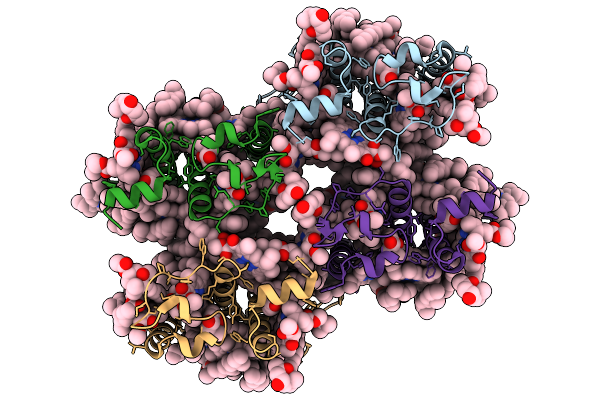
Cryo-Em Structure Of Streptococcus Thermophilus Foeab In Complex With Adp
Organism: Streptococcus thermophilus
Method: ELECTRON MICROSCOPY Release Date: 2026-06-03 Classification: TRANSPORT PROTEIN Ligands: ADP, MG |
Organism: Streptococcus thermophilus
Method: ELECTRON MICROSCOPY
Release Date: 2026-06-03
Ligands: ADP, MG
 |
Cryo-Em Structure Of Nucleotide-Free Streptococcus Thermophilus Foeab 1
Organism: Streptococcus thermophilus
Method: ELECTRON MICROSCOPY Release Date: 2026-06-03 Classification: TRANSPORT PROTEIN |
Organism: Streptococcus thermophilus
Method: ELECTRON MICROSCOPY
Release Date: 2026-06-03
 |
Cryo-Em Structure Of Nucleotide-Free Streptococcus Thermophilus Foeab 2
Organism: Streptococcus thermophilus
Method: ELECTRON MICROSCOPY Release Date: 2026-06-03 Classification: TRANSPORT PROTEIN |
Organism: Streptococcus thermophilus
Method: ELECTRON MICROSCOPY
Release Date: 2026-06-03
 |
Cryo-Em Structure Of Nucleotide-Free Streptococcus Thermophilus Foeab 3
Organism: Streptococcus thermophilus
Method: ELECTRON MICROSCOPY Release Date: 2026-06-03 Classification: TRANSPORT PROTEIN |
Organism: Streptococcus thermophilus
Method: ELECTRON MICROSCOPY
Release Date: 2026-06-03
 |
Cryo-Em Structure Of Streptococcus Thermophilus Foeab E504Q Mutant In Complex With Atp
Organism: Streptococcus thermophilus
Method: ELECTRON MICROSCOPY Release Date: 2026-06-03 Classification: TRANSPORT PROTEIN Ligands: ATP, MG |
Organism: Streptococcus thermophilus
Method: ELECTRON MICROSCOPY
Release Date: 2026-06-03
Ligands: ATP, MG
 |
Cryo-Em Structure Of Streptococcus Thermophilus Foeab In Complex With Atp And Adp
Organism: Streptococcus thermophilus
Method: ELECTRON MICROSCOPY Release Date: 2026-06-03 Classification: TRANSPORT PROTEIN Ligands: ATP, MG, ADP |
Organism: Streptococcus thermophilus
Method: ELECTRON MICROSCOPY
Release Date: 2026-06-03
Ligands: ATP, MG, ADP
 |
Cryo-Em Structure Of Streptococcus Thermophilus Foeab In Complex With Amppnp In Peptidisc
Organism: Streptococcus thermophilus
Method: ELECTRON MICROSCOPY Release Date: 2026-06-03 Classification: TRANSPORT PROTEIN Ligands: ANP, MG |
Organism: Streptococcus thermophilus
Method: ELECTRON MICROSCOPY
Release Date: 2026-06-03
Ligands: ANP, MG
 |
Cryo-Em Structure Of Streptococcus Thermophilus Foeab In Complex With Amppnp
Organism: Streptococcus thermophilus
Method: ELECTRON MICROSCOPY Resolution:2.90 Å Release Date: 2026-06-03 Classification: TRANSPORT PROTEIN Ligands: ANP, MG |
Organism: Streptococcus thermophilus
Method: ELECTRON MICROSCOPY
Release Date: 2026-06-03
Ligands: ANP, MG
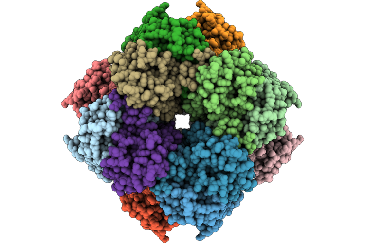 |
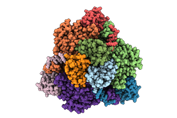
Cryoem Structure Of Arabidopsis Thaliana Col-0 Rubisco With D4 Symmetry
Organism: Arabidopsis thaliana
Method: ELECTRON MICROSCOPY Resolution:1.76 Å Release Date: 2026-05-20 Classification: PHOTOSYNTHESIS Ligands: SO4 |
Organism: Arabidopsis thaliana
Method: ELECTRON MICROSCOPY
Release Date: 2026-05-20
Ligands: SO4
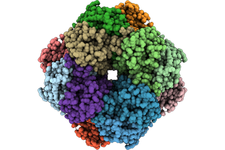 |
Cryoem Structure Of Arabidopsis Thaliana M309I Rubisco With D4 Symmetry
Organism: Arabidopsis thaliana
Method: ELECTRON MICROSCOPY Resolution:1.75 Å Release Date: 2026-05-20 Classification: PHOTOSYNTHESIS Ligands: SO4 |
Organism: Arabidopsis thaliana
Method: ELECTRON MICROSCOPY
Release Date: 2026-05-20
Ligands: SO4
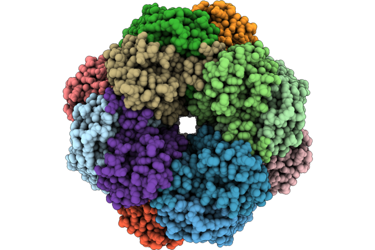 |
Cryoem Structure Of Arabidopsis Thaliana D397N Rubisco With D4 Symmetry
Organism: Arabidopsis thaliana
Method: ELECTRON MICROSCOPY Resolution:1.74 Å Release Date: 2026-05-20 Classification: PHOTOSYNTHESIS Ligands: SO4 |
Organism: Arabidopsis thaliana
Method: ELECTRON MICROSCOPY
Release Date: 2026-05-20
Ligands: SO4
 |
Subtomogram Average Of Apoferrtin (11X11) Using Cryo Arm 300Ii
Organism: Mus musculus
Method: ELECTRON MICROSCOPY Release Date: 2026-04-22 Classification: RECOMBINATION Ligands: FE, ZN |
Organism: Mus musculus
Method: ELECTRON MICROSCOPY
Release Date: 2026-04-22
Ligands: FE, ZN
 |
P Ring On Polyrod-P Ring Complex From Salmonella Th26292 Strain
Organism: Salmonella enterica subsp. enterica serovar typhimurium
Method: ELECTRON MICROSCOPY Resolution:2.33 Å Release Date: 2026-03-04 Classification: MOTOR PROTEIN |
Organism: Salmonella enterica subsp. enterica serovar typhimurium
Method: ELECTRON MICROSCOPY
Release Date: 2026-03-04
 |
Polyrod Formed By Flgg (G65V) From The Salmonella Th26292 Strain
Organism: Salmonella enterica subsp. enterica serovar typhimurium
Method: ELECTRON MICROSCOPY Release Date: 2026-02-04 Classification: MOTOR PROTEIN |
Organism: Salmonella enterica subsp. enterica serovar typhimurium
Method: ELECTRON MICROSCOPY
Release Date: 2026-02-04
 |
Cryo-Em Structure Of Violaxanthin-Chlorophyll-A-Binding Protein With Red Shifted Chl A (Rvcp) From Trachydiscus Minutus At 2.4 Angstrom
Organism: Trachydiscus minutus
Method: ELECTRON MICROSCOPY Resolution:2.42 Å Release Date: 2025-12-31 Classification: PHOTOSYNTHESIS Ligands: CLA, A1L1F, XAT |
Organism: Trachydiscus minutus
Method: ELECTRON MICROSCOPY
Release Date: 2025-12-31
Ligands: CLA, A1L1F, XAT
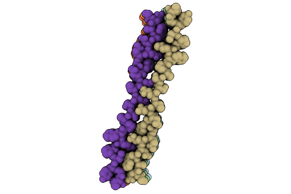 |
Cryo-Em Structure Of The Tia-1 Prion-Like Domain Amyloid Fibril, Wt
Organism: Homo sapiens
Method: ELECTRON MICROSCOPY Release Date: 2025-12-03 Classification: PROTEIN FIBRIL |
Organism: Homo sapiens
Method: ELECTRON MICROSCOPY
Release Date: 2025-12-03
 |
Cryo-Em Structure Of The Tia-1 Prion-Like Domain Amyloid Fibril, G355R
Organism: Homo sapiens
Method: ELECTRON MICROSCOPY Release Date: 2025-12-03 Classification: PROTEIN FIBRIL |
Organism: Homo sapiens
Method: ELECTRON MICROSCOPY
Release Date: 2025-12-03
 |
Cryo-Em Structure Of Urease From Ureaplasma Parvum
Organism: Ureaplasma parvum serovar 3 (strain atcc 700970)
Method: ELECTRON MICROSCOPY Release Date: 2025-08-20 Classification: HYDROLASE Ligands: BME, NI |
Organism: Ureaplasma parvum serovar 3 (strain atcc 700970)
Method: ELECTRON MICROSCOPY
Release Date: 2025-08-20
Ligands: BME, NI
 |
Cryo-Em Structure Of Linker-Extended Biparatopic Antibody Ba1-Gp4 In Complex With Tnfr2
Organism: Homo sapiens, Mus musculus
Method: ELECTRON MICROSCOPY Release Date: 2025-08-13 Classification: IMMUNE SYSTEM |
Organism: Homo sapiens, Mus musculus
Method: ELECTRON MICROSCOPY
Release Date: 2025-08-13