

Search Count: 22
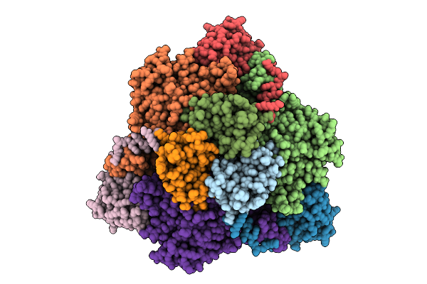 |
Cryo-Em Structure Of Urease From Ureaplasma Parvum
Organism: Ureaplasma parvum serovar 3 (strain atcc 700970)
Method: ELECTRON MICROSCOPY Release Date: 2025-08-20 Classification: HYDROLASE Ligands: BME, NI |
Organism: Ureaplasma parvum serovar 3 (strain atcc 700970)
Method: ELECTRON MICROSCOPY
Release Date: 2025-08-20
Ligands: BME, NI
 |
Structure Of The Sars-Cov-2 Ba.2.86 Spike Glycoprotein (Closed State)
Organism: Severe acute respiratory syndrome coronavirus 2
Method: ELECTRON MICROSCOPY Release Date: 2024-10-09 Classification: VIRAL PROTEIN Ligands: NAG |
Organism: Severe acute respiratory syndrome coronavirus 2
Method: ELECTRON MICROSCOPY
Release Date: 2024-10-09
Ligands: NAG
 |
Structure Of The Sars-Cov-2 Ba.2.86 Spike Protein (1-Up State)
Organism: Severe acute respiratory syndrome coronavirus 2
Method: ELECTRON MICROSCOPY Release Date: 2024-10-09 Classification: VIRAL PROTEIN Ligands: NAG |
Organism: Severe acute respiratory syndrome coronavirus 2
Method: ELECTRON MICROSCOPY
Release Date: 2024-10-09
Ligands: NAG
 |
Structure Of Sars-Cov-2 Ba.2.86 Spike Glycoprotein In Complex With Ace2 (2-Up State)
Organism: Severe acute respiratory syndrome coronavirus 2, Homo sapiens
Method: ELECTRON MICROSCOPY Release Date: 2024-10-09 Classification: VIRAL PROTEIN/PROTEIN BINDING Ligands: NAG |
Organism: Severe acute respiratory syndrome coronavirus 2, Homo sapiens
Method: ELECTRON MICROSCOPY
Release Date: 2024-10-09
Ligands: NAG
 |
Structure Of Sars-Cov-2 Ba.2.86 Spike Glycoprotein In Complex With Ace2 (2-Up And 1-Down State)
Organism: Severe acute respiratory syndrome coronavirus 2, Homo sapiens
Method: ELECTRON MICROSCOPY Release Date: 2024-10-09 Classification: VIRAL PROTEIN/PROTEIN BINDING Ligands: NAG |
Organism: Severe acute respiratory syndrome coronavirus 2, Homo sapiens
Method: ELECTRON MICROSCOPY
Release Date: 2024-10-09
Ligands: NAG
 |
Structure Of Sars-Cov-2 Ba.2.86 Spike Rbd In Complex With Ace2 (Up State)
Organism: Homo sapiens, Severe acute respiratory syndrome coronavirus 2
Method: ELECTRON MICROSCOPY Release Date: 2024-10-09 Classification: VIRAL PROTEIN/PROTEIN BINDING Ligands: NAG |
Organism: Homo sapiens, Severe acute respiratory syndrome coronavirus 2
Method: ELECTRON MICROSCOPY
Release Date: 2024-10-09
Ligands: NAG
 |
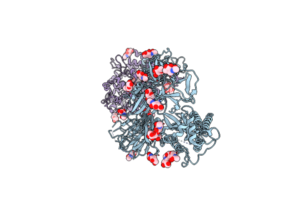
Structure Of Sars-Cov-2 Ba.2.86 Spike Rbd In Complex With Ace2 (Down State)
Organism: Severe acute respiratory syndrome coronavirus 2, Homo sapiens
Method: ELECTRON MICROSCOPY Release Date: 2024-10-09 Classification: VIRAL PROTEIN/PROTEIN BINDING Ligands: NAG |
Organism: Severe acute respiratory syndrome coronavirus 2, Homo sapiens
Method: ELECTRON MICROSCOPY
Release Date: 2024-10-09
Ligands: NAG
 |
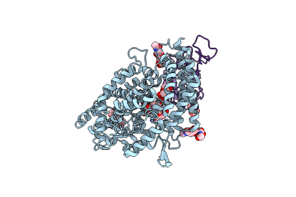
Structure Of Sars-Cov-2 Ba.2.86 Spike Glycoprotein In Complex With Ace2 (3-Up State)
Organism: Severe acute respiratory syndrome coronavirus 2, Homo sapiens
Method: ELECTRON MICROSCOPY Release Date: 2024-10-09 Classification: VIRAL PROTEIN/PROTEIN BINDING Ligands: NAG |
Organism: Severe acute respiratory syndrome coronavirus 2, Homo sapiens
Method: ELECTRON MICROSCOPY
Release Date: 2024-10-09
Ligands: NAG
 |
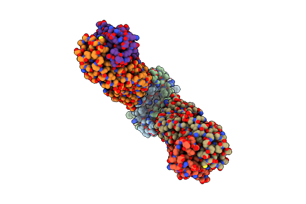
Structure Of Sars-Cov-2 Jn.1 Spike Rbd In Complex With Ace2 (Up State)
Organism: Homo sapiens, Severe acute respiratory syndrome coronavirus 2
Method: ELECTRON MICROSCOPY Release Date: 2024-10-09 Classification: VIRAL PROTEIN/PROTEIN BINDING Ligands: NAG |
Organism: Homo sapiens, Severe acute respiratory syndrome coronavirus 2
Method: ELECTRON MICROSCOPY
Release Date: 2024-10-09
Ligands: NAG
 |
Cryo-Em Structure Of The Mgte Mg2+ Channel Under Mg2+-Free Conditions
Organism: Thermus thermophilus hb8, Mus musculus
Method: ELECTRON MICROSCOPY Release Date: 2021-04-14 Classification: TRANSPORT PROTEIN/IMMUNE SYSTEM |
Organism: Thermus thermophilus hb8, Mus musculus
Method: ELECTRON MICROSCOPY
Release Date: 2021-04-14
 |
Crystal Structure Of The The Cd9
Organism: Homo sapiens
Method: X-RAY DIFFRACTION Resolution:2.70 Å Release Date: 2020-05-13 Classification: MEMBRANE PROTEIN Ligands: PLM, OLC, NI |
Organism: Homo sapiens
Method: X-RAY DIFFRACTION
Release Date: 2020-05-13
Ligands: PLM, OLC, NI
 |
Crystal Structure Of E.Coli Multidrug/H+ Antiporter Mdfa In Outward Open Conformation With Bound Fab Fragment
Organism: Escherichia coli k-12, Mus musculus
Method: X-RAY DIFFRACTION Resolution:3.40 Å Release Date: 2018-10-03 Classification: TRANSPORT PROTEIN Ligands: SO4 |
Organism: Escherichia coli k-12, Mus musculus
Method: X-RAY DIFFRACTION
Release Date: 2018-10-03
Ligands: SO4
 |
Crystal Structure Of Zebrafish Lysophosphatidic Acid Receptor Lpa6
Organism: Danio rerio, Enterobacteria phage t4
Method: X-RAY DIFFRACTION Resolution:3.20 Å Release Date: 2017-08-16 Classification: MEMBRANE PROTEIN Ligands: OLC |
Organism: Danio rerio, Enterobacteria phage t4
Method: X-RAY DIFFRACTION
Release Date: 2017-08-16
Ligands: OLC
 |
Crystal Structures Of The Tric Trimeric Intracellular Cation Channel Orthologue From Sulfolobus Solfataricus
Organism: Mus musculus, Sulfolobus solfataricus
Method: X-RAY DIFFRACTION Resolution:2.64 Å Release Date: 2017-01-11 Classification: IMMUNE SYSTEM/MEMBRANE PROTEIN Ligands: GOL, NA, PX4 |
Organism: Mus musculus, Sulfolobus solfataricus
Method: X-RAY DIFFRACTION
Release Date: 2017-01-11
Ligands: GOL, NA, PX4
 |
Crystal Structures Of The Tric Trimeric Intracellular Cation Channel Orthologue From Rhodobacter Sphaeroides
Organism: Rhodobacter sphaeroides (strain atcc 17023 / 2.4.1 / ncib 8253 / dsm 158)
Method: X-RAY DIFFRACTION Resolution:3.41 Å Release Date: 2017-01-11 Classification: MEMBRANE PROTEIN Ligands: PX4 |
Organism: Rhodobacter sphaeroides (strain atcc 17023 / 2.4.1 / ncib 8253 / dsm 158)
Method: X-RAY DIFFRACTION
Release Date: 2017-01-11
Ligands: PX4
 |
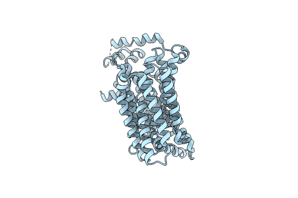
Crystal Structure Of An Invertebrate P2X Receptor From The Gulf Coast Tick In The Presence Of Atp And Zn2+ Ion At 2.9 Angstroms
Organism: Amblyomma maculatum
Method: X-RAY DIFFRACTION Resolution:2.90 Å Release Date: 2016-03-16 Classification: MEMBRANE PROTEIN Ligands: NAG, ZN, ATP |
Organism: Amblyomma maculatum
Method: X-RAY DIFFRACTION
Release Date: 2016-03-16
Ligands: NAG, ZN, ATP
 |
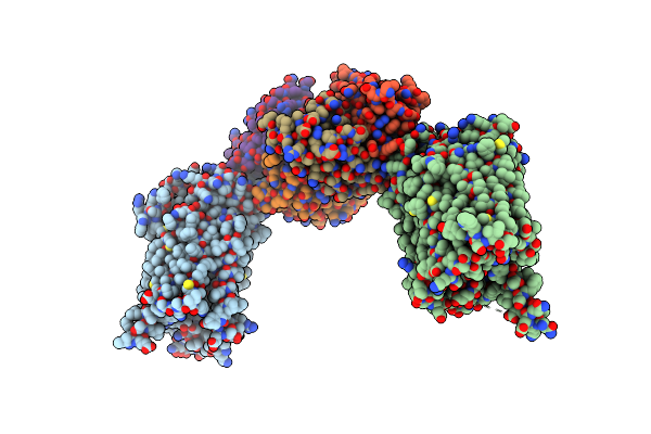
Crystal Structure Of The Bovine Fructose Transporter Glut5 In An Open Inward-Facing Conformation
Organism: Bos taurus
Method: X-RAY DIFFRACTION Resolution:3.20 Å Release Date: 2015-10-14 Classification: TRANSPORT PROTEIN |
Organism: Bos taurus
Method: X-RAY DIFFRACTION
Release Date: 2015-10-14
 |
Rat Glut5 With Fv In The Outward-Open Form
Organism: Rattus norvegicus, Mus musculus
Method: X-RAY DIFFRACTION Resolution:3.27 Å Release Date: 2015-10-07 Classification: TRANSPORT PROTEIN/IMMUNE SYSTEM |
Organism: Rattus norvegicus, Mus musculus
Method: X-RAY DIFFRACTION
Release Date: 2015-10-07
 |
Crystal Structure Of Yidc From Bacillus Halodurans (Form I)
Organism: Bacillus halodurans
Method: X-RAY DIFFRACTION Resolution:2.40 Å Release Date: 2014-04-23 Classification: MEMBRANE PROTEIN Ligands: CD, OLC |
Organism: Bacillus halodurans
Method: X-RAY DIFFRACTION
Release Date: 2014-04-23
Ligands: CD, OLC
 |
Crystal Structure Of Yidc From Bacillus Halodurans (Form Ii)
Organism: Bacillus halodurans
Method: X-RAY DIFFRACTION Resolution:3.20 Å Release Date: 2014-04-23 Classification: MEMBRANE PROTEIN Ligands: CU |
Organism: Bacillus halodurans
Method: X-RAY DIFFRACTION
Release Date: 2014-04-23
Ligands: CU