

Search Count: 25
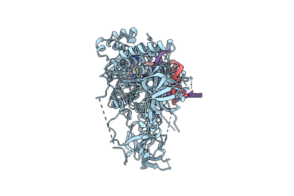 |
Cryo-Em Structure Of Arabidopsis Thaliana Met1 (Aa:621-1534) In Complex With Hemimethylated Dna Analog
Organism: Arabidopsis thaliana, Synthetic construct
Method: ELECTRON MICROSCOPY Resolution:2.74 Å Release Date: 2025-08-20 Classification: STRUCTURAL PROTEIN/DNA Ligands: SAH, ZN |
Organism: Arabidopsis thaliana, Synthetic construct
Method: ELECTRON MICROSCOPY
Release Date: 2025-08-20
Ligands: SAH, ZN
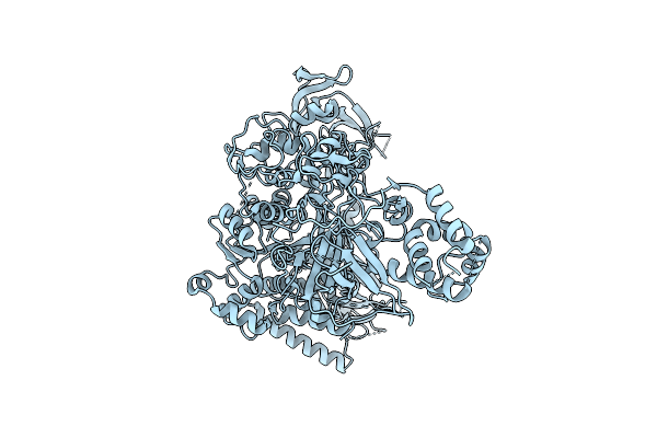 |
Cryo-Em Structure Of Arabidopsis Thaliana Met1
Organism: Arabidopsis thaliana
Method: ELECTRON MICROSCOPY Resolution:3.21 Å Release Date: 2025-08-20 Classification: STRUCTURAL PROTEIN Ligands: ZN |
Organism: Arabidopsis thaliana
Method: ELECTRON MICROSCOPY
Release Date: 2025-08-20
Ligands: ZN
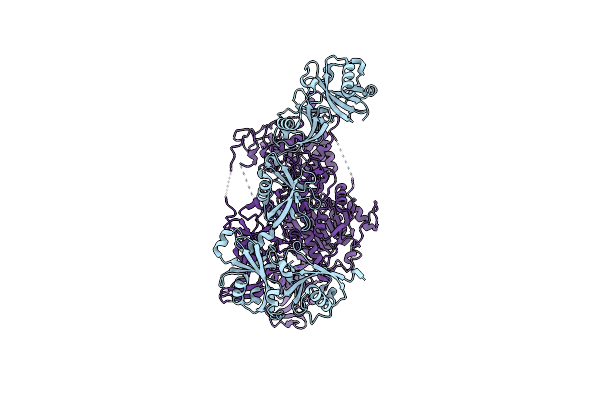 |
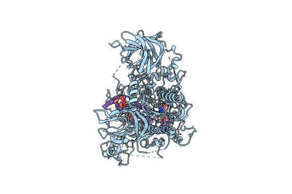
Cryo-Em Structure Of Usp7:Dnmt1 Complex; Closed Conformation
Organism: Homo sapiens
Method: ELECTRON MICROSCOPY Resolution:3.54 Å Release Date: 2025-06-25 Classification: STRUCTURAL PROTEIN Ligands: ZN |
Organism: Homo sapiens
Method: ELECTRON MICROSCOPY
Release Date: 2025-06-25
Ligands: ZN
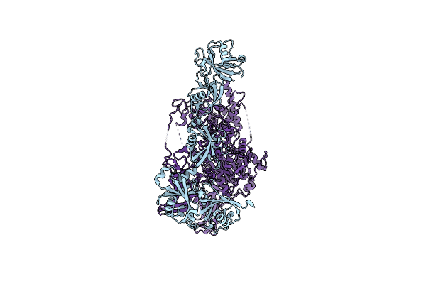 |
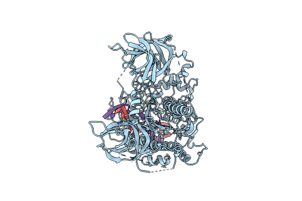
Cryo-Em Structure Of Usp7:Dnmt1 Complex; Open Conformation
Organism: Homo sapiens
Method: ELECTRON MICROSCOPY Resolution:3.75 Å Release Date: 2025-06-25 Classification: STRUCTURAL PROTEIN |
Organism: Homo sapiens
Method: ELECTRON MICROSCOPY
Release Date: 2025-06-25
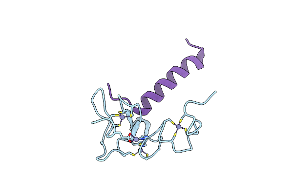 |
Crystal Structure Of Human Dppa3 In Complex With Human Uhrf1 Phd Domain
Organism: Homo sapiens
Method: X-RAY DIFFRACTION Resolution:2.40 Å Release Date: 2024-07-31 Classification: GENE REGULATION Ligands: ZN |
Organism: Homo sapiens
Method: X-RAY DIFFRACTION
Release Date: 2024-07-31
Ligands: ZN
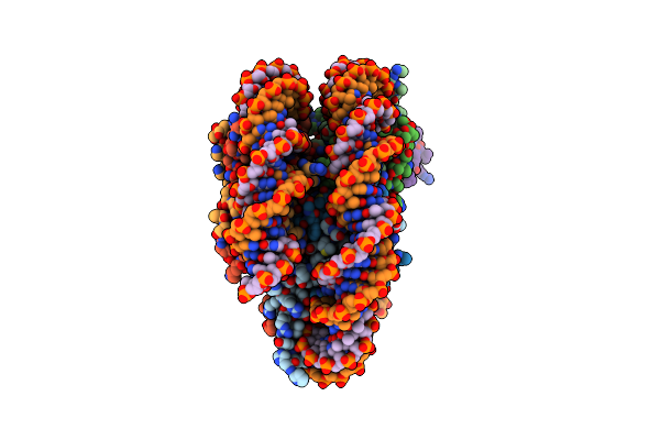 |
Cryo-Em Structure Of Cdca7 Bound To Nucleosome Including Hemimethylated Cpg Site In Widom601 Positioning Sequence.
Organism: Homo sapiens, Synthetic construct
Method: ELECTRON MICROSCOPY Release Date: 2024-07-31 Classification: DNA BINDING PROTEIN Ligands: ZN |
Organism: Homo sapiens, Synthetic construct
Method: ELECTRON MICROSCOPY
Release Date: 2024-07-31
Ligands: ZN
 |
Nmr Strucutre Of Chimeric Protein For Model Of Phd-Stella Complex
Organism: Mus musculus
Method: SOLUTION NMR Release Date: 2022-12-07 Classification: TRANSCRIPTION Ligands: ZN |
Organism: Mus musculus
Method: SOLUTION NMR
Release Date: 2022-12-07
Ligands: ZN
 |
Cryo-Em Structure Of Human Dnmt1 (Aa:351-1616) In Complex With Ubiquitinated H3 And Hemimethylated Dna Analog (Cxxc-Ordered Form)
Organism: Homo sapiens, Synthetic construct
Method: ELECTRON MICROSCOPY Release Date: 2022-11-30 Classification: TRANSFERASE/DNA Ligands: ZN, SAH |
Organism: Homo sapiens, Synthetic construct
Method: ELECTRON MICROSCOPY
Release Date: 2022-11-30
Ligands: ZN, SAH
 |
Cryo-Em Structure Of Human Dnmt1 (Aa:351-1616) In Complex With Ubiquitinated H3 And Hemimethylated Dna Analog (Cxxc-Disordered Form)
Organism: Homo sapiens, Synthetic construct
Method: ELECTRON MICROSCOPY Release Date: 2022-11-30 Classification: TRANSFERASE/DNA Ligands: ZN |
Organism: Homo sapiens, Synthetic construct
Method: ELECTRON MICROSCOPY
Release Date: 2022-11-30
Ligands: ZN
 |
Crystal Structure Of Human Uhrf1 Ttd In Complex With 5-Amino-2,4-Dimethylpyridine
Organism: Homo sapiens
Method: X-RAY DIFFRACTION Resolution:1.45 Å Release Date: 2022-01-05 Classification: DNA BINDING PROTEIN Ligands: MPD, 8NF, DMS, SO4 |
Organism: Homo sapiens
Method: X-RAY DIFFRACTION
Release Date: 2022-01-05
Ligands: MPD, 8NF, DMS, SO4
 |
Crystal Structure Of Human Uhrf1 Phd Finger In Complex With Paf15
Organism: Homo sapiens
Method: X-RAY DIFFRACTION Resolution:1.70 Å Release Date: 2019-10-09 Classification: LIGASE Ligands: ZN, EPE |
Organism: Homo sapiens
Method: X-RAY DIFFRACTION
Release Date: 2019-10-09
Ligands: ZN, EPE
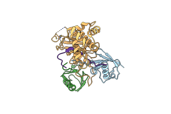 |
Crystal Structure Of Dnmt1 Rfts Domain In Complex With K18/K23 Mono-Ubiquitylated Histone H3
Organism: Homo sapiens
Method: X-RAY DIFFRACTION Resolution:2.00 Å Release Date: 2017-11-15 Classification: SIGNALING PROTEIN/TRANSFERASE Ligands: ZN |
Organism: Homo sapiens
Method: X-RAY DIFFRACTION
Release Date: 2017-11-15
Ligands: ZN
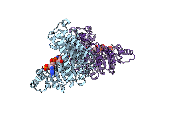 |
Crystal Structure Of The Ternary Complex Of An Isomerase
Organism: Plasmodium falciparum
Method: X-RAY DIFFRACTION Resolution:1.86 Å Release Date: 2011-08-10 Classification: ISOMERASE Ligands: NDP, MN |
Organism: Plasmodium falciparum
Method: X-RAY DIFFRACTION
Release Date: 2011-08-10
Ligands: NDP, MN
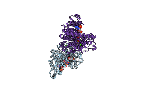 |
Crystal Structure Of The Quaternary Complex-1 Of An Isomerase
Organism: Plasmodium falciparum
Method: X-RAY DIFFRACTION Resolution:1.90 Å Release Date: 2011-08-10 Classification: ISOMERASE/ISOMERASE INHIBITOR Ligands: NDP, MG, FOM, CA |
Organism: Plasmodium falciparum
Method: X-RAY DIFFRACTION
Release Date: 2011-08-10
Ligands: NDP, MG, FOM, CA
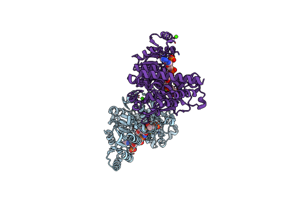 |
Crystal Structure Of The Quaternary Complex-2 Of An Isomerase
Organism: Plasmodium falciparum
Method: X-RAY DIFFRACTION Resolution:2.15 Å Release Date: 2011-08-10 Classification: ISOMERASE/ISOMERASE INHIBITOR Ligands: NDP, MG, F98, CA |
Organism: Plasmodium falciparum
Method: X-RAY DIFFRACTION
Release Date: 2011-08-10
Ligands: NDP, MG, F98, CA
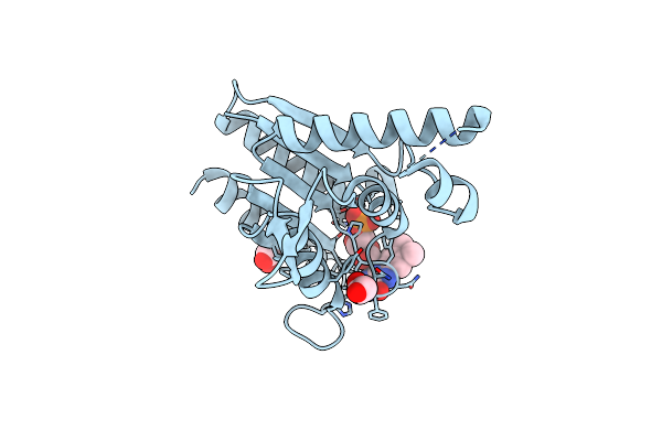 |
The Crystal Structure Of Azor (Azoreductase) From Escherichia Coli: Oxidized Azor In Tetragonal Crystals (The Resolution Has Improved From 1.8 (1V4B) To 1.4 Angstrom)
Organism: Escherichia coli
Method: X-RAY DIFFRACTION Resolution:1.40 Å Release Date: 2008-03-11 Classification: OXIDOREDUCTASE Ligands: FMN, EDO |
Organism: Escherichia coli
Method: X-RAY DIFFRACTION
Release Date: 2008-03-11
Ligands: FMN, EDO
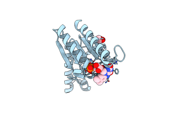 |
The Crystal Structure Of Azor (Azoreductase) From Escherichia Coli: Reduced Azor In Tetragonal Crystals
Organism: Escherichia coli
Method: X-RAY DIFFRACTION Resolution:1.70 Å Release Date: 2008-03-11 Classification: OXIDOREDUCTASE Ligands: FMN, EDO |
Organism: Escherichia coli
Method: X-RAY DIFFRACTION
Release Date: 2008-03-11
Ligands: FMN, EDO
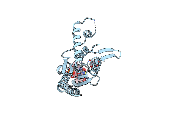 |
The Crystal Structure Of Azor (Azoreductase) From Escherichia Coli: Azor In Complex With Dicoumarol
Organism: Escherichia coli
Method: X-RAY DIFFRACTION Resolution:2.30 Å Release Date: 2008-03-11 Classification: OXIDOREDUCTASE Ligands: FMN, DTC, GOL |
Organism: Escherichia coli
Method: X-RAY DIFFRACTION
Release Date: 2008-03-11
Ligands: FMN, DTC, GOL
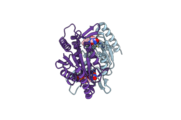 |
The Crystal Structure Of Azor (Azoreductase) From Escherichia Coli: Oxidized Azor In Orthorhombic Crystals
Organism: Escherichia coli
Method: X-RAY DIFFRACTION Resolution:2.10 Å Release Date: 2008-03-11 Classification: OXIDOREDUCTASE Ligands: FMN |
Organism: Escherichia coli
Method: X-RAY DIFFRACTION
Release Date: 2008-03-11
Ligands: FMN
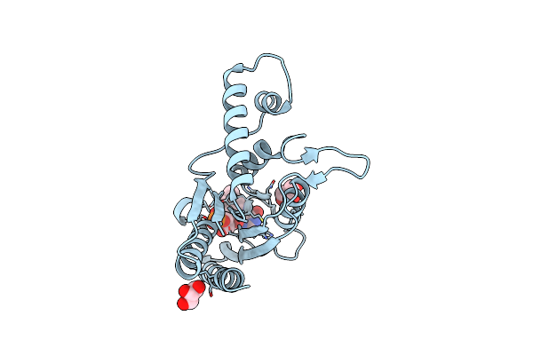 |
The Crystal Structure Of Azor (Azo Reductase) From Escherichia Coli
Organism: Escherichia coli
Method: X-RAY DIFFRACTION Resolution:2.20 Å Release Date: 2006-05-16 Classification: OXIDOREDUCTASE Ligands: FMN, GOL |
Organism: Escherichia coli
Method: X-RAY DIFFRACTION
Release Date: 2006-05-16
Ligands: FMN, GOL