

Search Count: 21
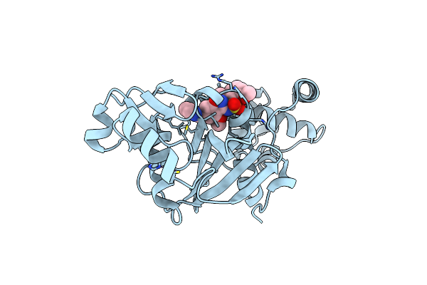 |
Crystal Structure Of Kat6A In Complex With Inhibitor Ctx-648 (Pf-9363)
Organism: Homo sapiens
Method: X-RAY DIFFRACTION Resolution:2.58 Å Release Date: 2023-07-05 Classification: TRANSFERASE/TRANSFERASE INHIBITOR Ligands: R7L, ZN |
Organism: Homo sapiens
Method: X-RAY DIFFRACTION
Release Date: 2023-07-05
Ligands: R7L, ZN
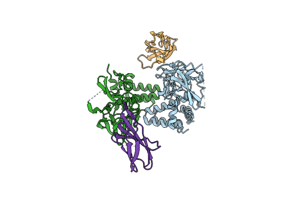 |
Crystal Structure Of Monobody Mb(S4) Bound To Prdm14 In Complex With Mtgr1
Organism: Mus musculus, Homo sapiens
Method: X-RAY DIFFRACTION Resolution:3.05 Å Release Date: 2015-12-02 Classification: GENE REGULATION/TRANSCRIPTION |
Organism: Mus musculus, Homo sapiens
Method: X-RAY DIFFRACTION
Release Date: 2015-12-02
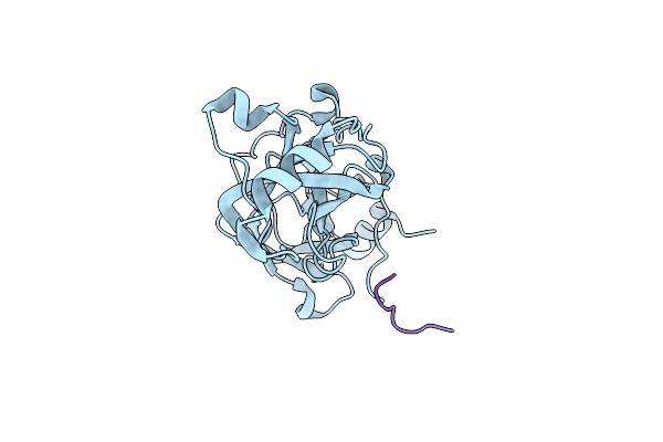 |
The Mbt Repeats Of Human Scml2 In A Complex With Histone H2A Peptide
Organism: Homo sapiens
Method: X-RAY DIFFRACTION Resolution:2.58 Å Release Date: 2012-09-19 Classification: TRANSCRIPTION |
Organism: Homo sapiens
Method: X-RAY DIFFRACTION
Release Date: 2012-09-19
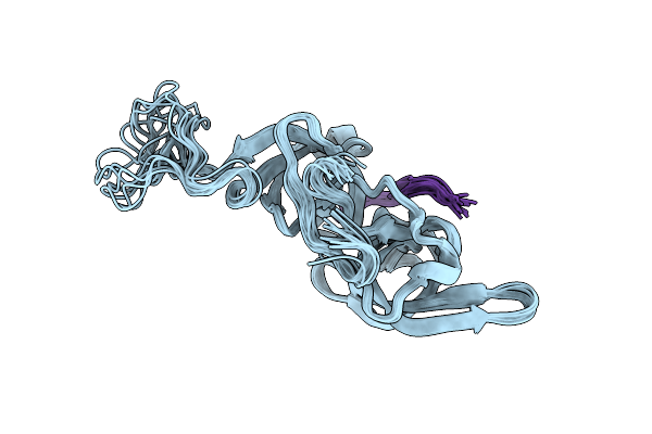 |
Nmr Structure Of Uhrf1 Tandem Tudor Domains In A Complex With Histone H3 Peptide
Organism: Homo sapiens
Method: SOLUTION NMR Release Date: 2011-04-13 Classification: DNA BINDING PROTEIN/GENE REGULATION |
Organism: Homo sapiens
Method: SOLUTION NMR
Release Date: 2011-04-13
 |
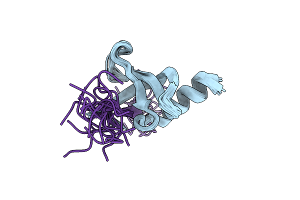
Solution Nmr Structure Of The Chromobox Protein Cbx7 With H3K27Me3
Organism: Homo sapiens, Xenopus laevis
Method: SOLUTION NMR Release Date: 2010-08-25 Classification: TRANSCRIPTION REGULATOR |
Organism: Homo sapiens, Xenopus laevis
Method: SOLUTION NMR
Release Date: 2010-08-25
 |
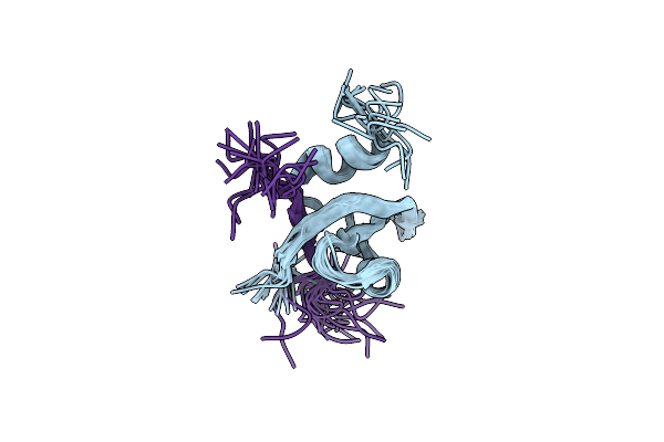
Solution Nmr Structure Of The Cbx3 In Complex With H3K9Me3 Peptide
Organism: Homo sapiens, Xenopus laevis
Method: SOLUTION NMR Release Date: 2010-08-04 Classification: TRANSCRIPTION REGULATOR |
Organism: Homo sapiens, Xenopus laevis
Method: SOLUTION NMR
Release Date: 2010-08-04
 |
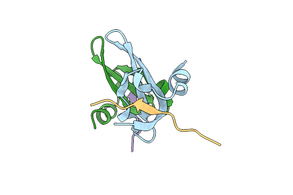
Solution Nmr Structure Of The Chromobox Protein 7 With H3K9Me3
Organism: Homo sapiens, Xenopus laevis
Method: SOLUTION NMR Release Date: 2010-08-04 Classification: TRANSCRIPTION REGULATOR |
Organism: Homo sapiens, Xenopus laevis
Method: SOLUTION NMR
Release Date: 2010-08-04
 |
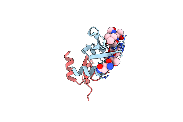
Crystal Structure Of Human Chromobox Homolog 6 (Cbx6) With H3K27 Peptide
Organism: Homo sapiens
Method: X-RAY DIFFRACTION Resolution:2.00 Å Release Date: 2009-09-08 Classification: TRANSCRIPTION |
Organism: Homo sapiens
Method: X-RAY DIFFRACTION
Release Date: 2009-09-08
 |
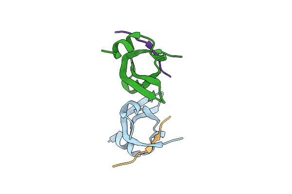
Crystal Structure Of Human Chromobox Homolog 8 (Cbx8) With H3K9 Peptide
Organism: Homo sapiens
Method: X-RAY DIFFRACTION Resolution:1.55 Å Release Date: 2009-09-08 Classification: TRANSCRIPTION |
Organism: Homo sapiens
Method: X-RAY DIFFRACTION
Release Date: 2009-09-08
 |
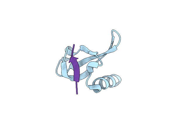
Crystal Structure Of The Complex Of Human Chromobox Homolog 2 (Cbx2) And H3K27 Peptide
Organism: Homo sapiens
Method: X-RAY DIFFRACTION Resolution:1.50 Å Release Date: 2009-08-18 Classification: TRANSCRIPTION |
Organism: Homo sapiens
Method: X-RAY DIFFRACTION
Release Date: 2009-08-18
 |
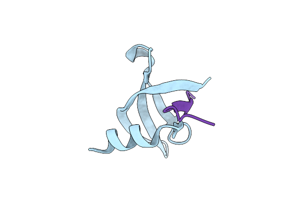
Crystal Structure Of Human Chromobox Homolog 6 (Cbx6) With H3K9 Peptide
Organism: Homo sapiens, Xenopus laevis
Method: X-RAY DIFFRACTION Resolution:1.76 Å Release Date: 2009-04-21 Classification: Transcription/DNA binding protein |
Organism: Homo sapiens, Xenopus laevis
Method: X-RAY DIFFRACTION
Release Date: 2009-04-21
 |
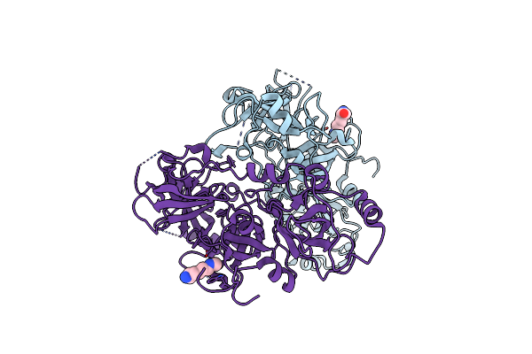
Crystal Structure Of The Complex Of Human Chromobox Homolog 5 (Cbx5) With H3K9(Me)3 Peptide
Organism: Homo sapiens, Synthetic construct
Method: X-RAY DIFFRACTION Resolution:2.00 Å Release Date: 2009-01-13 Classification: PROTEIN BINDING |
Organism: Homo sapiens, Synthetic construct
Method: X-RAY DIFFRACTION
Release Date: 2009-01-13
 |
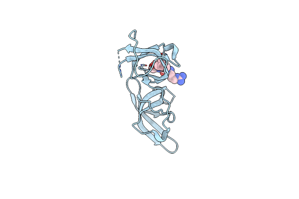
Crystal Structure Of L3Mbtl2-H4K20Me1 Complex
Organism: Homo sapiens
Method: X-RAY DIFFRACTION Resolution:2.10 Å Release Date: 2009-01-06 Classification: TRANSCRIPTION REGULATOR Ligands: MLZ |
Organism: Homo sapiens
Method: X-RAY DIFFRACTION
Release Date: 2009-01-06
Ligands: MLZ
 |
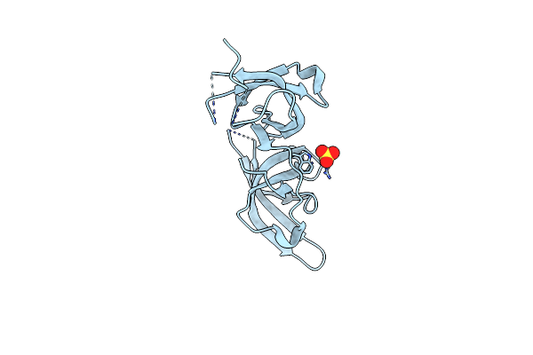
Crystal Structure Of The Tandem Tudor Domains Of The E3 Ubiquitin-Protein Ligase Uhrf1 In Complex With Trimethylated Histone H3-K9 Peptide
Organism: Homo sapiens
Method: X-RAY DIFFRACTION Resolution:2.40 Å Release Date: 2008-09-16 Classification: LIGASE |
Organism: Homo sapiens
Method: X-RAY DIFFRACTION
Release Date: 2008-09-16
 |
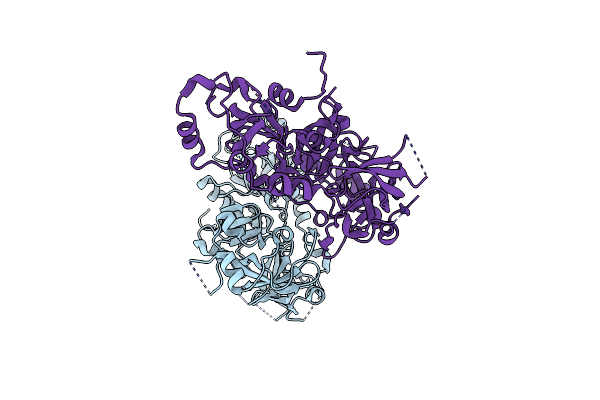
Crystal Structure Of The Tandem Tudor Domains Of The E3 Ubiquitin-Protein Ligase Uhrf1
Organism: Homo sapiens
Method: X-RAY DIFFRACTION Resolution:2.40 Å Release Date: 2008-09-16 Classification: LIGASE Ligands: SO4 |
Organism: Homo sapiens
Method: X-RAY DIFFRACTION
Release Date: 2008-09-16
Ligands: SO4
 |
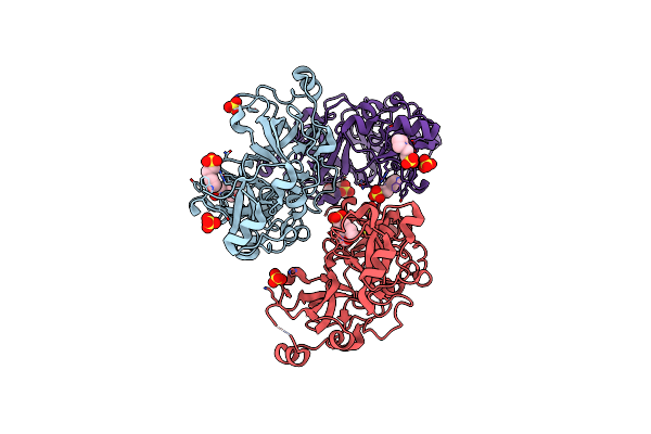
Crystal Structure Of L3Mbtl2
Organism: Homo sapiens
Method: X-RAY DIFFRACTION Resolution:2.20 Å Release Date: 2008-05-06 Classification: TRANSCRIPTION REGULATOR |
Organism: Homo sapiens
Method: X-RAY DIFFRACTION
Release Date: 2008-05-06
 |
Crystal Structure Of L3Mbtl1 Protein In Complex With Mes
Organism: Homo sapiens
Method: X-RAY DIFFRACTION Resolution:2.00 Å Release Date: 2007-10-30 Classification: TRANSCRIPTION Ligands: SO4, MES |
Organism: Homo sapiens
Method: X-RAY DIFFRACTION
Release Date: 2007-10-30
Ligands: SO4, MES
 |
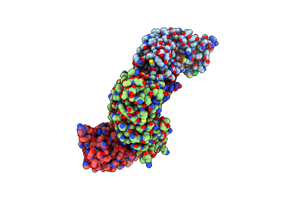
Crystal Structure Of L3Mbtl1 Protein
Organism: Homo sapiens
Method: X-RAY DIFFRACTION Resolution:1.65 Å Release Date: 2007-10-30 Classification: TRANSCRIPTION |
Organism: Homo sapiens
Method: X-RAY DIFFRACTION
Release Date: 2007-10-30
 |
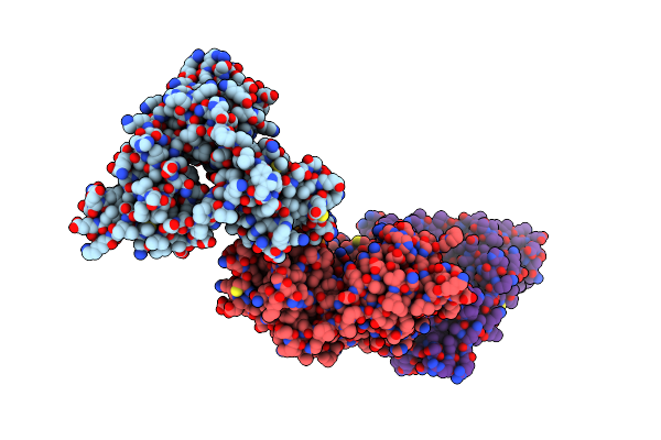
Crystal Structure Of L3Mbtl1 In Complex With H4K20Me2 (Residues 17-25), Orthorhombic Form Ii
Organism: Homo sapiens
Method: X-RAY DIFFRACTION Resolution:1.86 Å Release Date: 2007-10-30 Classification: TRANSCRIPTION Ligands: CL |
Organism: Homo sapiens
Method: X-RAY DIFFRACTION
Release Date: 2007-10-30
Ligands: CL
 |
Crystal Structure Of L3Mbtl1 In Complex With H4K20Me2 (Residues 12-30), Orthorhombic Form I
Organism: Homo sapiens
Method: X-RAY DIFFRACTION Resolution:2.05 Å Release Date: 2007-10-30 Classification: TRANSCRIPTION |
Organism: Homo sapiens
Method: X-RAY DIFFRACTION
Release Date: 2007-10-30