
Search Count: 839
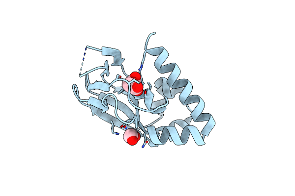 |
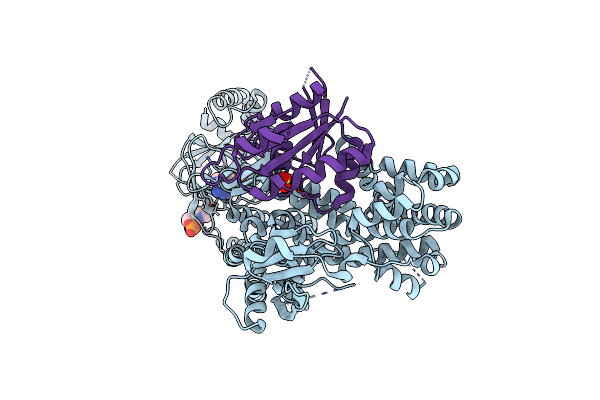
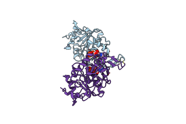
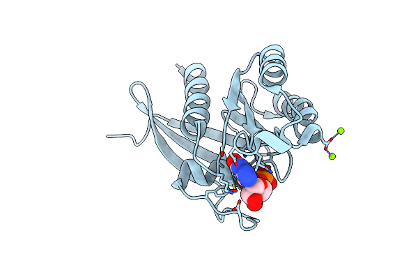
Crystal Structure Of A Putative Cyclic Nucleotide-Binding Protein (Gmet_1532) From Geobacter Metallireducens Gs-15 At 1.90 A Resolution
Organism: Geobacter metallireducens
Method: X-RAY DIFFRACTION Resolution:1.90 Å Release Date: 2010-05-19 Classification: Nucleotide Binding Protein Ligands: SIN, EDO |
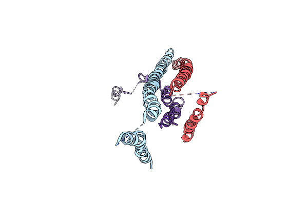 |
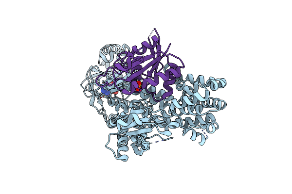
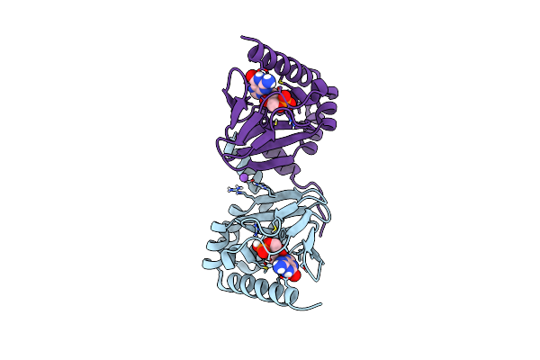
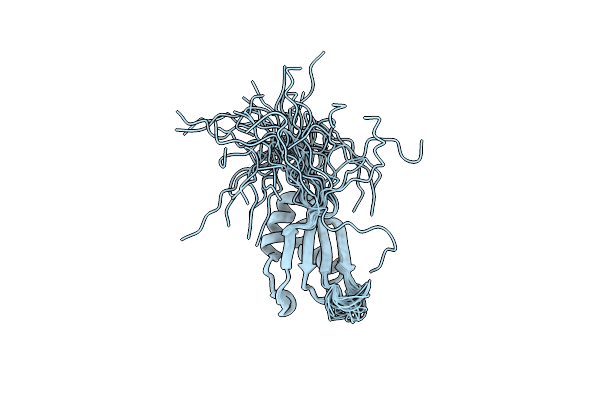
Expression, Purification, Spectroscopical And Crystallographical Studies Of Segments Of The Nucleotide Binding Domain Of The Reticulocyte Binding Protein Py235 Of Plasmodium Yoelii
Organism: Plasmodium yoelii yoelii
Method: X-RAY DIFFRACTION Resolution:4.00 Å Release Date: 2010-02-23 Classification: NUCLEOTIDE BINDING PROTEIN |
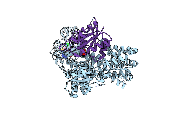 |
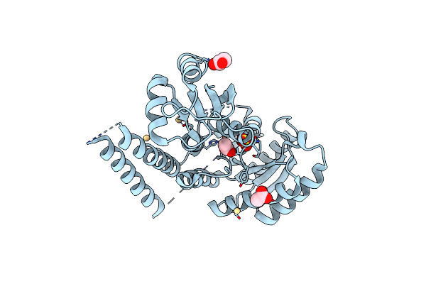
Organism: Mus musculus, Homo sapiens
Method: X-RAY DIFFRACTION Resolution:2.60 Å Release Date: 2014-09-03 Classification: SIGNALING PROTEIN/GTP-BINDING PROTEIN Ligands: H07, SO4 |
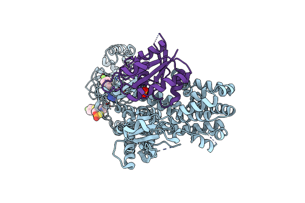 |
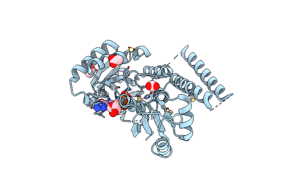
Organism: Mus musculus, Homo sapiens
Method: X-RAY DIFFRACTION Resolution:2.40 Å Release Date: 2014-09-03 Classification: SIGNALING PROTEIN/GTP-BINDING PROTEIN Ligands: HR4, SO4 |
 |
Organism: Mus musculus, Homo sapiens
Method: X-RAY DIFFRACTION Resolution:3.00 Å Release Date: 2014-09-03 Classification: SIGNALING PROTEIN/GTP-BINDING PROTEIN Ligands: HR6, SO4 |
 |
Organism: Mus musculus, Homo sapiens
Method: X-RAY DIFFRACTION Resolution:2.80 Å Release Date: 2014-09-03 Classification: SIGNALING PROTEIN/GTP-BINDING PROTEIN Ligands: CMP, SO4 |
 |
Organism: Mus musculus, Homo sapiens
Method: X-RAY DIFFRACTION Resolution:2.70 Å Release Date: 2014-09-03 Classification: SIGNALING PROTEIN/GTP-BINDING PROTEIN Ligands: CMP, SO4 |
 |
Organism: Homo sapiens
Method: X-RAY DIFFRACTION Resolution:1.82 Å Release Date: 2008-03-04 Classification: NUCLEOTIDE BINDING PROTEIN Ligands: MG, GDP, EDO, UNX |
 |
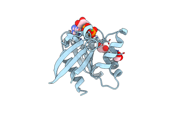
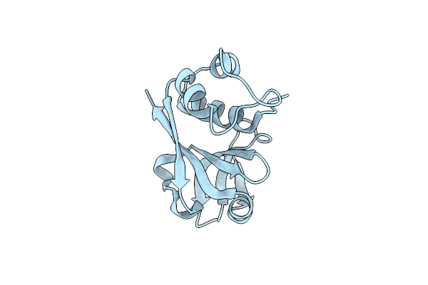
Crystal Structure Of A Putative Cyclic Nucleotide Binding Protein (Spoa0323) From Ruegeria Pomeroyi Dss-3 At 2.35 A Resolution
Organism: Ruegeria pomeroyi dss-3
Method: X-RAY DIFFRACTION Resolution:2.35 Å Release Date: 2009-05-05 Classification: cNMP-BINDING PROTEIN Ligands: CL |
 |
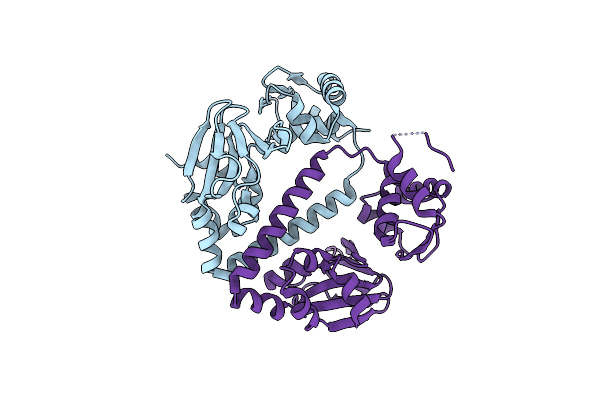
Crystal Structure Of The Nucleotide-Binding Protein Af_226 In Complex With Adp From Archaeoglobus Fulgidus, Northeast Structural Genomics Consortium Target Gr157
Organism: Archaeoglobus fulgidus
Method: X-RAY DIFFRACTION Resolution:2.90 Å Release Date: 2009-10-27 Classification: Nucleotide binding protein Ligands: ADP, ZN |
 |
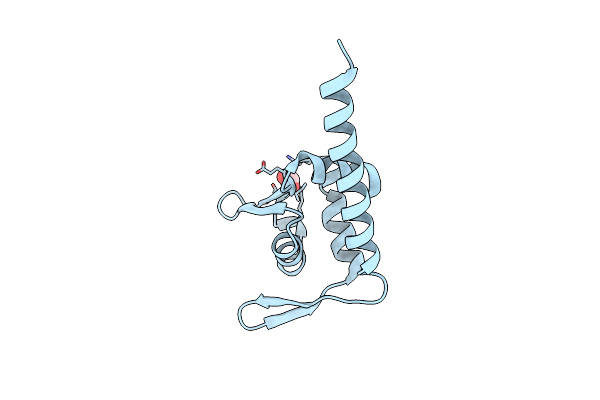
M. Loti Cyclic-Nucleotide Binding Domain Mutant Displaying Inverted Ligand Selectivity, Cyclic-Gmp Bound
Organism: Mesorhizobium loti
Method: X-RAY DIFFRACTION Resolution:1.25 Å Release Date: 2014-06-25 Classification: NUCLEOTIDE BINDING PROTEIN Ligands: PCG, NA |
 |
Crystal Structure Of A Gtp-Binding Protein From The Hyperthermophilic Archaeon Sulfolobus Solfataricus
Organism: Sulfolobus solfataricus p2
Method: X-RAY DIFFRACTION Resolution:2.00 Å Release Date: 2008-08-19 Classification: NUCLEOTIDE BINDING PROTEIN Ligands: CD, SO4, ACT |
 |
Crystal Structure Of A Gtp-Binding Protein From The Hyperthermophilic Archaeon Sulfolobus Solfataricus In Complex With Gdp
Organism: Sulfolobus solfataricus p2
Method: X-RAY DIFFRACTION Resolution:2.00 Å Release Date: 2008-08-19 Classification: NUCLEOTIDE BINDING PROTEIN Ligands: CD, MG, ACT, GDP |
 |
Crystal Structure Of The N-Terminal Peptidase C39 Like Domain Of The Toxin Secretion Atp-Binding Protein From Vibrio Parahaemolyticus
Organism: Vibrio parahaemolyticus rimd 2210633
Method: X-RAY DIFFRACTION Resolution:1.37 Å Release Date: 2007-11-13 Classification: NUCLEOTIDE BINDING PROTEIN |
 |
Crystal Structure Of The C-Terminal Domain Of A Possible Atp-Binding Protein From Methanocaldococcus Jannaschii Dsm 2661
Organism: Methanocaldococcus jannaschii dsm 2661
Method: X-RAY DIFFRACTION Resolution:2.05 Å Release Date: 2007-12-18 Classification: NUCLEOTIDE BINDING PROTEIN Ligands: FMT |
 |
Organism: Escherichia coli k-12
Method: X-RAY DIFFRACTION Resolution:2.80 Å Release Date: 2009-08-25 Classification: Nucleotide binding protein Ligands: GDP, SO4, TRS |
 |
The Crystal Structure Of The Human Rnd1 Gtpase In The Active Gtp Bound State
Organism: Homo sapiens
Method: X-RAY DIFFRACTION Resolution:2.31 Å Release Date: 2006-05-04 Classification: NUCLEOTIDE BINDING PROTEIN Ligands: GTP, MG |
 |
Crystal Structure Of N-Terminal Domain Of Putative Atp/Gtp Binding Protein From Clostridium Difficile 630
Organism: Clostridium difficile
Method: X-RAY DIFFRACTION Resolution:1.76 Å Release Date: 2009-03-10 Classification: NUCLEOTIDE BINDING PROTEIN Ligands: PGE, GOL |
 |
Organism: Homo sapiens
Method: SOLUTION NMR Release Date: 2005-11-19 Classification: RNA BINDING PROTEIN |
 |
Organism: Cryptosporidium parvum
Method: X-RAY DIFFRACTION Resolution:2.06 Å Release Date: 2007-10-23 Classification: NUCLEOTIDE BINDING PROTEIN Ligands: MG, GDP |