

Search Count: 61
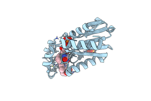 |
Heme-Bound Flavodoxin Fldh From Fusobacterium Nucleatum
Organism: Fusobacterium nucleatum
Method: X-RAY DIFFRACTION Resolution:1.60 Å Release Date: 2023-08-09 Classification: FLAVOPROTEIN Ligands: FMN, HEM, SO4 |
Organism: Fusobacterium nucleatum
Method: X-RAY DIFFRACTION
Release Date: 2023-08-09
Ligands: FMN, HEM, SO4
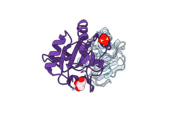 |
Membrane-Associated Thioredoxin Oxidoreductase Fete From Campylobacter Jejuni
Organism: Campylobacter jejuni subsp. jejuni
Method: X-RAY DIFFRACTION Resolution:1.50 Å Release Date: 2023-08-09 Classification: OXIDOREDUCTASE Ligands: SO4, GOL |
Organism: Campylobacter jejuni subsp. jejuni
Method: X-RAY DIFFRACTION
Release Date: 2023-08-09
Ligands: SO4, GOL
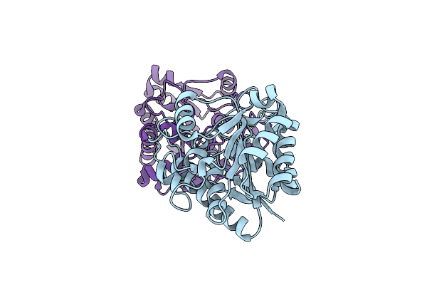 |
Arginase Domain Of Ornithine Decarboxylase/Arginase From Fusobacterium Nucleatum
Organism: Fusobacterium nucleatum subsp. nucleatum atcc 25586
Method: X-RAY DIFFRACTION Resolution:2.25 Å Release Date: 2022-07-06 Classification: HYDROLASE |
Organism: Fusobacterium nucleatum subsp. nucleatum atcc 25586
Method: X-RAY DIFFRACTION
Release Date: 2022-07-06
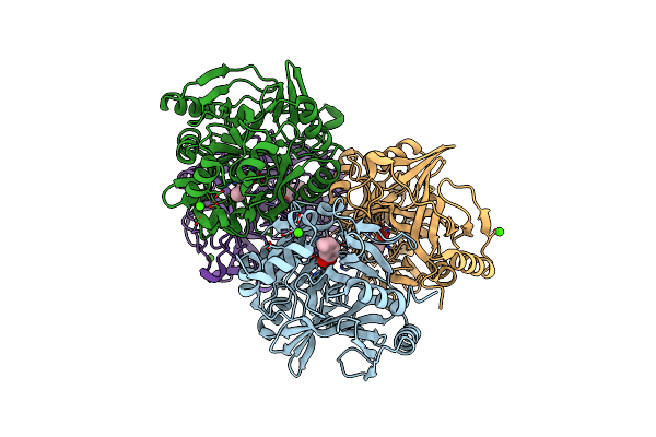 |
Crystal Structure Of Aphc In Complex With 4-Ethylcatechol
Organism: Rhodococcus jostii (strain rha1)
Method: X-RAY DIFFRACTION Resolution:1.60 Å Release Date: 2022-04-06 Classification: OXIDOREDUCTASE Ligands: FE, CA, 8RU |
Organism: Rhodococcus jostii (strain rha1)
Method: X-RAY DIFFRACTION
Release Date: 2022-04-06
Ligands: FE, CA, 8RU
 |
Crystal Structure Of Cobalt-Bound Lsd4 From Sphingobium Sp. Strain Syk-6
Organism: Sphingobium sp. (strain nbrc 103272 / syk-6)
Method: X-RAY DIFFRACTION Resolution:1.45 Å Release Date: 2021-05-12 Classification: OXIDOREDUCTASE Ligands: CO |
Organism: Sphingobium sp. (strain nbrc 103272 / syk-6)
Method: X-RAY DIFFRACTION
Release Date: 2021-05-12
Ligands: CO
 |
Crystal Structure Of Dca-S Bound To Co-Lsd4 From Sphingobium Sp. Strain Syk-6
Organism: Sphingobium sp. (strain nbrc 103272 / syk-6)
Method: X-RAY DIFFRACTION Resolution:1.45 Å Release Date: 2021-05-12 Classification: OXIDOREDUCTASE Ligands: V5M, DMS, CO, MG |
Organism: Sphingobium sp. (strain nbrc 103272 / syk-6)
Method: X-RAY DIFFRACTION
Release Date: 2021-05-12
Ligands: V5M, DMS, CO, MG
 |
Crystal Structure Of Lignostilbene Bound To Co-Lsd4 From Sphingobium Sp. Strain Syk-6
Organism: Sphingobium sp. (strain nbrc 103272 / syk-6)
Method: X-RAY DIFFRACTION Resolution:1.85 Å Release Date: 2021-05-12 Classification: OXIDOREDUCTASE Ligands: CO, V5P, DMS, MG |
Organism: Sphingobium sp. (strain nbrc 103272 / syk-6)
Method: X-RAY DIFFRACTION
Release Date: 2021-05-12
Ligands: CO, V5P, DMS, MG
 |
Crystal Structure Of Vanillin Bound To Co-Lsd4 From Sphingobium Sp. Strain Syk-6
Organism: Sphingobium sp. (strain nbrc 103272 / syk-6)
Method: X-RAY DIFFRACTION Resolution:1.65 Å Release Date: 2021-05-12 Classification: OXIDOREDUCTASE Ligands: V55, DMS, ACT, CO, MG |
Organism: Sphingobium sp. (strain nbrc 103272 / syk-6)
Method: X-RAY DIFFRACTION
Release Date: 2021-05-12
Ligands: V55, DMS, ACT, CO, MG
 |
Crystal Structure Of Iron-Bound Lsd4 From Sphingobium Sp. Strain Syk-6
Organism: Sphingobium sp. (strain nbrc 103272 / syk-6)
Method: X-RAY DIFFRACTION Resolution:1.45 Å Release Date: 2021-05-12 Classification: OXIDOREDUCTASE Ligands: SO4, FE |
Organism: Sphingobium sp. (strain nbrc 103272 / syk-6)
Method: X-RAY DIFFRACTION
Release Date: 2021-05-12
Ligands: SO4, FE
 |
Crystal Structure Of The Helical Cell Shape Determining Protein Pgp2 From Campylobacter Jejuni
Organism: Campylobacter jejuni subsp. jejuni
Method: X-RAY DIFFRACTION Resolution:1.50 Å Release Date: 2021-03-17 Classification: HYDROLASE |
Organism: Campylobacter jejuni subsp. jejuni
Method: X-RAY DIFFRACTION
Release Date: 2021-03-17
 |
Crystal Structure Of The Helical Cell Shape Determining Protein Pgp2 (K307A Mutant) From Campylobacter Jejuni
Organism: Campylobacter jejuni subsp. jejuni
Method: X-RAY DIFFRACTION Resolution:1.85 Å Release Date: 2021-03-17 Classification: HYDROLASE |
Organism: Campylobacter jejuni subsp. jejuni
Method: X-RAY DIFFRACTION
Release Date: 2021-03-17
 |
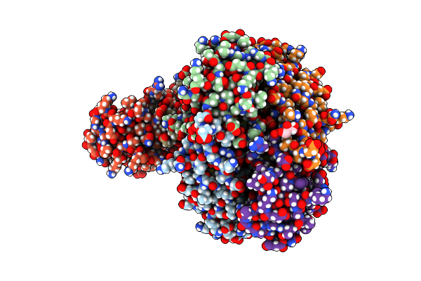
Copper-Bound E44Q Variant Of Campylobacter Jejuni P19
Organism: Campylobacter jejuni subsp. jejuni serotype o:23/36 (strain 81-176)
Method: X-RAY DIFFRACTION Resolution:1.90 Å Release Date: 2021-02-10 Classification: TRANSPORT PROTEIN Ligands: CU, SO4 |
Organism: Campylobacter jejuni subsp. jejuni serotype o:23/36 (strain 81-176)
Method: X-RAY DIFFRACTION
Release Date: 2021-02-10
Ligands: CU, SO4
 |
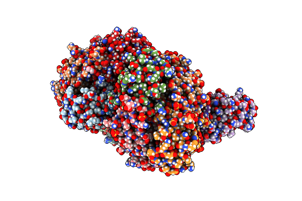
Copper-Bound M88I Variant Of Campylobacter Jejuni P19
Organism: Campylobacter jejuni subsp. jejuni serotype o:23/36 (strain 81-176)
Method: X-RAY DIFFRACTION Resolution:2.30 Å Release Date: 2021-02-10 Classification: TRANSPORT PROTEIN Ligands: CU, GOL, SO4, TRS |
Organism: Campylobacter jejuni subsp. jejuni serotype o:23/36 (strain 81-176)
Method: X-RAY DIFFRACTION
Release Date: 2021-02-10
Ligands: CU, GOL, SO4, TRS
 |
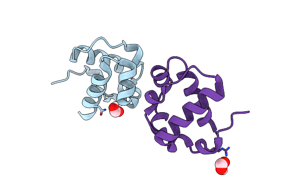
Copper-Bound D92H Variant Of Campylobacter Jejuni P19
Organism: Campylobacter jejuni subsp. jejuni serotype o:23/36 (strain 81-176)
Method: X-RAY DIFFRACTION Resolution:2.50 Å Release Date: 2021-02-10 Classification: TRANSPORT PROTEIN Ligands: CU, SO4, GOL |
Organism: Campylobacter jejuni subsp. jejuni serotype o:23/36 (strain 81-176)
Method: X-RAY DIFFRACTION
Release Date: 2021-02-10
Ligands: CU, SO4, GOL
 |
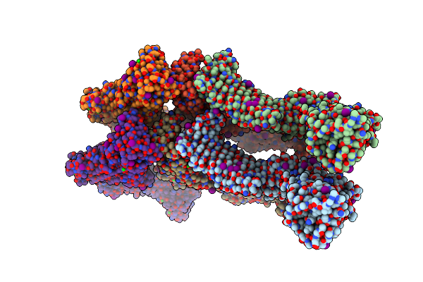
Crystal Structure Of The Monomeric Etv6 Pnt Domain
Organism: Homo sapiens
Method: X-RAY DIFFRACTION Resolution:1.85 Å Release Date: 2021-01-20 Classification: ONCOPROTEIN Ligands: FMT |
Organism: Homo sapiens
Method: X-RAY DIFFRACTION
Release Date: 2021-01-20
Ligands: FMT
 |
Surface-Layer (S-Layer) Rsaa Protein From Caulobacter Crescentus Bound To Strontium And Iodide
Organism: Caulobacter vibrioides
Method: X-RAY DIFFRACTION Resolution:2.10 Å Release Date: 2020-01-15 Classification: MEMBRANE PROTEIN Ligands: IOD, SR |
Organism: Caulobacter vibrioides
Method: X-RAY DIFFRACTION
Release Date: 2020-01-15
Ligands: IOD, SR
 |
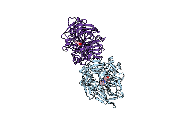
Crystal Structure Of Sphingomonas Paucimobilis Tmy1009 Apo-Lsda
Organism: Sphingomonas paucimobilis
Method: X-RAY DIFFRACTION Resolution:2.30 Å Release Date: 2019-07-24 Classification: OXIDOREDUCTASE Ligands: GOL, MG |
Organism: Sphingomonas paucimobilis
Method: X-RAY DIFFRACTION
Release Date: 2019-07-24
Ligands: GOL, MG
 |
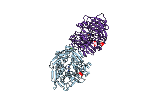
Crystal Structure Of Sphingomonas Paucimobilis Tmy1009 Lsda Phenylazophenol Complex
Organism: Sphingomonas paucimobilis
Method: X-RAY DIFFRACTION Resolution:3.00 Å Release Date: 2019-07-24 Classification: OXIDOREDUCTASE Ligands: NSL, FE |
Organism: Sphingomonas paucimobilis
Method: X-RAY DIFFRACTION
Release Date: 2019-07-24
Ligands: NSL, FE
 |
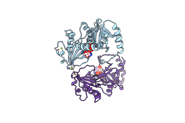
Crystal Structure Of Sphingomonas Paucimobilis Tmy1009 Holo-Lsda
Organism: Sphingomonas paucimobilis
Method: X-RAY DIFFRACTION Resolution:2.60 Å Release Date: 2019-07-24 Classification: OXIDOREDUCTASE Ligands: FE, GOL, MG |
Organism: Sphingomonas paucimobilis
Method: X-RAY DIFFRACTION
Release Date: 2019-07-24
Ligands: FE, GOL, MG
 |
Crystal Structure Of Staphylococcus Pseudintermedius Sbni In Complex With Adp
Organism: Staphylococcus pseudintermedius
Method: X-RAY DIFFRACTION Resolution:1.90 Å Release Date: 2019-06-19 Classification: BIOSYNTHETIC PROTEIN Ligands: MG, ADP |
Organism: Staphylococcus pseudintermedius
Method: X-RAY DIFFRACTION
Release Date: 2019-06-19
Ligands: MG, ADP