

Search Count: 21
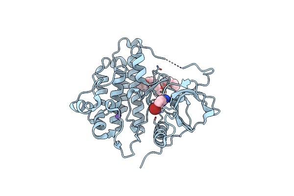 |
Crystal Structure Of Egfr_Dmx In Complex With Covalently Bound Fragment 1
Organism: Homo sapiens
Method: X-RAY DIFFRACTION Resolution:2.40 Å Release Date: 2023-12-13 Classification: TRANSFERASE Ligands: PE5, N6N, NA |
Organism: Homo sapiens
Method: X-RAY DIFFRACTION
Release Date: 2023-12-13
Ligands: PE5, N6N, NA
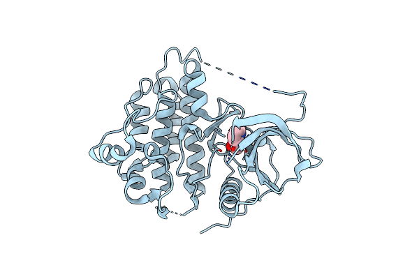 |
Crystal Structure Of Egfr_Wt In Complex With Covalently Bound Fragment 4
Organism: Homo sapiens
Method: X-RAY DIFFRACTION Resolution:2.80 Å Release Date: 2023-12-13 Classification: TRANSFERASE Ligands: MWU |
Organism: Homo sapiens
Method: X-RAY DIFFRACTION
Release Date: 2023-12-13
Ligands: MWU
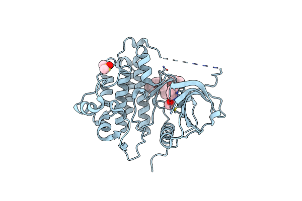 |
Crystal Structure Of Egfr_Dmx In Complex With Covalently Bound Fragment 4
Organism: Homo sapiens
Method: X-RAY DIFFRACTION Resolution:2.40 Å Release Date: 2023-12-13 Classification: TRANSFERASE Ligands: PE5, DMS, MWU |
Organism: Homo sapiens
Method: X-RAY DIFFRACTION
Release Date: 2023-12-13
Ligands: PE5, DMS, MWU
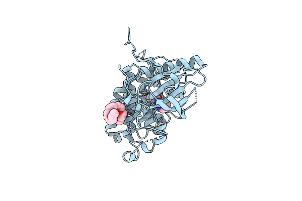 |
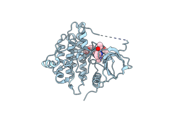
Crystal Structure Of Egfr_Tmx In Complex With Covalently Bound Fragment 4
Organism: Homo sapiens
Method: X-RAY DIFFRACTION Resolution:2.20 Å Release Date: 2023-12-13 Classification: TRANSFERASE Ligands: PE5, MWU, DMS, CL |
Organism: Homo sapiens
Method: X-RAY DIFFRACTION
Release Date: 2023-12-13
Ligands: PE5, MWU, DMS, CL
 |
Crystal Structure Of Egfr_Dmx In Complex With Compound 7
Organism: Homo sapiens
Method: X-RAY DIFFRACTION Resolution:2.20 Å Release Date: 2023-12-13 Classification: TRANSFERASE Ligands: PE5, N7C |
Organism: Homo sapiens
Method: X-RAY DIFFRACTION
Release Date: 2023-12-13
Ligands: PE5, N7C
 |
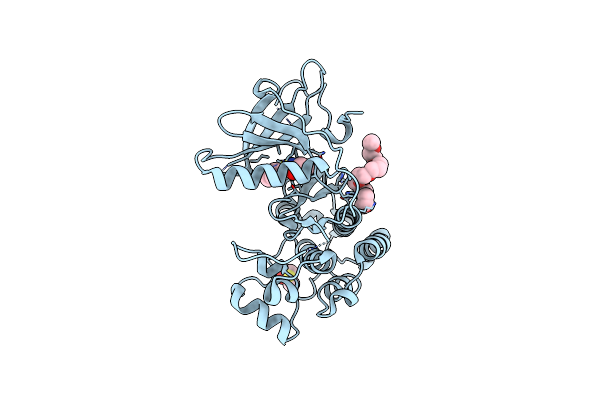
Crystal Structure Of Egfr_Tmx In Complex With Covalently Bound Fragment 8
Organism: Homo sapiens
Method: X-RAY DIFFRACTION Resolution:2.20 Å Release Date: 2023-12-13 Classification: TRANSFERASE Ligands: PE5, N86, DMS |
Organism: Homo sapiens
Method: X-RAY DIFFRACTION
Release Date: 2023-12-13
Ligands: PE5, N86, DMS
 |
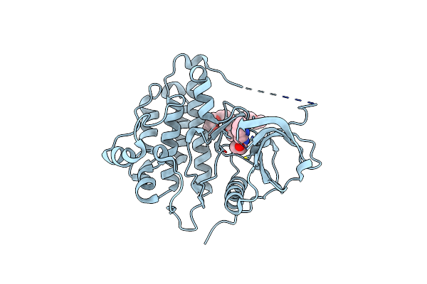
Crystal Structure Of Egfr_Tmx In Complex With Covalently Bound Fragment 9
Organism: Homo sapiens
Method: X-RAY DIFFRACTION Resolution:2.69 Å Release Date: 2023-12-13 Classification: TRANSFERASE Ligands: PE5, N8O, DMS |
Organism: Homo sapiens
Method: X-RAY DIFFRACTION
Release Date: 2023-12-13
Ligands: PE5, N8O, DMS
 |
Crystal Structure Of Egfr_Tmx In Complex With Covalently Bound Fragment 10
Organism: Homo sapiens
Method: X-RAY DIFFRACTION Resolution:2.40 Å Release Date: 2023-12-13 Classification: TRANSFERASE Ligands: PE5, N99 |
Organism: Homo sapiens
Method: X-RAY DIFFRACTION
Release Date: 2023-12-13
Ligands: PE5, N99
 |
Crystal Structure Of Egfr_Tmx In Complex With Covalently Bound Fragment 12
Organism: Homo sapiens
Method: X-RAY DIFFRACTION Resolution:2.50 Å Release Date: 2023-12-13 Classification: TRANSFERASE Ligands: PE5, N9L, DMS |
Organism: Homo sapiens
Method: X-RAY DIFFRACTION
Release Date: 2023-12-13
Ligands: PE5, N9L, DMS
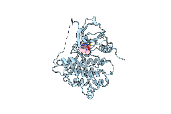 |
Crystal Structure Of Egfr_Tmx In Complex With Covalently Bound Compound 14
Organism: Homo sapiens
Method: X-RAY DIFFRACTION Resolution:2.77 Å Release Date: 2023-12-13 Classification: TRANSFERASE Ligands: N9R |
Organism: Homo sapiens
Method: X-RAY DIFFRACTION
Release Date: 2023-12-13
Ligands: N9R
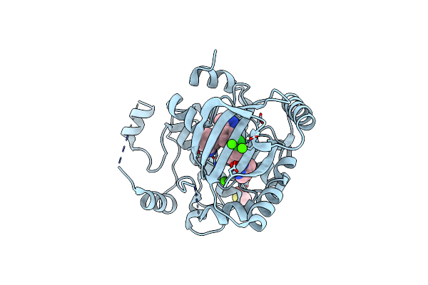 |
Mek1 Bound To Ds03090629
Organism: Homo sapiens
Method: X-RAY DIFFRACTION Resolution:2.10 Å Release Date: 2023-03-01 Classification: TRANSFERASE/TRANSFERASE INHIBITOR Ligands: FZC, DMS, CA |
Organism: Homo sapiens
Method: X-RAY DIFFRACTION
Release Date: 2023-03-01
Ligands: FZC, DMS, CA
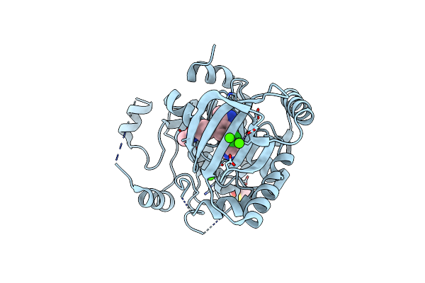 |
Mek1 Bound To Ds94070624
Organism: Homo sapiens
Method: X-RAY DIFFRACTION Resolution:2.10 Å Release Date: 2023-03-01 Classification: TRANSFERASE/TRANSFERASE INHIBITOR Ligands: GIK, DMS, CA |
Organism: Homo sapiens
Method: X-RAY DIFFRACTION
Release Date: 2023-03-01
Ligands: GIK, DMS, CA
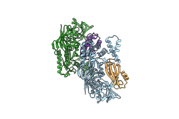 |
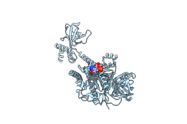
Crystal Structure Of The Archaeal Ef1A-Ef1B Complex
Organism: Pyrococcus horikoshii ot-3
Method: X-RAY DIFFRACTION Resolution:2.00 Å Release Date: 2021-06-23 Classification: TRANSLATION |
Organism: Pyrococcus horikoshii ot-3
Method: X-RAY DIFFRACTION
Release Date: 2021-06-23
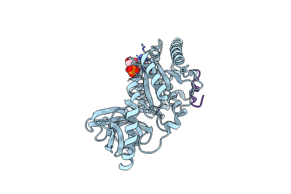 |
Crystal Structure Of The Complex Of Archaeal Ribosomal Stalk Protein Ap1 And Archaeal Translation Initiation Factor Aif5B
Organism: Aeropyrum pernix k1
Method: X-RAY DIFFRACTION Resolution:1.89 Å Release Date: 2018-06-27 Classification: TRANSLATION Ligands: GDP |
Organism: Aeropyrum pernix k1
Method: X-RAY DIFFRACTION
Release Date: 2018-06-27
Ligands: GDP
 |
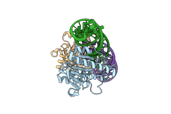
Rhodobacter Sphaeroides Argonaute In Complex With Guide Rna/Target Dna Heteroduplex
Organism: Rhodobacter sphaeroides, Synthetic construct
Method: X-RAY DIFFRACTION Resolution:2.00 Å Release Date: 2016-07-06 Classification: RNA BINDING PROTEIN/DNA/RNA Ligands: MG |
Organism: Rhodobacter sphaeroides, Synthetic construct
Method: X-RAY DIFFRACTION
Release Date: 2016-07-06
Ligands: MG
 |
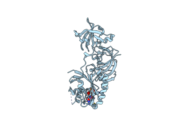
Crystal Structure Of Gdp-Bound Aif5B From Aeropyrum Pernix
Organism: Aeropyrum pernix (strain atcc 700893 / dsm 11879 / jcm 9820 / nbrc 100138 / k1)
Method: X-RAY DIFFRACTION Resolution:1.90 Å Release Date: 2016-05-04 Classification: TRANSLATION Ligands: GDP |
Organism: Aeropyrum pernix (strain atcc 700893 / dsm 11879 / jcm 9820 / nbrc 100138 / k1)
Method: X-RAY DIFFRACTION
Release Date: 2016-05-04
Ligands: GDP
 |
Crystal Structure Of A Complex Of The Archaeal Ribosomal Stalk Protein Ap1 And The Gdp-Bound Archaeal Elongation Factor Aef1Alpha
Organism: Pyrococcus horikoshii ot3
Method: X-RAY DIFFRACTION Resolution:2.30 Å Release Date: 2014-12-24 Classification: TRANSLATION/RIBOSOMAL PROTEIN Ligands: GDP |
Organism: Pyrococcus horikoshii ot3
Method: X-RAY DIFFRACTION
Release Date: 2014-12-24
Ligands: GDP
 |
Crystal Structure Of Gdp-Bound Ef1Alpha From Pyrococcus Horikoshii
Organism: Pyrococcus horikoshii ot3
Method: X-RAY DIFFRACTION Resolution:2.35 Å Release Date: 2014-12-24 Classification: TRANSLATION Ligands: GDP |
Organism: Pyrococcus horikoshii ot3
Method: X-RAY DIFFRACTION
Release Date: 2014-12-24
Ligands: GDP
 |
Crystal Structure Of Peptidyl-Trna Hydrolase From Escherichia Coli In Complex With The Cca-Acceptor-T[Psi]C Domain Of Trna
Organism: Escherichia coli
Method: X-RAY DIFFRACTION Resolution:2.40 Å Release Date: 2012-09-12 Classification: hydrolase/RNA |
Organism: Escherichia coli
Method: X-RAY DIFFRACTION
Release Date: 2012-09-12
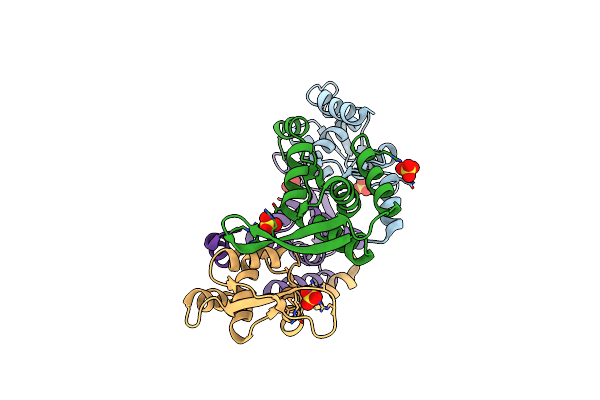 |
Crystal Structure Of Pex From Synechococcus Sp. (Strain Pcc 7942) (Anacystis Nidulans R2)
Organism: Synechococcus sp.
Method: X-RAY DIFFRACTION Resolution:2.90 Å Release Date: 2009-01-20 Classification: CIRCADIAN CLOCK PROTEIN Ligands: SO4 |
Organism: Synechococcus sp.
Method: X-RAY DIFFRACTION
Release Date: 2009-01-20
Ligands: SO4