

Search Count: 25
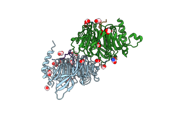 |
Crystal Structure Of Rbbp4 Bound To Znf512B Peptide
Organism: Homo sapiens
Method: X-RAY DIFFRACTION Resolution:2.20 Å Release Date: 2024-11-06 Classification: TRANSCRIPTION Ligands: FMT, EDO, ACT, OXM |
Organism: Homo sapiens
Method: X-RAY DIFFRACTION
Release Date: 2024-11-06
Ligands: FMT, EDO, ACT, OXM
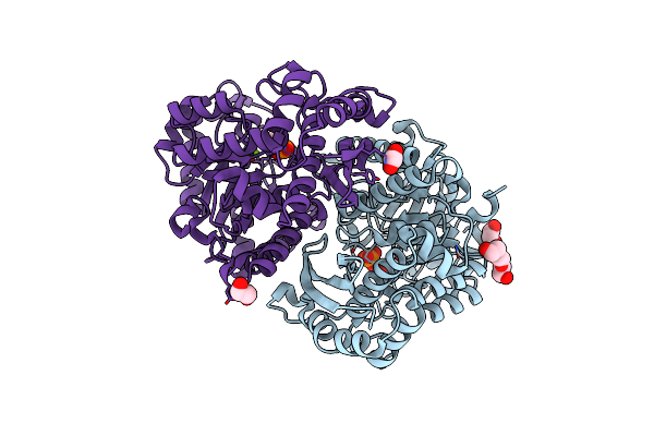 |
Structure Of Human Enolase 2 In Complex With Phosphoserine
Organism: Homo sapiens
Method: X-RAY DIFFRACTION Resolution:2.10 Å Release Date: 2022-11-09 Classification: LYASE Ligands: PO4, MG, ACT, EDO, PGE, SEP |
Organism: Homo sapiens
Method: X-RAY DIFFRACTION
Release Date: 2022-11-09
Ligands: PO4, MG, ACT, EDO, PGE, SEP
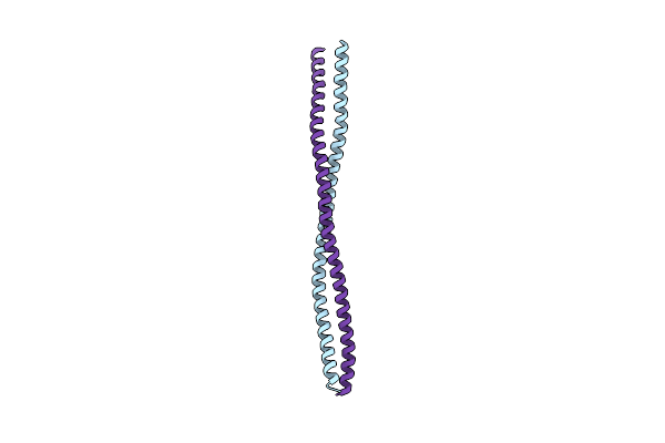 |
Crystal Structure Of The N-Terminal Parallel Dimeric Coiled-Coil Region Of The Human Kinetochore Associated Protein Spindly
Organism: Homo sapiens
Method: X-RAY DIFFRACTION Resolution:2.80 Å Release Date: 2022-09-07 Classification: CELL CYCLE |
Organism: Homo sapiens
Method: X-RAY DIFFRACTION
Release Date: 2022-09-07
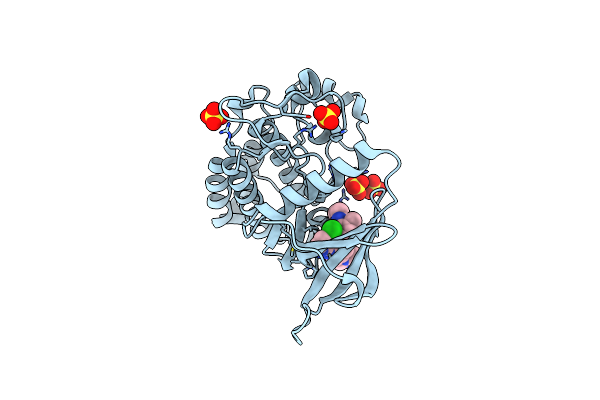 |
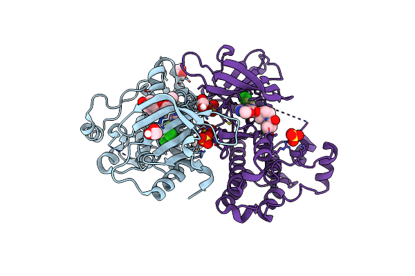
Crystal Structure Of Egfr-T790M/V948R In Complex With Ldc8201
Organism: Homo sapiens
Method: X-RAY DIFFRACTION Resolution:2.40 Å Release Date: 2022-02-23 Classification: TRANSFERASE Ligands: SO4, R1W |
Organism: Homo sapiens
Method: X-RAY DIFFRACTION
Release Date: 2022-02-23
Ligands: SO4, R1W
 |
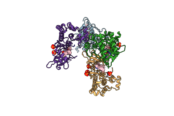
Crystal Structure Of Egfr-T790M/V948R In Complex With Poziotinib
Organism: Homo sapiens
Method: X-RAY DIFFRACTION Resolution:2.00 Å Release Date: 2022-02-23 Classification: TRANSFERASE Ligands: R2E, SO4, EDO |
Organism: Homo sapiens
Method: X-RAY DIFFRACTION
Release Date: 2022-02-23
Ligands: R2E, SO4, EDO
 |
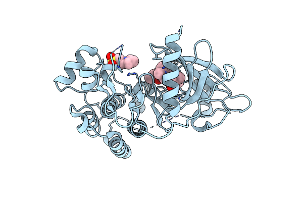
Crystal Structure Of Egfr-T790M/V948R In Complex With Tak-788
Organism: Homo sapiens
Method: X-RAY DIFFRACTION Resolution:2.00 Å Release Date: 2022-02-23 Classification: TRANSFERASE Ligands: R28, SO4 |
Organism: Homo sapiens
Method: X-RAY DIFFRACTION
Release Date: 2022-02-23
Ligands: R28, SO4
 |
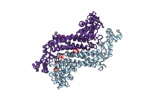
Crystal Structure Of Egfr-Wt In Complex With Tak-788
Organism: Homo sapiens
Method: X-RAY DIFFRACTION Resolution:2.50 Å Release Date: 2022-02-23 Classification: TRANSFERASE Ligands: R28, MES |
Organism: Homo sapiens
Method: X-RAY DIFFRACTION
Release Date: 2022-02-23
Ligands: R28, MES
 |
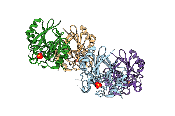
Crystal Structure Of Recombinant Human Fumarase In Complex With D-2-Amino-3-Phosphono-Propionic Acid
Organism: Homo sapiens
Method: X-RAY DIFFRACTION Resolution:2.15 Å Release Date: 2022-02-23 Classification: LYASE Ligands: APO, GOL |
Organism: Homo sapiens
Method: X-RAY DIFFRACTION
Release Date: 2022-02-23
Ligands: APO, GOL
 |
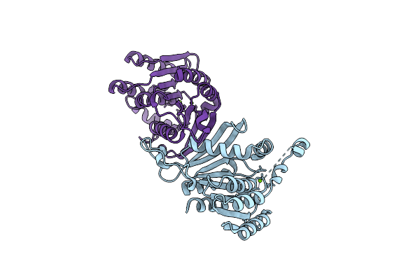
Crystal Structure Of Hydroxyacyl Glutathione Hydrolase (Glob) From Staphylococcus Aureus, Apoenzyme
Organism: Staphylococcus aureus
Method: X-RAY DIFFRACTION Resolution:1.65 Å Release Date: 2020-12-30 Classification: HYDROLASE Ligands: ZN, SO4 |
Organism: Staphylococcus aureus
Method: X-RAY DIFFRACTION
Release Date: 2020-12-30
Ligands: ZN, SO4
 |
Crystal Structure Of S-Formylglutathione Hydrolase (Frmb) From Staphylococcus Aureus, Apoenzyme
Organism: Staphylococcus aureus
Method: X-RAY DIFFRACTION Resolution:1.60 Å Release Date: 2020-12-23 Classification: HYDROLASE Ligands: MG |
Organism: Staphylococcus aureus
Method: X-RAY DIFFRACTION
Release Date: 2020-12-23
Ligands: MG
 |
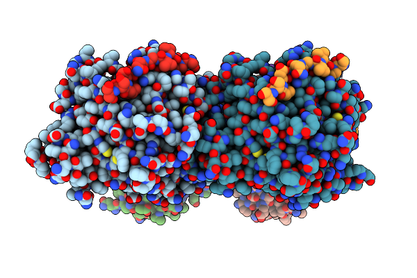
Clathrin Heavy Chain N-Terminal Domain Bound To Gtse1 Lidl Motif
Organism: Homo sapiens
Method: X-RAY DIFFRACTION Resolution:2.03 Å Release Date: 2019-08-28 Classification: CELL CYCLE |
Organism: Homo sapiens
Method: X-RAY DIFFRACTION
Release Date: 2019-08-28
 |
Clathrin Heavy Chain N-Terminal Domain Bound To Gtse1 Lidl Motif
Organism: Homo sapiens
Method: X-RAY DIFFRACTION Resolution:2.70 Å Release Date: 2019-08-28 Classification: CELL CYCLE |
Organism: Homo sapiens
Method: X-RAY DIFFRACTION
Release Date: 2019-08-28
 |
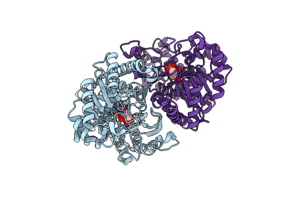
Crystal Structure Of 6-Phosphogluconate Dehydrogenase With ((4R,5R)-5-(Hydroxycarbamoyl)-2,2-Dimethyl-1,3-Dioxolan-4-Yl)Methyl Dihydrogen Phosphate
Organism: Homo sapiens
Method: X-RAY DIFFRACTION Resolution:3.00 Å Release Date: 2018-08-22 Classification: OXIDOREDUCTASE Ligands: 8HS |
Organism: Homo sapiens
Method: X-RAY DIFFRACTION
Release Date: 2018-08-22
Ligands: 8HS
 |
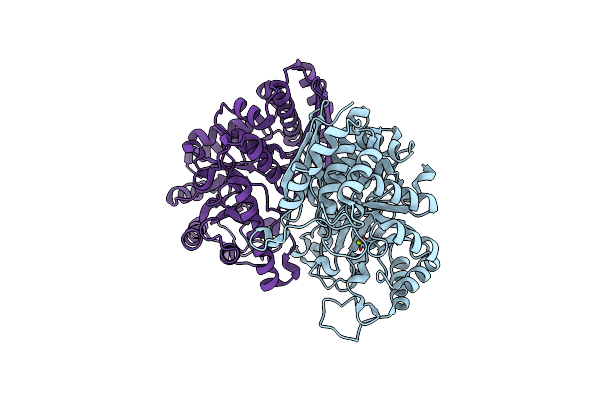
Structure Of Human Enolase 2 With ((3S,5S)-1,5-Dihydroxy-3-Methyl-2-Oxopyrrolidin-3-Yl)Phosphonate (Purified Enantiomer)
Organism: Homo sapiens
Method: X-RAY DIFFRACTION Resolution:2.63 Å Release Date: 2017-10-18 Classification: LYASE/LYASE INHIBITOR Ligands: 5TX, MG |
Organism: Homo sapiens
Method: X-RAY DIFFRACTION
Release Date: 2017-10-18
Ligands: 5TX, MG
 |
Structure Of Human Enolase 2
Organism: Homo sapiens
Method: X-RAY DIFFRACTION Resolution:2.32 Å Release Date: 2017-09-27 Classification: LYASE Ligands: MG, CL |
Organism: Homo sapiens
Method: X-RAY DIFFRACTION
Release Date: 2017-09-27
Ligands: MG, CL
 |
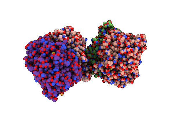
Structure Of Human Enolase 2 In Complex With (S)-(1-Hydroxy-2-Oxopiperidin-3-Yl)Phosphonate
Organism: Homo sapiens
Method: X-RAY DIFFRACTION Resolution:2.63 Å Release Date: 2017-02-22 Classification: LYASE/LYASE INHIBITOR Ligands: MG, 6BM, PGE |
Organism: Homo sapiens
Method: X-RAY DIFFRACTION
Release Date: 2017-02-22
Ligands: MG, 6BM, PGE
 |
Structure Of Human Enolase 2 In Complex With ((3S,5S)-1,5-Dihydroxy-3-Methyl-2-Oxopyrrolidin-3-Yl)Phosphonic Acid
Organism: Homo sapiens
Method: X-RAY DIFFRACTION Resolution:2.05 Å Release Date: 2016-11-23 Classification: LYASE/LYASE INHIBITOR Ligands: MG, 5TX, PGE |
Organism: Homo sapiens
Method: X-RAY DIFFRACTION
Release Date: 2016-11-23
Ligands: MG, 5TX, PGE
 |
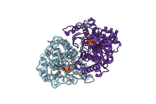
Structure Of Human Enolase 2 In Complex With Sf2312
Organism: Homo sapiens
Method: X-RAY DIFFRACTION Resolution:1.99 Å Release Date: 2016-07-20 Classification: LYASE/LYASE INHIBITOR Ligands: 4NG, MG |
Organism: Homo sapiens
Method: X-RAY DIFFRACTION
Release Date: 2016-07-20
Ligands: 4NG, MG
 |
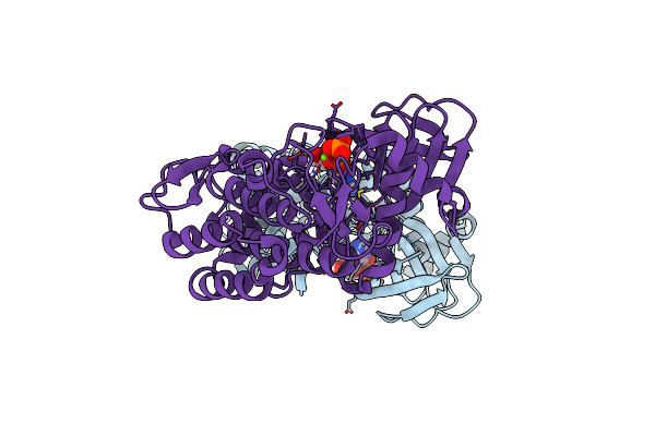
Structure Of Human Enolase 2 In Complex With Phosphonoacetohydroxamate
Organism: Homo sapiens
Method: X-RAY DIFFRACTION Resolution:2.31 Å Release Date: 2016-04-13 Classification: LYASE/LYASE INHIBITOR Ligands: MG, PAH |
Organism: Homo sapiens
Method: X-RAY DIFFRACTION
Release Date: 2016-04-13
Ligands: MG, PAH
 |
Structure Of Human Bub1 Kinase Domain Phosphorylated At Ser969
Organism: Homo sapiens
Method: X-RAY DIFFRACTION Resolution:2.40 Å Release Date: 2015-12-16 Classification: TRANSFERASE Ligands: ADP, MG |
Organism: Homo sapiens
Method: X-RAY DIFFRACTION
Release Date: 2015-12-16
Ligands: ADP, MG