

Search Count: 13
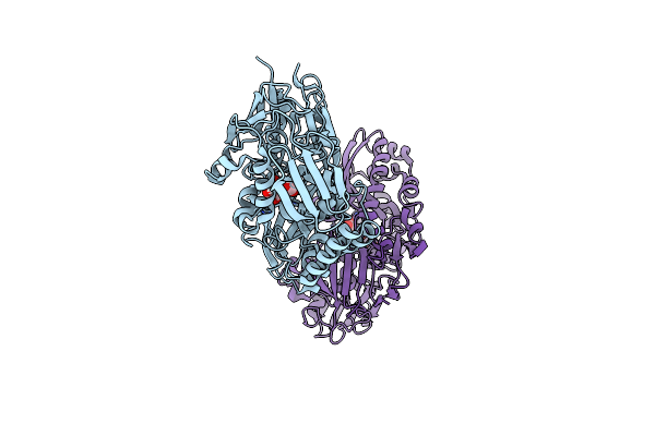 |
Crystal Structure Of Sialic Acid Binding Protein From Haemophilus Ducreyi With Neu5Gc
Organism: Haemophilus ducreyi 35000hp
Method: X-RAY DIFFRACTION Resolution:2.48 Å Release Date: 2018-10-24 Classification: SUGAR BINDING PROTEIN Ligands: NGE |
Organism: Haemophilus ducreyi 35000hp
Method: X-RAY DIFFRACTION
Release Date: 2018-10-24
Ligands: NGE
 |
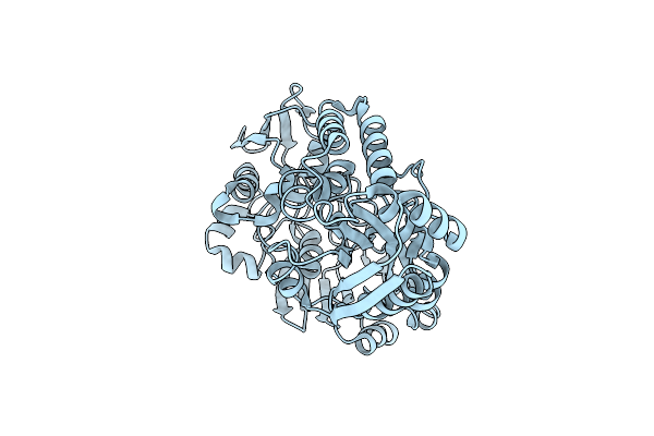
Crystal Structure Of Sialic Acid Binding Protein From Haemophilus Ducreyi With Neu5Ac
Organism: Haemophilus ducreyi 35000hp
Method: X-RAY DIFFRACTION Resolution:1.49 Å Release Date: 2018-10-24 Classification: SUGAR BINDING PROTEIN Ligands: SLB |
Organism: Haemophilus ducreyi 35000hp
Method: X-RAY DIFFRACTION
Release Date: 2018-10-24
Ligands: SLB
 |
Crystal Structure Of Sialic Acid Binding Protein From Haemophilus Ducreyi
Organism: Haemophilus ducreyi
Method: X-RAY DIFFRACTION Resolution:2.19 Å Release Date: 2018-10-24 Classification: SUGAR BINDING PROTEIN |
Organism: Haemophilus ducreyi
Method: X-RAY DIFFRACTION
Release Date: 2018-10-24
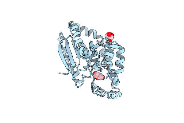 |
Crystal Structure Of The Ech2 Decarboxylase Domain Of Curf From Lyngbya Majuscula
Organism: Lyngbya majuscula
Method: X-RAY DIFFRACTION Resolution:2.00 Å Release Date: 2007-10-09 Classification: LYASE Ligands: GOL |
Organism: Lyngbya majuscula
Method: X-RAY DIFFRACTION
Release Date: 2007-10-09
Ligands: GOL
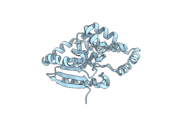 |
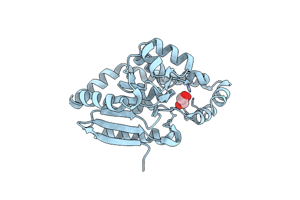
Crystal Structure Of The Ech2 Decarboxylase Domain Of Curf From Lyngbya Majuscula, Rhombohedral Crystal Form
Organism: Lyngbya majuscula
Method: X-RAY DIFFRACTION Resolution:1.85 Å Release Date: 2007-10-09 Classification: LYASE |
Organism: Lyngbya majuscula
Method: X-RAY DIFFRACTION
Release Date: 2007-10-09
 |
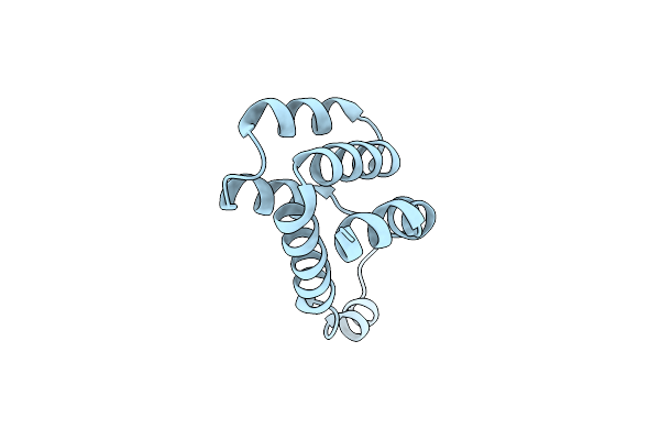
Crystal Structure Of The Y82F Variant Of Ech2 Decarboxylase Domain Of Curf From Lyngbya Majuscula
Organism: Lyngbya majuscula
Method: X-RAY DIFFRACTION Resolution:1.65 Å Release Date: 2007-10-09 Classification: LYASE Ligands: CL, GOL |
Organism: Lyngbya majuscula
Method: X-RAY DIFFRACTION
Release Date: 2007-10-09
Ligands: CL, GOL
 |
Crystal Structure Of Caspace Activation And Recruitment Domain (Card) Of Nod1
Organism: Homo sapiens
Method: X-RAY DIFFRACTION Resolution:2.00 Å Release Date: 2006-12-19 Classification: APOPTOSIS |
Organism: Homo sapiens
Method: X-RAY DIFFRACTION
Release Date: 2006-12-19
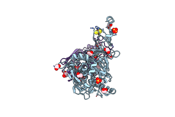 |
Crystal Structure Of The Naphthalene 1,2-Dioxygenase Phe-352-Val Mutant.
Organism: Pseudomonas sp.
Method: X-RAY DIFFRACTION Resolution:1.50 Å Release Date: 2006-10-10 Classification: OXIDOREDUCTASE Ligands: FE, SO4, FES, EDO |
Organism: Pseudomonas sp.
Method: X-RAY DIFFRACTION
Release Date: 2006-10-10
Ligands: FE, SO4, FES, EDO
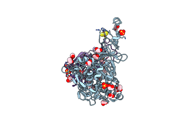 |
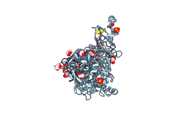
Crystal Structure Of Naphthalene 1,2-Dioxygenase Bound To Phenanthrene
Organism: Pseudomonas sp.
Method: X-RAY DIFFRACTION Resolution:1.65 Å Release Date: 2006-10-10 Classification: OXIDOREDUCTASE Ligands: FE, SO4, FES, PEY, EDO |
Organism: Pseudomonas sp.
Method: X-RAY DIFFRACTION
Release Date: 2006-10-10
Ligands: FE, SO4, FES, PEY, EDO
 |
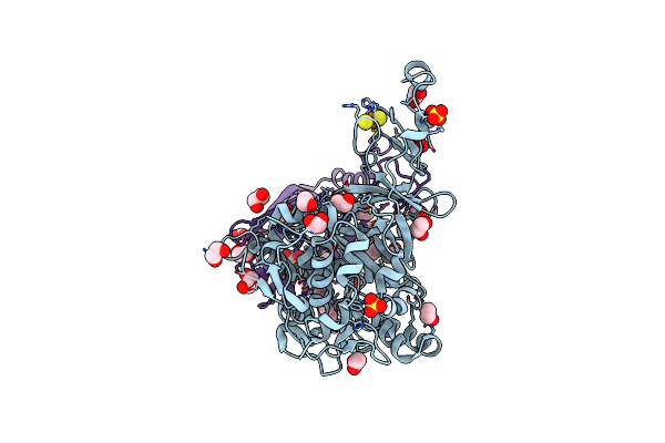
Crystal Structure Of The Naphthalene 1,2-Dioxygenase F352V Mutant Bound To Phenanthrene.
Organism: Pseudomonas sp.
Method: X-RAY DIFFRACTION Resolution:1.80 Å Release Date: 2006-10-10 Classification: OXIDOREDUCTASE Ligands: FE, SO4, FES, PEY, EDO |
Organism: Pseudomonas sp.
Method: X-RAY DIFFRACTION
Release Date: 2006-10-10
Ligands: FE, SO4, FES, PEY, EDO
 |
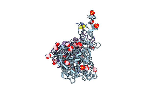
Crystal Structure Of Naphthalene 1,2-Dioxygenase Bound To Anthracene
Organism: Pseudomonas sp.
Method: X-RAY DIFFRACTION Resolution:1.60 Å Release Date: 2006-10-10 Classification: OXIDOREDUCTASE Ligands: FE, SO4, FES, AN3, EDO |
Organism: Pseudomonas sp.
Method: X-RAY DIFFRACTION
Release Date: 2006-10-10
Ligands: FE, SO4, FES, AN3, EDO
 |
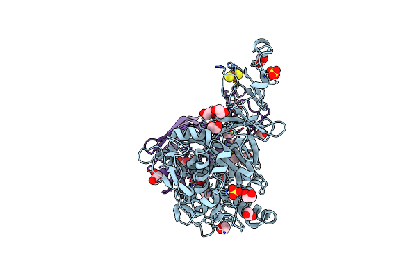
Crystal Structure Of The Naphthalene 1,2-Dioxygenase F352V Mutant Bound To Anthracene.
Organism: Pseudomonas sp.
Method: X-RAY DIFFRACTION Resolution:1.70 Å Release Date: 2006-10-10 Classification: OXIDOREDUCTASE Ligands: FE, SO4, FES, AN3, EDO |
Organism: Pseudomonas sp.
Method: X-RAY DIFFRACTION
Release Date: 2006-10-10
Ligands: FE, SO4, FES, AN3, EDO
 |
Crystal Structure Of Naphthalene 1,2-Dioxygenase Bound To 3-Nitrotoluene.
Organism: Pseudomonas sp.
Method: X-RAY DIFFRACTION Resolution:1.60 Å Release Date: 2006-10-10 Classification: OXIDOREDUCTASE Ligands: FE, SO4, FES, 3NT, EDO |
Organism: Pseudomonas sp.
Method: X-RAY DIFFRACTION
Release Date: 2006-10-10
Ligands: FE, SO4, FES, 3NT, EDO