

Search Count: 62
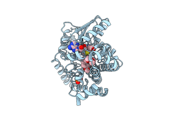 |
Tetrahydroprotoberberine N-Methyltransferase Complex With N-Methylcanadine And Sah
Organism: Glaucium flavum
Method: X-RAY DIFFRACTION Resolution:1.75 Å Release Date: 2025-09-17 Classification: TRANSFERASE Ligands: SAH, A1A4I, SO4 |
Organism: Glaucium flavum
Method: X-RAY DIFFRACTION
Release Date: 2025-09-17
Ligands: SAH, A1A4I, SO4
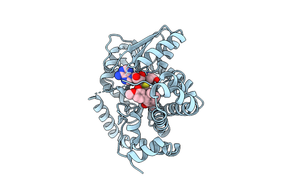 |
Tetrahydroprotoberberine N-Methyltransferase E204A Mutant In Complex With R-Reticuline And Sam
Organism: Glaucium flavum
Method: X-RAY DIFFRACTION Resolution:2.50 Å Release Date: 2025-09-17 Classification: TRANSFERASE Ligands: SAM, A1A4H |
Organism: Glaucium flavum
Method: X-RAY DIFFRACTION
Release Date: 2025-09-17
Ligands: SAM, A1A4H
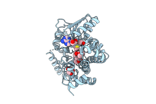 |
Tetrahydroprotoberberine N-Methyltransferase E204A Mutant In Complex With (S)-Reticuline And Sam
Organism: Glaucium flavum
Method: X-RAY DIFFRACTION Resolution:2.20 Å Release Date: 2025-09-17 Classification: TRANSFERASE Ligands: SAM, REN, GOL |
Organism: Glaucium flavum
Method: X-RAY DIFFRACTION
Release Date: 2025-09-17
Ligands: SAM, REN, GOL
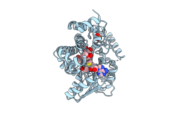 |
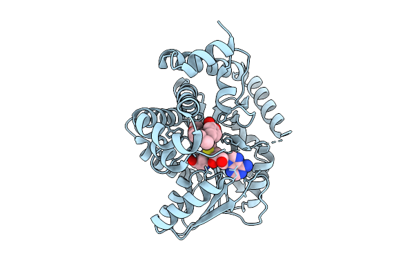
Tetrahydroprotoberberine N-Methyltransferase E207A Mutant In Complex With R-Reticuline And Sam
Organism: Glaucium flavum
Method: X-RAY DIFFRACTION Resolution:1.60 Å Release Date: 2025-09-17 Classification: TRANSFERASE Ligands: SAM, A1A4H, SO4, GOL, CL |
Organism: Glaucium flavum
Method: X-RAY DIFFRACTION
Release Date: 2025-09-17
Ligands: SAM, A1A4H, SO4, GOL, CL
 |
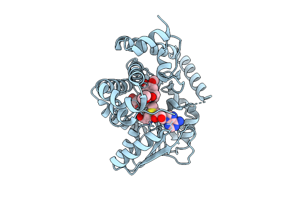
Tetrahydroprotoberberine N-Methyltransferase In Complex With (R)-Reticuline And Sam
Organism: Glaucium flavum
Method: X-RAY DIFFRACTION Resolution:2.00 Å Release Date: 2025-09-17 Classification: TRANSFERASE Ligands: SAM, A1A4H |
Organism: Glaucium flavum
Method: X-RAY DIFFRACTION
Release Date: 2025-09-17
Ligands: SAM, A1A4H
 |
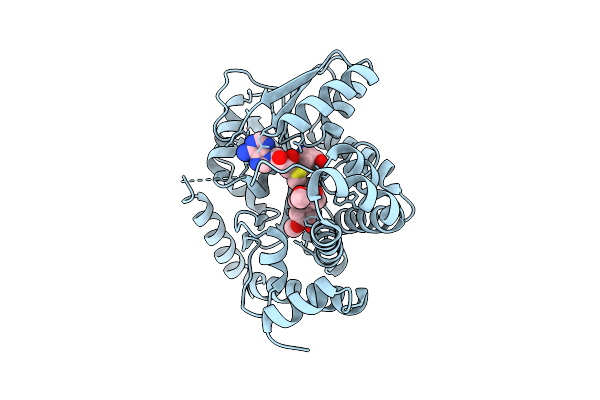
Tetrahydroprotoberberine N-Methyltransferase In Complex With (S)-Reticuline And Sam
Organism: Glaucium flavum
Method: X-RAY DIFFRACTION Resolution:1.60 Å Release Date: 2025-09-17 Classification: TRANSFERASE Ligands: SAM, REN, GOL |
Organism: Glaucium flavum
Method: X-RAY DIFFRACTION
Release Date: 2025-09-17
Ligands: SAM, REN, GOL
 |
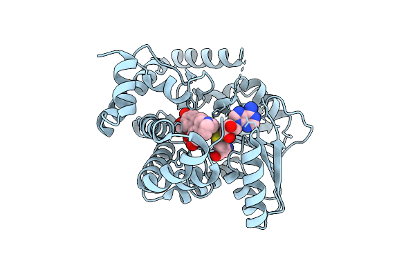
Tetrahydroprotoberberine N-Methyltransferase E207A Mutant In Complex With (S)-Reticuline And Sam
Organism: Glaucium flavum
Method: X-RAY DIFFRACTION Resolution:2.00 Å Release Date: 2025-09-17 Classification: TRANSFERASE Ligands: SAM, REN |
Organism: Glaucium flavum
Method: X-RAY DIFFRACTION
Release Date: 2025-09-17
Ligands: SAM, REN
 |
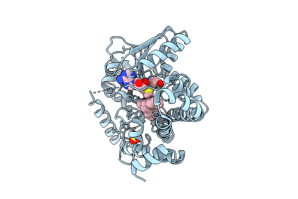
Tetrahydroprotoberberine N-Methyltransferase In Complex With Boldine And Sah
Organism: Glaucium flavum
Method: X-RAY DIFFRACTION Resolution:1.80 Å Release Date: 2025-09-17 Classification: TRANSFERASE Ligands: SAH, GHT |
Organism: Glaucium flavum
Method: X-RAY DIFFRACTION
Release Date: 2025-09-17
Ligands: SAH, GHT
 |
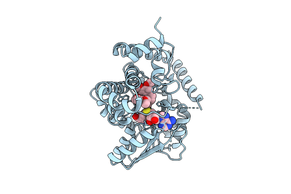
Tetrahydroprotoberberine N-Methyltransferase Product Complex With Dextromethorphan And Sam
Organism: Glaucium flavum
Method: X-RAY DIFFRACTION Resolution:1.60 Å Release Date: 2025-09-17 Classification: TRANSFERASE Ligands: SAM, 4NQ, SO4 |
Organism: Glaucium flavum
Method: X-RAY DIFFRACTION
Release Date: 2025-09-17
Ligands: SAM, 4NQ, SO4
 |
Tetrahydroprotoberberine N-Methyltransferase Product Complex With N-Methyltetrahydropalmatine And Sah
Organism: Glaucium flavum
Method: X-RAY DIFFRACTION Resolution:1.85 Å Release Date: 2025-09-17 Classification: TRANSFERASE Ligands: SAH, A1A4J |
Organism: Glaucium flavum
Method: X-RAY DIFFRACTION
Release Date: 2025-09-17
Ligands: SAH, A1A4J
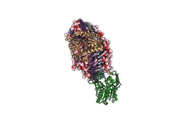 |
Lecithin:Cholesterol Acyltransferase Bound To Apolipoprotein A-I Dimer In Hdl
Organism: Homo sapiens
Method: ELECTRON MICROSCOPY Release Date: 2025-04-02 Classification: LIPID TRANSPORT Ligands: 6PL |
Organism: Homo sapiens
Method: ELECTRON MICROSCOPY
Release Date: 2025-04-02
Ligands: 6PL
 |
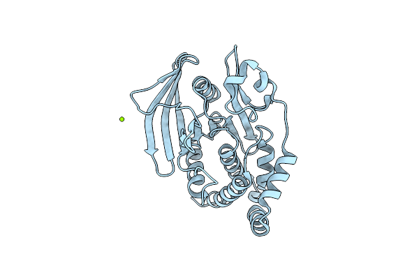
Crystal Structure Of Wild-Type Human Ptp1B (Ptpn1) At Room Temperature (298 K)
Organism: Homo sapiens
Method: X-RAY DIFFRACTION Resolution:1.94 Å Release Date: 2024-08-21 Classification: HYDROLASE Ligands: MG |
Organism: Homo sapiens
Method: X-RAY DIFFRACTION
Release Date: 2024-08-21
Ligands: MG
 |
Crystal Structure Of I19V Mutant Human Ptp1B (Ptpn1) At Room Temperature (298 K)
Organism: Homo sapiens
Method: X-RAY DIFFRACTION Resolution:1.99 Å Release Date: 2024-08-21 Classification: HYDROLASE Ligands: MG |
Organism: Homo sapiens
Method: X-RAY DIFFRACTION
Release Date: 2024-08-21
Ligands: MG
 |
Crystal Structure Of Q78R Mutant Human Ptp1B (Ptpn1) At Room Temperature (298 K)
Organism: Homo sapiens
Method: X-RAY DIFFRACTION Resolution:2.30 Å Release Date: 2024-08-21 Classification: HYDROLASE Ligands: MG |
Organism: Homo sapiens
Method: X-RAY DIFFRACTION
Release Date: 2024-08-21
Ligands: MG
 |
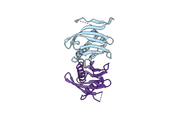
Crystal Structure Of D245G Mutant Human Ptp1B (Ptpn1) At Room Temperature (298 K)
Organism: Homo sapiens
Method: X-RAY DIFFRACTION Resolution:1.65 Å Release Date: 2024-08-21 Classification: HYDROLASE Ligands: MG |
Organism: Homo sapiens
Method: X-RAY DIFFRACTION
Release Date: 2024-08-21
Ligands: MG
 |
Pathogenesis Related 10-10 App From
Organism: Papaver somniferum
Method: X-RAY DIFFRACTION Resolution:1.90 Å Release Date: 2023-03-01 Classification: BIOSYNTHETIC PROTEIN |
Organism: Papaver somniferum
Method: X-RAY DIFFRACTION
Release Date: 2023-03-01
 |
Pathogenesis Related 10-10 Papaverine Complex
Organism: Papaver somniferum
Method: X-RAY DIFFRACTION Resolution:1.80 Å Release Date: 2023-03-01 Classification: BIOSYNTHETIC PROTEIN Ligands: EV1 |
Organism: Papaver somniferum
Method: X-RAY DIFFRACTION
Release Date: 2023-03-01
Ligands: EV1
 |
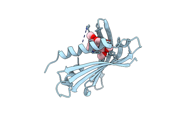
Pathogenesis Related 10-10 Noscapine Complex
Organism: Papaver somniferum
Method: X-RAY DIFFRACTION Resolution:1.70 Å Release Date: 2023-03-01 Classification: BIOSYNTHETIC PROTEIN Ligands: 08N |
Organism: Papaver somniferum
Method: X-RAY DIFFRACTION
Release Date: 2023-03-01
Ligands: 08N
 |
Pathogenesis Related 10-10 (S)-Tetrahydropapaverine Complex
Organism: Papaver somniferum
Method: X-RAY DIFFRACTION Resolution:1.75 Å Release Date: 2023-03-01 Classification: BIOSYNTHETIC PROTEIN Ligands: S9T |
Organism: Papaver somniferum
Method: X-RAY DIFFRACTION
Release Date: 2023-03-01
Ligands: S9T
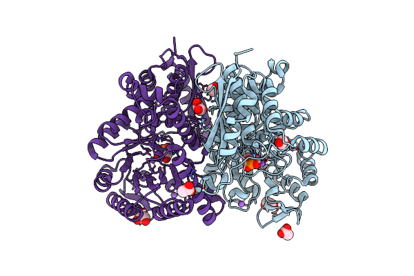 |
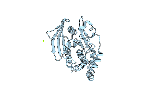
Crystal Structure Of Enolase Family Protein From Naegleria Fowleri With Bound 2-Phosphoglyceric Acid
Organism: Naegleria fowleri
Method: X-RAY DIFFRACTION Resolution:1.95 Å Release Date: 2022-04-13 Classification: LYASE Ligands: 2PG, EDO, ACT, NA |
Organism: Naegleria fowleri
Method: X-RAY DIFFRACTION
Release Date: 2022-04-13
Ligands: 2PG, EDO, ACT, NA