

Search Count: 42
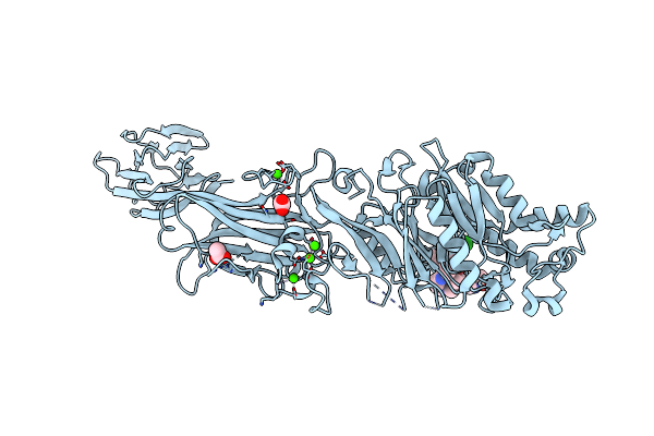 |
Crystal Structure Of The Human Pad2 Protein Bound To Inhibitor
Organism: Homo sapiens
Method: X-RAY DIFFRACTION Resolution:1.64 Å Release Date: 2024-10-09 Classification: HYDROLASE Ligands: A1AJA, ACT, CA |
Organism: Homo sapiens
Method: X-RAY DIFFRACTION
Release Date: 2024-10-09
Ligands: A1AJA, ACT, CA
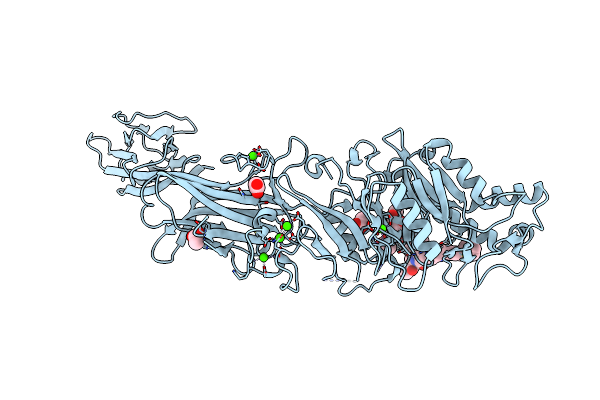 |
Crystal Structure Of The Human Pad2 Protein Bound To Small Molecule
Organism: Homo sapiens
Method: X-RAY DIFFRACTION Resolution:1.95 Å Release Date: 2024-10-09 Classification: HYDROLASE Ligands: MPD, A1AJB, ACT, CA |
Organism: Homo sapiens
Method: X-RAY DIFFRACTION
Release Date: 2024-10-09
Ligands: MPD, A1AJB, ACT, CA
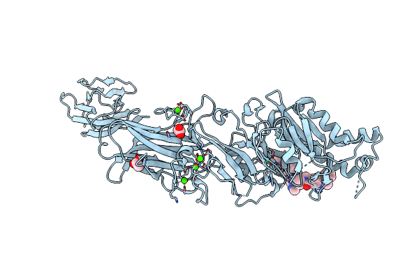 |
Crystal Structure Of The Human Pad2 Protein Bound To Small Molecule
Organism: Homo sapiens
Method: X-RAY DIFFRACTION Resolution:2.00 Å Release Date: 2024-10-09 Classification: HYDROLASE Ligands: ACT, CA, A1AJC |
Organism: Homo sapiens
Method: X-RAY DIFFRACTION
Release Date: 2024-10-09
Ligands: ACT, CA, A1AJC
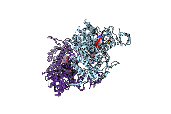 |
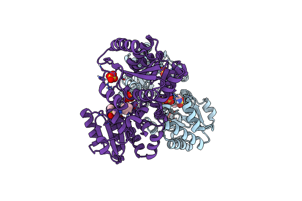
Cardiac Myosin Motor Domain In The Pre-Powerstroke State Co-Crystallized With The Inhibitor Aficamten
Organism: Bos taurus
Method: X-RAY DIFFRACTION Resolution:2.33 Å Release Date: 2024-09-04 Classification: MOTOR PROTEIN Ligands: SO4, MG, 6I6, ADP, VO4 |
Organism: Bos taurus
Method: X-RAY DIFFRACTION
Release Date: 2024-09-04
Ligands: SO4, MG, 6I6, ADP, VO4
 |
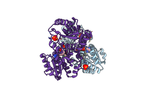
Pac-Fragmentdel: Photoactivated Covalent Capture Of Dna Encoded Fragments For Hit Discovery
Organism: Bacillus anthracis
Method: X-RAY DIFFRACTION Resolution:2.11 Å Release Date: 2022-09-14 Classification: ISOMERASE Ligands: M2U, SO4 |
Organism: Bacillus anthracis
Method: X-RAY DIFFRACTION
Release Date: 2022-09-14
Ligands: M2U, SO4
 |
Pac-Fragmentdel: Photoactivated Covalent Capture Of Dna Encoded Fragments For Hit Discovery
Organism: Bacillus anthracis
Method: X-RAY DIFFRACTION Resolution:2.27 Å Release Date: 2022-09-14 Classification: ISOMERASE Ligands: M3U, SO4 |
Organism: Bacillus anthracis
Method: X-RAY DIFFRACTION
Release Date: 2022-09-14
Ligands: M3U, SO4
 |
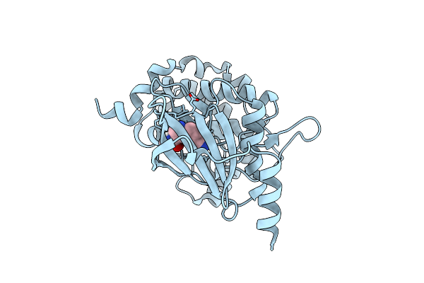
Pac-Fragmentdel: Photoactivated Covalent Capture Of Dna Encoded Fragments For Hit Discovery
Organism: Homo sapiens
Method: X-RAY DIFFRACTION Resolution:1.89 Å Release Date: 2022-09-14 Classification: TRANSFERASE Ligands: M4I |
Organism: Homo sapiens
Method: X-RAY DIFFRACTION
Release Date: 2022-09-14
Ligands: M4I
 |
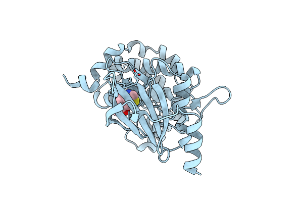
Pac Fragmentdel: Photoactivated Covalent Capture Of Dna Encoded Fragments For Hit Discovery
Organism: Homo sapiens
Method: X-RAY DIFFRACTION Resolution:2.04 Å Release Date: 2022-09-14 Classification: TRANSFERASE Ligands: M4X |
Organism: Homo sapiens
Method: X-RAY DIFFRACTION
Release Date: 2022-09-14
Ligands: M4X
 |
Pac-Fragmentdel: Photoactivated Covalent Capture Of Dna Encoded Fragments For Hit Discovery
Organism: Homo sapiens
Method: X-RAY DIFFRACTION Resolution:2.69 Å Release Date: 2022-09-14 Classification: TRANSFERASE Ligands: M56 |
Organism: Homo sapiens
Method: X-RAY DIFFRACTION
Release Date: 2022-09-14
Ligands: M56
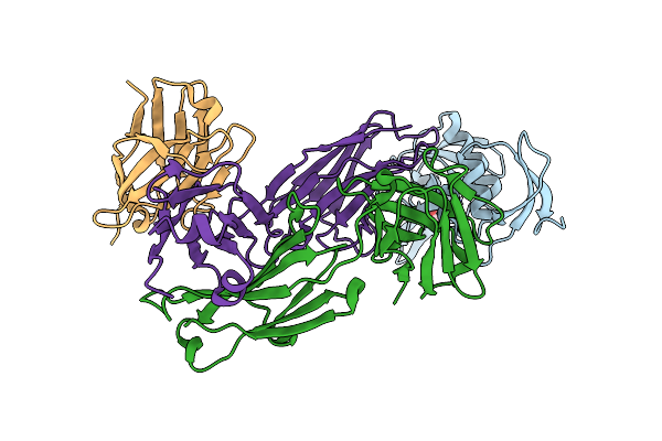 |
Complement C6 Fim1-2 Bound To Cp010 Antibody
Organism: Homo sapiens, Mus musculus, Lama glama
Method: X-RAY DIFFRACTION Resolution:2.29 Å Release Date: 2022-05-25 Classification: IMMUNE SYSTEM Ligands: ACT |
Organism: Homo sapiens, Mus musculus, Lama glama
Method: X-RAY DIFFRACTION
Release Date: 2022-05-25
Ligands: ACT
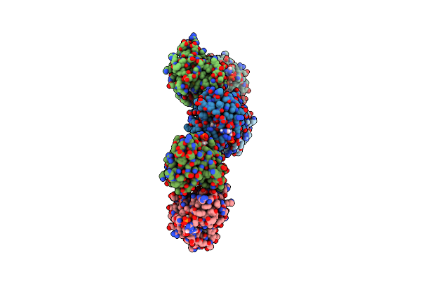 |
Crystal Structure Of Human Naa50 In Complex With A Cofactor Derived Inhibitor (Compound 1)
Organism: Homo sapiens, Synthetic construct
Method: X-RAY DIFFRACTION Resolution:2.29 Å Release Date: 2020-07-01 Classification: TRANSFERASE/INHIBITOR Ligands: COA |
Organism: Homo sapiens, Synthetic construct
Method: X-RAY DIFFRACTION
Release Date: 2020-07-01
Ligands: COA
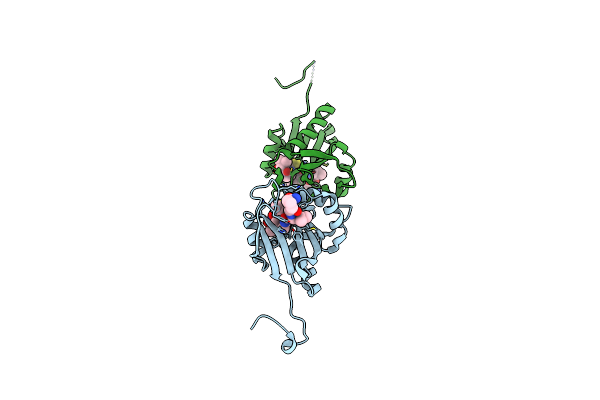 |
Crystal Structure Of Human Naa50 In Complex With A Truncated Cofactor Derived Inhibitor (Compound 2)
Organism: Homo sapiens, Synthetic construct
Method: X-RAY DIFFRACTION Resolution:2.04 Å Release Date: 2020-07-01 Classification: TRANSFERASE/INHIBITOR Ligands: U44 |
Organism: Homo sapiens, Synthetic construct
Method: X-RAY DIFFRACTION
Release Date: 2020-07-01
Ligands: U44
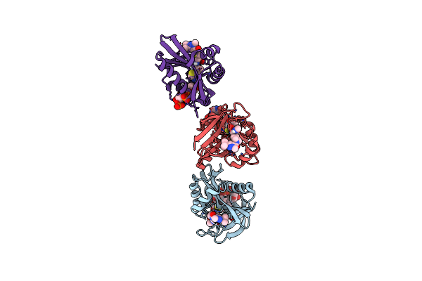 |
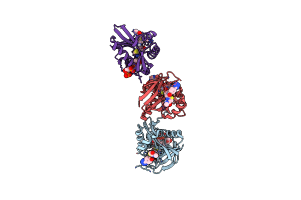
Crystal Structure Of Human Naa50 In Complex With An Inhibitor (Compound 3) Identified Using Dna Encoded Library Technology
Organism: Homo sapiens
Method: X-RAY DIFFRACTION Resolution:2.16 Å Release Date: 2020-07-01 Classification: TRANSFERASE/INHIBITOR Ligands: COA, U3V |
Organism: Homo sapiens
Method: X-RAY DIFFRACTION
Release Date: 2020-07-01
Ligands: COA, U3V
 |
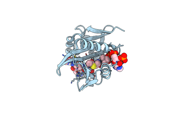
Crystal Structure Of Human Naa50 In Complex With Coa And An Inhibitor (Compound 4A) Identified Using Dna Encoded Library Technology
Organism: Homo sapiens
Method: X-RAY DIFFRACTION Resolution:1.87 Å Release Date: 2020-07-01 Classification: TRANSFERASE Ligands: COA, U2J |
Organism: Homo sapiens
Method: X-RAY DIFFRACTION
Release Date: 2020-07-01
Ligands: COA, U2J
 |
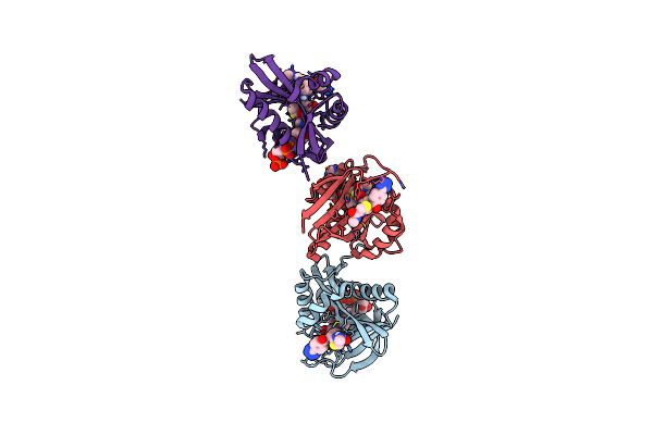
Crystal Structure Of Human Naa50 In Complex With Accoa And An Inhibitor (Compound 4A) Identified Using Dna Encoded Library Technology
Organism: Homo sapiens
Method: X-RAY DIFFRACTION Resolution:1.07 Å Release Date: 2020-07-01 Classification: TRANSFERASE/INHIBITOR Ligands: ACO, U2J |
Organism: Homo sapiens
Method: X-RAY DIFFRACTION
Release Date: 2020-07-01
Ligands: ACO, U2J
 |
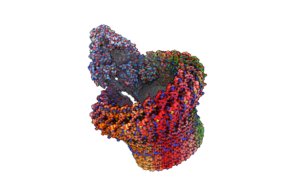
Crystal Structure Of Human Naa50 In Complex With Accoa And An Inhibitor (Compound 4B) Identified Using Dna Encoded Library Technology
Organism: Homo sapiens
Method: X-RAY DIFFRACTION Resolution:1.85 Å Release Date: 2020-07-01 Classification: TRANSFERASE/INHIBITOR Ligands: ACO, U3Y |
Organism: Homo sapiens
Method: X-RAY DIFFRACTION
Release Date: 2020-07-01
Ligands: ACO, U3Y
 |
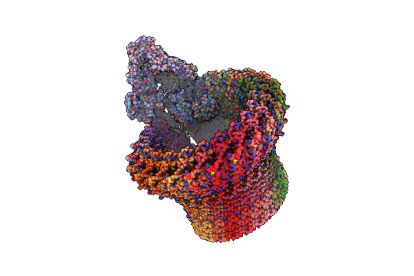
Open Conformation Of The Membrane Attack Complex
Organism: Homo sapiens
Method: ELECTRON MICROSCOPY Resolution:5.60 Å Release Date: 2018-12-19 Classification: IMMUNE SYSTEM Ligands: NAG |
Organism: Homo sapiens
Method: ELECTRON MICROSCOPY
Release Date: 2018-12-19
Ligands: NAG
 |
Closed Conformation Of The Membrane Attack Complex
Organism: Homo sapiens
Method: ELECTRON MICROSCOPY Resolution:5.60 Å Release Date: 2018-12-19 Classification: IMMUNE SYSTEM Ligands: NAG |
Organism: Homo sapiens
Method: ELECTRON MICROSCOPY
Release Date: 2018-12-19
Ligands: NAG
 |
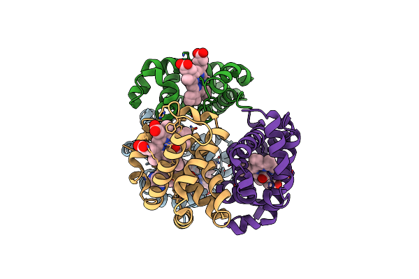
Crystal Structure Of Carbonmonoxy Hemoglobin S (Liganded Sickle Cell Hemoglobin) Complexed With Gbt Compound 31
Organism: Homo sapiens
Method: X-RAY DIFFRACTION Resolution:1.95 Å Release Date: 2017-02-22 Classification: OXYGEN TRANSPORT Ligands: HEM, CMO, 7SJ |
Organism: Homo sapiens
Method: X-RAY DIFFRACTION
Release Date: 2017-02-22
Ligands: HEM, CMO, 7SJ
 |
Crystal Structure Of Carbonmonoxy Hemoglobin S (Liganded Sickle Cell Hemoglobin) Complexed With Gbt Compound 6
Organism: Homo sapiens
Method: X-RAY DIFFRACTION Resolution:2.05 Å Release Date: 2017-02-22 Classification: OXYGEN TRANSPORT Ligands: HEM, CMO, 86M |
Organism: Homo sapiens
Method: X-RAY DIFFRACTION
Release Date: 2017-02-22
Ligands: HEM, CMO, 86M