
Search Count: 90
All
Selected
 |
Organism: Streptococcus sanguinis sk36
Method: X-RAY DIFFRACTION Resolution:1.80 Å Release Date: 2026-01-21 Classification: CELL ADHESION Ligands: EDO, PEG, PGE |
 |
Organism: Streptococcus sanguinis
Method: X-RAY DIFFRACTION Resolution:3.20 Å Release Date: 2026-01-21 Classification: CELL ADHESION Ligands: CD, GOL |
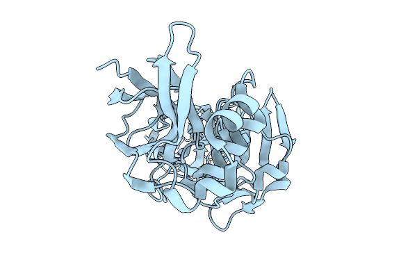 |
Crystal Structure Of Se-Met Ssa1633 From Streptococcus Sanguinis (Phase Determination Model)
Organism: Streptococcus sanguinis
Method: X-RAY DIFFRACTION Resolution:2.20 Å Release Date: 2026-01-21 Classification: CELL ADHESION |
 |
Structural Insight Into Determinants Of Endogenous Agonist Selectivity Of Aminergic Receptors From Octopamine B2 Receptor Structure
Organism: Ixodes scapularis, Homo sapiens, Mus musculus
Method: ELECTRON MICROSCOPY Release Date: 2025-12-17 Classification: SIGNALING PROTEIN Ligands: A1L8N |
 |
Structural Insight Into Determinants Of Endogenous Agonist Selectivity Of Aminergic Receptors From Octopamine B2 Receptor Structure
Organism: Ixodes scapularis, Homo sapiens, Mus musculus
Method: ELECTRON MICROSCOPY Release Date: 2025-12-17 Classification: SIGNALING PROTEIN Ligands: OTR |
 |
Organism: Homo sapiens
Method: X-RAY DIFFRACTION Resolution:2.70 Å Release Date: 2025-08-06 Classification: LIGASE Ligands: SO4, EDO |
 |
Cryo-Em Structure Of The Zeaxanthin-Bound Light-Driven Proton Pumping Rhodopsin, Nm-R1
Organism: Nonlabens marinus s1-08
Method: ELECTRON MICROSCOPY Resolution:2.46 Å Release Date: 2025-07-30 Classification: MEMBRANE PROTEIN Ligands: RET, R16, D12, K3I |
 |
Cryo-Em Structure Of The Myxol-Bound Light-Driven Proton Pumping Rhodopsin, Nm-R1
Organism: Nonlabens marinus s1-08
Method: ELECTRON MICROSCOPY Resolution:2.27 Å Release Date: 2025-07-30 Classification: MEMBRANE PROTEIN Ligands: RET, R16, D12, A1L4O |
 |
Cryo-Em Structure Of The Myxol-Bound Light-Driven Chloride Ion-Pumping Rhodopsin, Nm-R3
Organism: Nonlabens marinus s1-08
Method: ELECTRON MICROSCOPY Resolution:2.48 Å Release Date: 2025-07-30 Classification: MEMBRANE PROTEIN Ligands: RET, A1L4O, CL, PC1, PLC, R16, 8K6, D12, C14, D10 |
 |
Cryo-Em Structure Of The Light-Driven Chloride Ion-Pumping Rhodopsin, Nm-R3
Organism: Nonlabens marinus s1-08
Method: ELECTRON MICROSCOPY Resolution:2.50 Å Release Date: 2025-07-30 Classification: MEMBRANE PROTEIN Ligands: RET, CL, PC1, PLC, D12, R16, 8K6, C14 |
 |
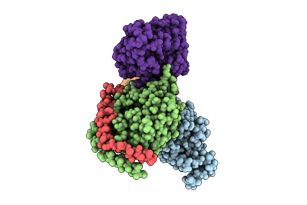
Structure Of Influenza A Virus Rna Polymerase Pb1 And Nuclear Import Host Factor Ranbp5 Complex
Organism: Homo sapiens, influenza a virus (strain a/puerto rico/8/1934 h1n1)
Method: ELECTRON MICROSCOPY Resolution:3.21 Å Release Date: 2024-11-27 Classification: NUCLEAR PROTEIN |
 |
Organism: Escherichia coli, Homo sapiens, Macaca mulatta, Synthetic construct
Method: ELECTRON MICROSCOPY Release Date: 2024-05-29 Classification: SIGNALING PROTEIN |
 |
Full Agonist- And Positive Allosteric Modulator-Bound Mu-Type Opioid Receptor-G Protein Complex
Organism: Escherichia coli, Homo sapiens, Macaca mulatta, Synthetic construct
Method: ELECTRON MICROSCOPY Release Date: 2024-05-29 Classification: SIGNALING PROTEIN Ligands: VV9 |
 |
Organism: Microchaete diplosiphon
Method: X-RAY DIFFRACTION Resolution:1.75 Å Release Date: 2024-05-15 Classification: SIGNALING PROTEIN Ligands: CYC |
 |
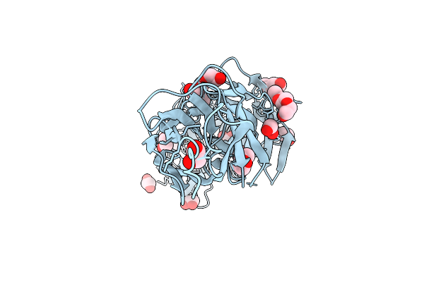
Crystal Structure Of Metallo-Beta-Lactamse, Imp-1, Complexed With A Quinolinone-Based Inhibitor
Organism: Serratia marcescens
Method: X-RAY DIFFRACTION Resolution:2.65 Å Release Date: 2024-05-08 Classification: HYDROLASE/HYDROLASE INHIBITOR Ligands: ZN, A1LXO |
 |
Organism: Chiba virus, Homo sapiens
Method: ELECTRON MICROSCOPY Release Date: 2024-04-17 Classification: VIRUS |
 |
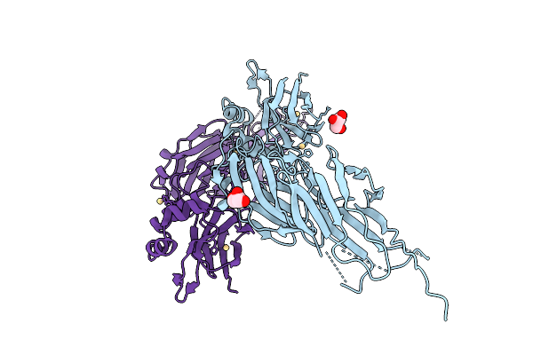
Crystal Structure Of P Domain From Norovirus Gi.4 Capsid Protein In Complex With Broad Specificity Antibody Single-Chain Fv Fragment Cv-2F5.
Organism: Chiba virus, Homo sapiens
Method: X-RAY DIFFRACTION Resolution:2.70 Å Release Date: 2024-01-31 Classification: VIRAL PROTEIN |
 |
Organism: Streptococcus pyogenes serotype m3
Method: X-RAY DIFFRACTION Resolution:2.80 Å Release Date: 2023-12-06 Classification: PROTEIN FIBRIL Ligands: GOL |
 |
The Sars-Cov-2 Receptor Binding Domain Bound With The Fab Fragment Of A Human Neutralizing Antibody Ab816
Organism: Severe acute respiratory syndrome coronavirus 2, Homo sapiens
Method: ELECTRON MICROSCOPY Release Date: 2023-06-21 Classification: VIRAL PROTEIN Ligands: NAG |
 |
The Sars-Cov-2 Receptor Binding Domain Bound With The Fab Fragment Of A Human Neutralizing Antibody Ab803
Organism: Severe acute respiratory syndrome coronavirus 2, Homo sapiens
Method: ELECTRON MICROSCOPY Release Date: 2023-06-21 Classification: VIRAL PROTEIN Ligands: NAG |