

Search Count: 666
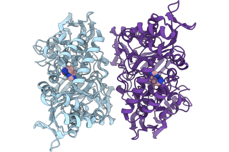 |
Crystal Structure Of The Peptide-Binding Protein Nika From Streptococcus Agalactiae In Complex With Zinc And L-Histidine.
Organism: Streptococcus agalactiae 2603v/r
Method: X-RAY DIFFRACTION Resolution:1.95 Å Release Date: 2026-07-15 Classification: TRANSPORT PROTEIN Ligands: NI, HIS |
Organism: Streptococcus agalactiae 2603v/r
Method: X-RAY DIFFRACTION
Release Date: 2026-07-15
Ligands: NI, HIS
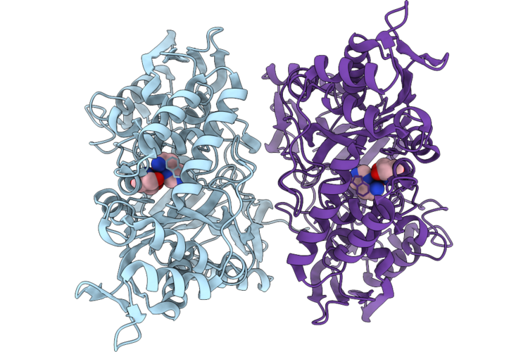 |
Crystal Structure Of The Peptide-Binding Protein Nika From Streptococcus Agalactiae In Complex With Zinc, L-Histidine And Tris Buffer.
Organism: Streptococcus agalactiae 2603v/r
Method: X-RAY DIFFRACTION Resolution:1.80 Å Release Date: 2026-07-15 Classification: TRANSPORT PROTEIN Ligands: NI, HIS, TRS |
Organism: Streptococcus agalactiae 2603v/r
Method: X-RAY DIFFRACTION
Release Date: 2026-07-15
Ligands: NI, HIS, TRS
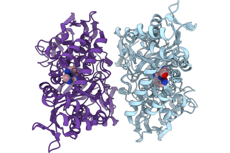 |
Crystal Structure Of The Peptide-Binding Protein Nika From Streptococcus Agalactiae In Complex With Zinc, L-Histidine, Imidazole And Ethylene Glycol.
Organism: Streptococcus agalactiae 2603v/r
Method: X-RAY DIFFRACTION Resolution:1.95 Å Release Date: 2026-07-15 Classification: TRANSPORT PROTEIN Ligands: NI, HIS, IMD, EDO |
Organism: Streptococcus agalactiae 2603v/r
Method: X-RAY DIFFRACTION
Release Date: 2026-07-15
Ligands: NI, HIS, IMD, EDO
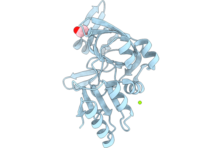 |
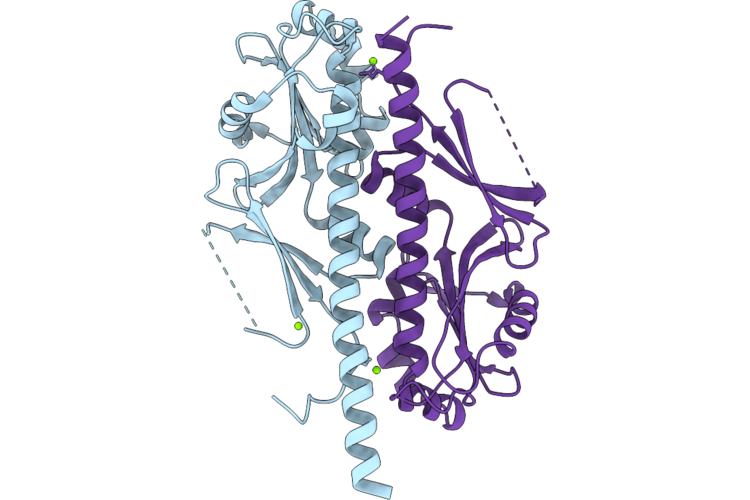
Nucleotide Binding Domain (Residues 475-720) Of Abc3 Transporter Permease From Clostridioides Difficile Strain 630
Organism: Clostridioides difficile 630
Method: X-RAY DIFFRACTION Resolution:2.00 Å Release Date: 2026-06-24 Classification: TRANSPORT PROTEIN Ligands: MG, CL, EDO |
Organism: Clostridioides difficile 630
Method: X-RAY DIFFRACTION
Release Date: 2026-06-24
Ligands: MG, CL, EDO
 |
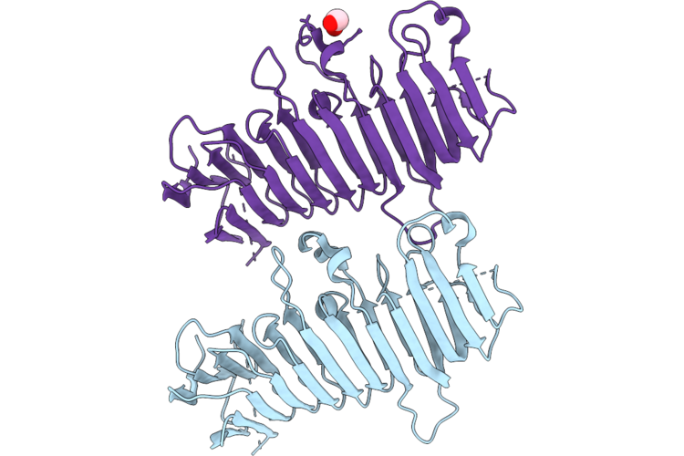
Crystal Structure Of The Ggdef Domain (Residues 31-260) Of Diguanylate Cyclase From Vibrio Cholerae Serotype O1
Organism: Vibrio cholerae o1 biovar el tor str. n16961
Method: X-RAY DIFFRACTION Resolution:2.00 Å Release Date: 2026-06-24 Classification: OXIDOREDUCTASE Ligands: MG, CL |
Organism: Vibrio cholerae o1 biovar el tor str. n16961
Method: X-RAY DIFFRACTION
Release Date: 2026-06-24
Ligands: MG, CL
 |
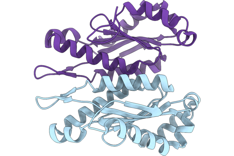
Crystal Structure Of Duf4097 Domain-Containing Protein From Clostridium Difficile Strain 630
Organism: Clostridioides difficile 630
Method: X-RAY DIFFRACTION Resolution:2.00 Å Release Date: 2026-06-24 Classification: UNKNOWN FUNCTION Ligands: EDO |
Organism: Clostridioides difficile 630
Method: X-RAY DIFFRACTION
Release Date: 2026-06-24
Ligands: EDO
 |
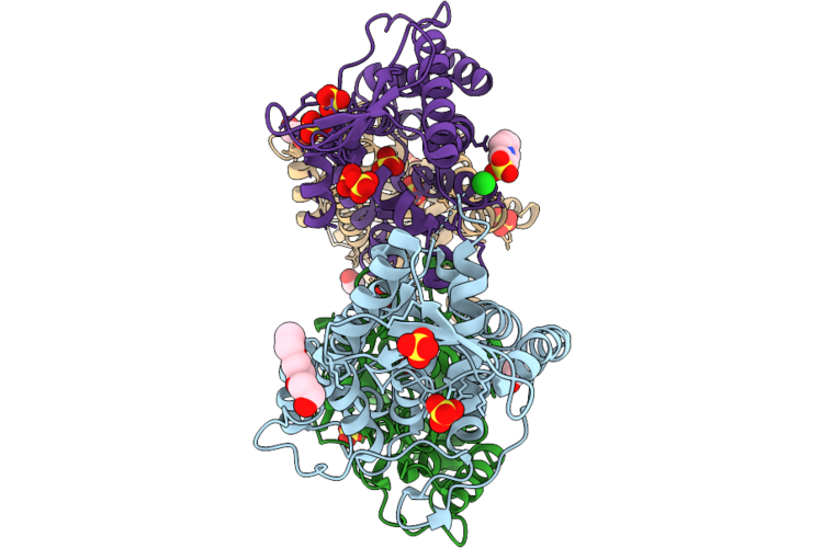
Crystal Structure Of The Ggdef Domain Of The Diguanylate Cyclase From Vibrio Vulnificus Cmcp6
Organism: Vibrio vulnificus cmcp6
Method: X-RAY DIFFRACTION Resolution:2.60 Å Release Date: 2026-06-24 Classification: TRANSFERASE |
Organism: Vibrio vulnificus cmcp6
Method: X-RAY DIFFRACTION
Release Date: 2026-06-24
 |
Crystal Structure Of Conserved Hypothetical Protein From Stenotrophomonas Maltophilia (Strain K279A)
Organism: Stenotrophomonas maltophilia k279a
Method: X-RAY DIFFRACTION Resolution:1.85 Å Release Date: 2026-06-17 Classification: UNKNOWN FUNCTION Ligands: SO4, PGE, CL, EDO, EPE |
Organism: Stenotrophomonas maltophilia k279a
Method: X-RAY DIFFRACTION
Release Date: 2026-06-17
Ligands: SO4, PGE, CL, EDO, EPE
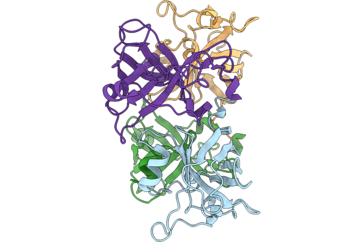 |
Crystal Structure Of Flagellar Assembly Protein Fliw From Campylobacter Jejuni Subsp. Jejuni 81-176-Drh212
Organism: Campylobacter jejuni subsp. jejuni 81-176-drh212
Method: X-RAY DIFFRACTION Resolution:2.95 Å Release Date: 2026-06-17 Classification: TRANSLATION Ligands: CL |
Organism: Campylobacter jejuni subsp. jejuni 81-176-drh212
Method: X-RAY DIFFRACTION
Release Date: 2026-06-17
Ligands: CL
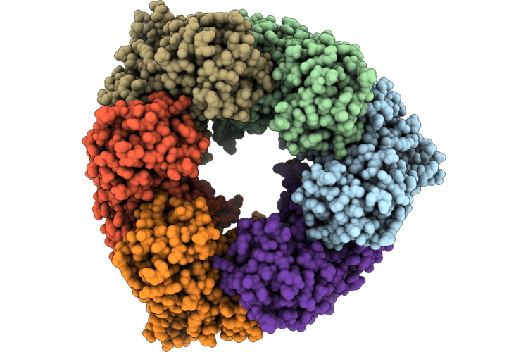 |
Single Particle Reconstruction Of Pilu From Vibrio Cholerae El Tor E7946, Form 1
Organism: Vibrio cholerae
Method: ELECTRON MICROSCOPY Release Date: 2026-05-20 Classification: MOTOR PROTEIN |
Organism: Vibrio cholerae
Method: ELECTRON MICROSCOPY
Release Date: 2026-05-20
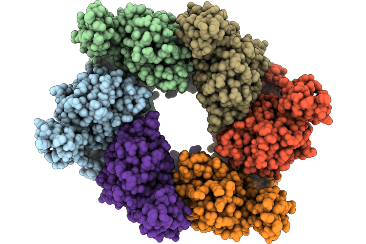 |
Single Particle Reconstruction Of Pilu From Vibrio Cholerae El Tor E7946, Form 2
Organism: Vibrio cholerae
Method: ELECTRON MICROSCOPY Release Date: 2026-05-20 Classification: MOTOR PROTEIN |
Organism: Vibrio cholerae
Method: ELECTRON MICROSCOPY
Release Date: 2026-05-20
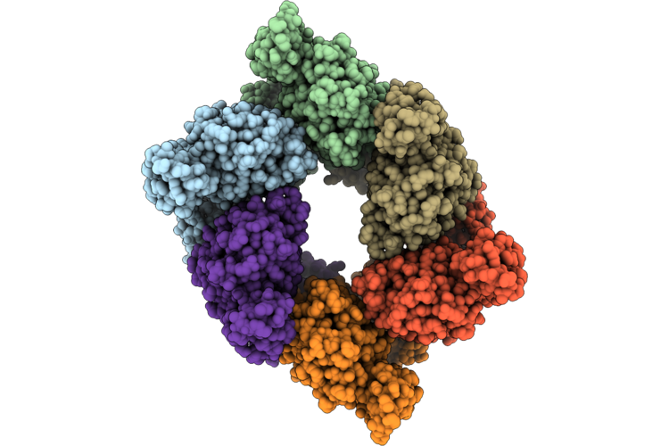 |
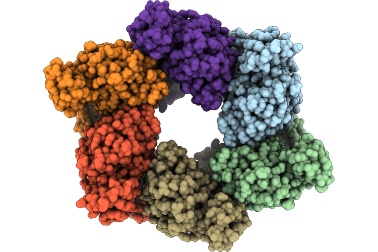
Single Particle Reconstruction Of Pilu From Vibrio Cholerae El Tor E7946, Form 3
Organism: Vibrio cholerae
Method: ELECTRON MICROSCOPY Release Date: 2026-05-20 Classification: MOTOR PROTEIN |
Organism: Vibrio cholerae
Method: ELECTRON MICROSCOPY
Release Date: 2026-05-20
 |
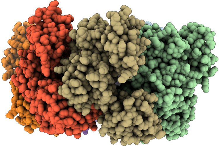
Single Particle Reconstruction Of Pilu From Vibrio Cholerae El Tor E7946, Form 4
Organism: Vibrio cholerae
Method: ELECTRON MICROSCOPY Release Date: 2026-05-20 Classification: MOTOR PROTEIN |
Organism: Vibrio cholerae
Method: ELECTRON MICROSCOPY
Release Date: 2026-05-20
 |
Crystal Structure Of Vibrio Cholerae Pilu, A Pilt-Dependent Retraction Atpase - Crystal Form 1.
Organism: Vibrio cholerae o1 biovar el tor str. n16961
Method: X-RAY DIFFRACTION Resolution:2.95 Å Release Date: 2026-05-20 Classification: MOTOR PROTEIN |
Organism: Vibrio cholerae o1 biovar el tor str. n16961
Method: X-RAY DIFFRACTION
Release Date: 2026-05-20
 |
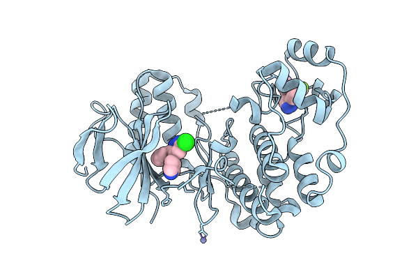
Crystal Structure Of Vibrio Cholerae Pilu, A Pilt-Dependent Retraction Atpase - Crystal Form 2
Organism: Vibrio cholerae o1 biovar el tor str. n16961
Method: X-RAY DIFFRACTION Resolution:2.70 Å Release Date: 2026-05-20 Classification: MOTOR PROTEIN Ligands: SO4 |
Organism: Vibrio cholerae o1 biovar el tor str. n16961
Method: X-RAY DIFFRACTION
Release Date: 2026-05-20
Ligands: SO4
 |
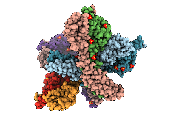
Crystal Structure Of Human P38 Alpha Mapk In Complex With Mw01-21-114Srm
Organism: Homo sapiens
Method: X-RAY DIFFRACTION Resolution:2.05 Å Release Date: 2025-09-17 Classification: TRANSFERASE Ligands: GG5, A1CRZ, ZN |
Organism: Homo sapiens
Method: X-RAY DIFFRACTION
Release Date: 2025-09-17
Ligands: GG5, A1CRZ, ZN
 |
Crystal Structure Of The Histidine Kinase Vc2136 From Vibrio Cholerae Serotype O1
Organism: Vibrio cholerae o1 biovar el tor str. n16961
Method: X-RAY DIFFRACTION Resolution:2.75 Å Release Date: 2025-08-20 Classification: TRANSFERASE Ligands: SO4, CL |
Organism: Vibrio cholerae o1 biovar el tor str. n16961
Method: X-RAY DIFFRACTION
Release Date: 2025-08-20
Ligands: SO4, CL
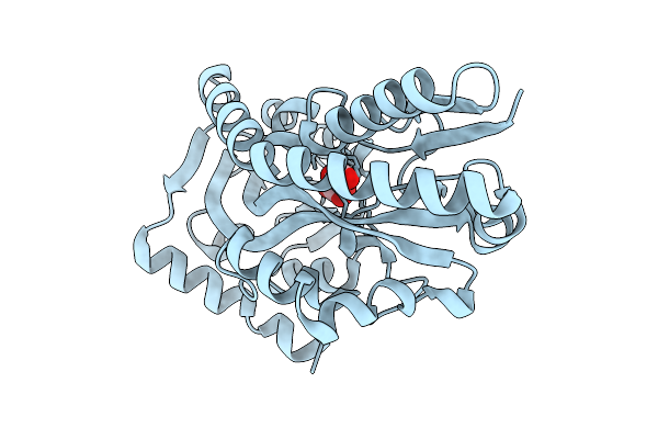 |
Crystal Structure Of C4-Dicarboxylate-Binding Protein (Pa0884) Of Tripartite Atp-Independent Periplasmic Transporter Family From Pseudomonas Aeruginosa Pao1 In Complex With L-Malate
Organism: Pseudomonas aeruginosa
Method: X-RAY DIFFRACTION Resolution:1.50 Å Release Date: 2025-07-09 Classification: TRANSPORT PROTEIN Ligands: LMR |
Organism: Pseudomonas aeruginosa
Method: X-RAY DIFFRACTION
Release Date: 2025-07-09
Ligands: LMR
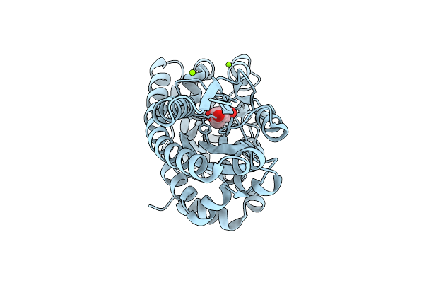 |
Crystal Structure Of C4-Dicarboxylate-Binding Protein (Pa0884) Of Tripartite Atp-Independent Periplasmic Transporter Family From Pseudomonas Aeruginosa Pao1 In Complex With Mesaconic Acid
Organism: Pseudomonas aeruginosa
Method: X-RAY DIFFRACTION Resolution:1.32 Å Release Date: 2025-07-09 Classification: TRANSPORT PROTEIN Ligands: MEZ, MG, CL |
Organism: Pseudomonas aeruginosa
Method: X-RAY DIFFRACTION
Release Date: 2025-07-09
Ligands: MEZ, MG, CL
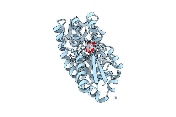 |
Crystal Structure Of C4-Dicarboxylate-Binding Periplasmic Protein (Pa5167) Of Tripartite Atp-Independent Periplasmic Transporter Family From Pseudomonas Aeruginosa Pao1 In Complex With L-Malate
Organism: Pseudomonas aeruginosa
Method: X-RAY DIFFRACTION Resolution:1.30 Å Release Date: 2025-07-09 Classification: TRANSPORT PROTEIN Ligands: LMR, ZN |
Organism: Pseudomonas aeruginosa
Method: X-RAY DIFFRACTION
Release Date: 2025-07-09
Ligands: LMR, ZN