

Search Count: 15
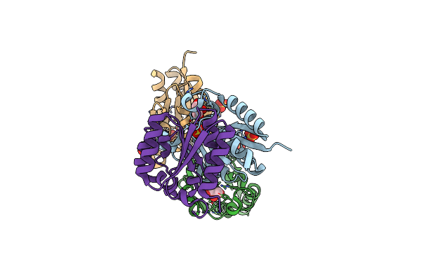 |
The Crystal Structure Of Type Ii Dehydroquinase From Propionibacterium Acnes
Organism: Cutibacterium acnes
Method: X-RAY DIFFRACTION Resolution:1.70 Å Release Date: 2020-07-08 Classification: BIOSYNTHETIC PROTEIN Ligands: SO4, GOL |
Organism: Cutibacterium acnes
Method: X-RAY DIFFRACTION
Release Date: 2020-07-08
Ligands: SO4, GOL
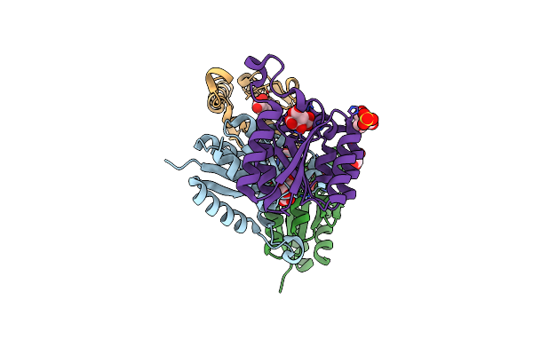 |
The Crystal Structure Of Type Ii Dehydroquinase From Zymomonas Mobilis
Organism: Zymomonas mobilis subsp. mobilis (strain atcc 31821 / zm4 / cp4)
Method: X-RAY DIFFRACTION Resolution:2.34 Å Release Date: 2020-07-08 Classification: BIOSYNTHETIC PROTEIN Ligands: FLC, TRS, SO4, GOL |
Organism: Zymomonas mobilis subsp. mobilis (strain atcc 31821 / zm4 / cp4)
Method: X-RAY DIFFRACTION
Release Date: 2020-07-08
Ligands: FLC, TRS, SO4, GOL
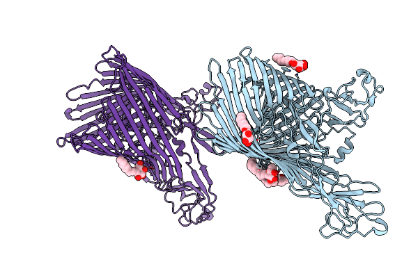 |
The Crystal Structure Of The Ferredoxin Receptor Fusa From Pectobacterium Atrosepticum Scri1043
Organism: Pectobacterium atrosepticum
Method: X-RAY DIFFRACTION Resolution:3.20 Å Release Date: 2016-08-31 Classification: TRANSPORT PROTEIN Ligands: LDA, BOG |
Organism: Pectobacterium atrosepticum
Method: X-RAY DIFFRACTION
Release Date: 2016-08-31
Ligands: LDA, BOG
 |
The Crystal Structure Of Arabidopsis Ferredoxin 2 With 2Fe-2S Cluster
Organism: Arabidopsis thaliana
Method: X-RAY DIFFRACTION Resolution:2.34 Å Release Date: 2016-08-31 Classification: ELECTRON TRANSPORT Ligands: FES, CL |
Organism: Arabidopsis thaliana
Method: X-RAY DIFFRACTION
Release Date: 2016-08-31
Ligands: FES, CL
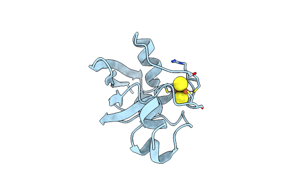 |
The Crystal Structure Of Potato Ferredoxin I With 2Fe-2S Cluster
Organism: Solanum tuberosum
Method: X-RAY DIFFRACTION Resolution:2.46 Å Release Date: 2016-08-31 Classification: ELECTRON TRANSPORT Ligands: FES |
Organism: Solanum tuberosum
Method: X-RAY DIFFRACTION
Release Date: 2016-08-31
Ligands: FES
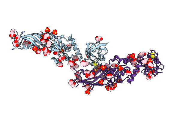 |
Crystal Structure Of Pectocin M2 At 1.86 Angstroms
Organism: Pectobacterium carotovorum subsp. brasiliensis pbr1692
Method: X-RAY DIFFRACTION Resolution:1.86 Å Release Date: 2014-06-04 Classification: HYDROLASE Ligands: FES, SO4, GOL, MPD, CL |
Organism: Pectobacterium carotovorum subsp. brasiliensis pbr1692
Method: X-RAY DIFFRACTION
Release Date: 2014-06-04
Ligands: FES, SO4, GOL, MPD, CL
 |
The Crystal Structure Of Pectocin M2 At 2.3 Angstroms
Organism: Pectobacterium carotovorum subsp. brasiliensis
Method: X-RAY DIFFRACTION Resolution:2.30 Å Release Date: 2014-06-04 Classification: HYDROLASE Ligands: FES, SO4, CL |
Organism: Pectobacterium carotovorum subsp. brasiliensis
Method: X-RAY DIFFRACTION
Release Date: 2014-06-04
Ligands: FES, SO4, CL
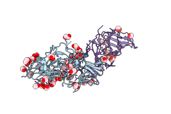 |
The Crystal Structure Of Pyocin L1 At 2.09 Angstroms
Organism: Pseudomonas aeruginosa
Method: X-RAY DIFFRACTION Resolution:2.09 Å Release Date: 2014-02-19 Classification: SUGAR BINDING PROTEIN Ligands: EDO, CL |
Organism: Pseudomonas aeruginosa
Method: X-RAY DIFFRACTION
Release Date: 2014-02-19
Ligands: EDO, CL
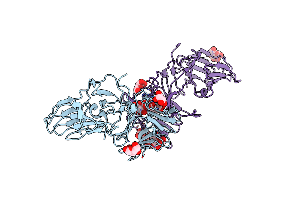 |
The Crystal Structure Of Pyocin L1 Bound To D-Mannose At 2.55 Angstroms
Organism: Pseudomonas aeruginosa
Method: X-RAY DIFFRACTION Resolution:2.55 Å Release Date: 2014-02-19 Classification: SUGAR BINDING PROTEIN Ligands: BMA |
Organism: Pseudomonas aeruginosa
Method: X-RAY DIFFRACTION
Release Date: 2014-02-19
Ligands: BMA
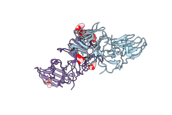 |
The Crystal Structure Of Pyocin L1 Bound To D-Rhamnose At 2.37 Angstroms
Organism: Pseudomonas aeruginosa
Method: X-RAY DIFFRACTION Resolution:2.37 Å Release Date: 2014-02-19 Classification: SUGAR BINDING PROTEIN Ligands: XXR |
Organism: Pseudomonas aeruginosa
Method: X-RAY DIFFRACTION
Release Date: 2014-02-19
Ligands: XXR
 |
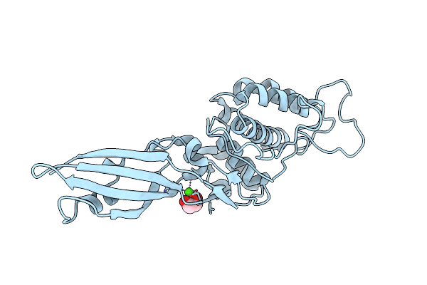
High Resolution Structure Of Truncated Bacteriocin Syringacin M From Pseudomonas Syringae Pv. Tomato Dc3000
Organism: Pseudomonas syringae pv. tomato
Method: X-RAY DIFFRACTION Resolution:1.46 Å Release Date: 2012-10-03 Classification: ANTIMICROBIAL PROTEIN Ligands: CA, EDO, CL, MG |
Organism: Pseudomonas syringae pv. tomato
Method: X-RAY DIFFRACTION
Release Date: 2012-10-03
Ligands: CA, EDO, CL, MG
 |
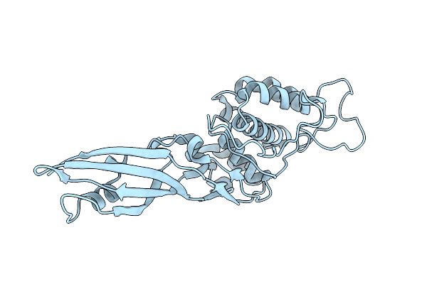
Crystal Structure Of The Bacteriocin Syringacin M From Pseudomonas Syringae Pv. Tomato Dc3000
Organism: Pseudomonas syringae pv. tomato
Method: X-RAY DIFFRACTION Resolution:2.83 Å Release Date: 2012-10-03 Classification: ANTIMICROBIAL PROTEIN Ligands: CA, EDO |
Organism: Pseudomonas syringae pv. tomato
Method: X-RAY DIFFRACTION
Release Date: 2012-10-03
Ligands: CA, EDO
 |
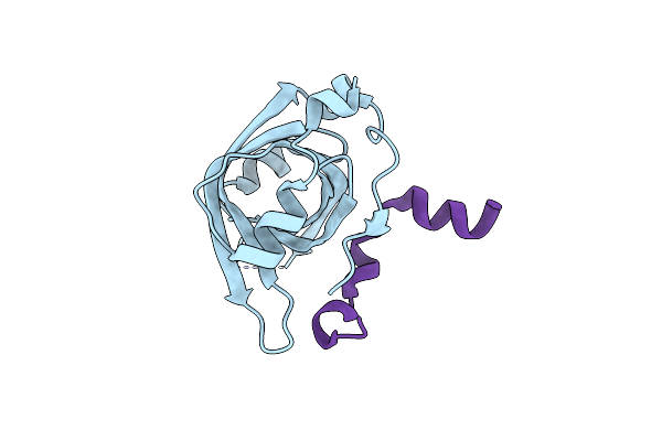
Crystal Structure Of Syringacin M Mutant D232A From Pseudomonas Syringae Pv. Tomato Dc3000
Organism: Pseudomonas syringae pv. tomato
Method: X-RAY DIFFRACTION Resolution:3.12 Å Release Date: 2012-10-03 Classification: ANTIMICROBIAL PROTEIN |
Organism: Pseudomonas syringae pv. tomato
Method: X-RAY DIFFRACTION
Release Date: 2012-10-03
 |
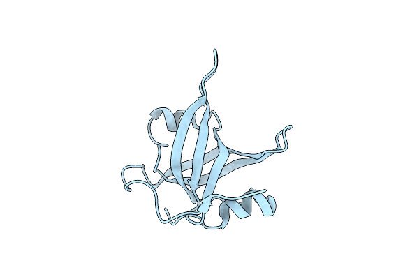
P53N (Fragment 33-60) Bound To Rpa70N
Organism: Homo sapiens
Method: X-RAY DIFFRACTION Resolution:1.60 Å Release Date: 2005-10-11 Classification: REPLICATION |
Organism: Homo sapiens
Method: X-RAY DIFFRACTION
Release Date: 2005-10-11
 |
N-Terminal Domain Of The Rpa70 Subunit Of Human Replication Protein A.
Organism: Homo sapiens
Method: X-RAY DIFFRACTION Resolution:1.60 Å Release Date: 2005-10-04 Classification: REPLICATION |
Organism: Homo sapiens
Method: X-RAY DIFFRACTION
Release Date: 2005-10-04