
Search Count: 15
All
Selected
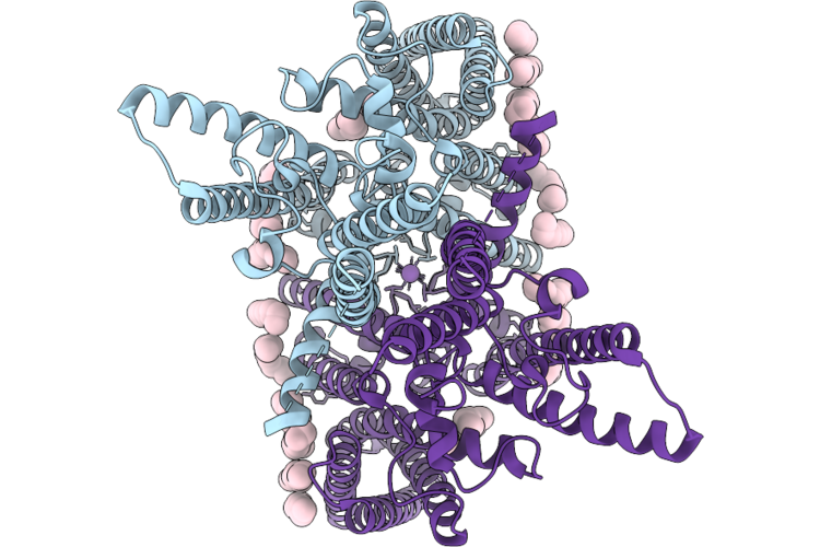 |
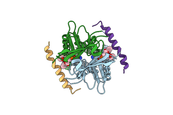
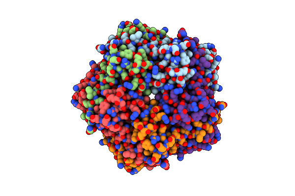
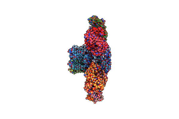
Structure Of The Two-Pore Domain, Outwardly Rectifying Potassium (Tok1) From Candida Albicans, Overall Structure
Organism: Candida albicans
Method: ELECTRON MICROSCOPY Release Date: 2026-04-29 Classification: METAL TRANSPORT Ligands: K, LMT |
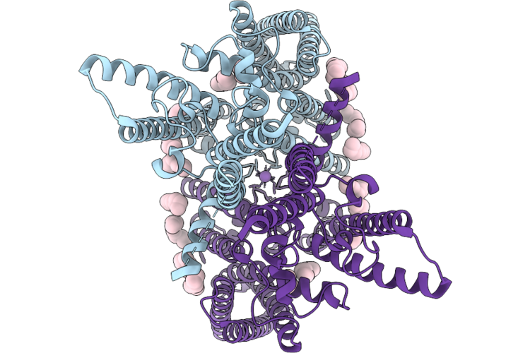 |
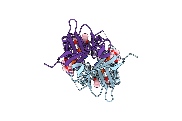
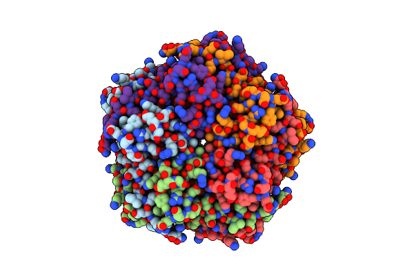
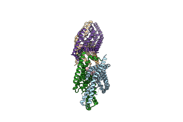
Structure Of The Two-Pore Domain, Outwardly Rectifying Potassium (Tok1) From Candida Albicans, Up Conformation
Organism: Candida albicans
Method: ELECTRON MICROSCOPY Release Date: 2026-04-29 Classification: METAL TRANSPORT Ligands: K, LMT |
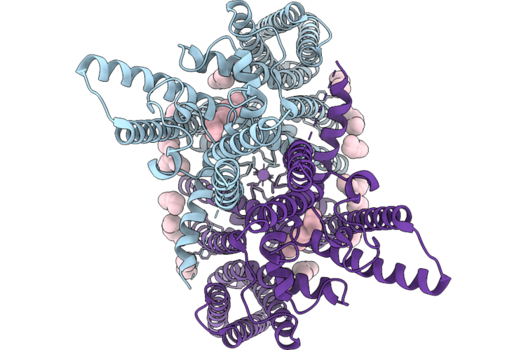 |
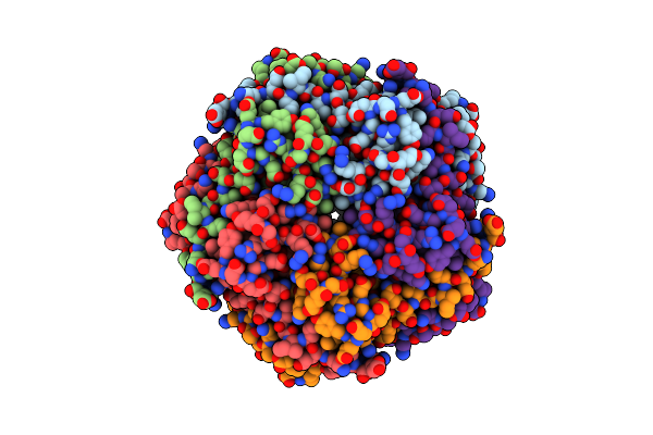
Structure Of The Two-Pore Domain, Outwardly Rectifying Potassium (Tok1) From Candida Albicans, Down Conformation
Organism: Candida albicans
Method: ELECTRON MICROSCOPY Release Date: 2026-04-29 Classification: METAL TRANSPORT Ligands: K, LMT |
 |
Structure Of Sars-Cov-2 Orf3A In Late Endosome/Lysosome-Like Membrane Environment, Msp1D1 Nanodisc
Organism: Severe acute respiratory syndrome coronavirus 2
Method: ELECTRON MICROSCOPY Release Date: 2023-02-08 Classification: VIRAL PROTEIN Ligands: PEE |
 |
Structure Of Sars-Cov-1 Orf3A In Late Endosome/Lysosome-Like Environment, Msp1D1 Nanodisc
Organism: Severe acute respiratory syndrome coronavirus, Homo sapiens
Method: ELECTRON MICROSCOPY Release Date: 2023-02-08 Classification: VIRAL PROTEIN Ligands: PEE |
 |
Structure Of Sars-Cov-2 Orf3A In Plasma Membrane-Like Environment, Msp1D1 Nanodisc
Organism: Severe acute respiratory syndrome coronavirus 2
Method: ELECTRON MICROSCOPY Release Date: 2023-02-08 Classification: VIRAL PROTEIN Ligands: PEE |
 |
Structure Of Sars-Cov-2 Orf3A In Late Endosome/Lysosome-Like Environment, Saposin A Nanodisc
Organism: Severe acute respiratory syndrome coronavirus 2, Homo sapiens
Method: ELECTRON MICROSCOPY Release Date: 2023-02-08 Classification: VIRAL PROTEIN Ligands: PEE |
 |
Organism: Gallus gallus
Method: ELECTRON MICROSCOPY Release Date: 2019-01-23 Classification: MEMBRANE PROTEIN Ligands: CA |
 |
Organism: Gallus gallus
Method: ELECTRON MICROSCOPY Resolution:3.00 Å Release Date: 2019-01-23 Classification: MEMBRANE PROTEIN |
 |
Organism: Gallus gallus
Method: ELECTRON MICROSCOPY Release Date: 2019-01-23 Classification: MEMBRANE PROTEIN Ligands: CA |
 |
Organism: Gallus gallus
Method: ELECTRON MICROSCOPY Resolution:3.00 Å Release Date: 2019-01-23 Classification: MEMBRANE PROTEIN |
 |
Organism: Gallus gallus
Method: ELECTRON MICROSCOPY Release Date: 2019-01-23 Classification: MEMBRANE PROTEIN Ligands: CA |
 |
Organism: Gallus gallus
Method: ELECTRON MICROSCOPY Release Date: 2019-01-23 Classification: MEMBRANE PROTEIN Ligands: CA |
 |
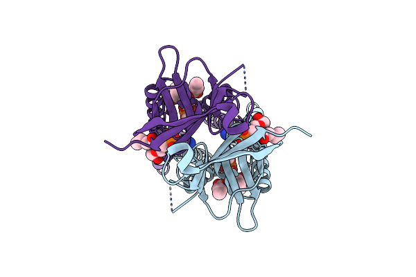
Calcium-Activated Chloride Channel Bestrophin-1 (Best1), Triple Mutant: I76A, F80A, F84A; In Complex With An Fab Antibody Fragment, Chloride, And Calcium
Organism: Gallus gallus, Mus musculus
Method: X-RAY DIFFRACTION Resolution:3.10 Å Release Date: 2016-11-30 Classification: MEMBRANE PROTEIN Ligands: CL, K, GOL, CA |
 |
Crystal Structure Of The Human Two Pore Domain Potassium Ion Channel K2P1 (Twik-1)
Organism: Homo sapiens
Method: X-RAY DIFFRACTION Resolution:3.40 Å Release Date: 2012-02-08 Classification: MEMBRANE PROTEIN Ligands: UND, K |