Deposition Date
2011-11-09
Release Date
2012-02-08
Last Version Date
2024-10-30
Entry Detail
PDB ID:
3UKM
Keywords:
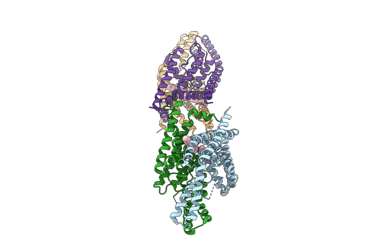
Title:
Crystal structure of the human two pore domain potassium ion channel K2P1 (TWIK-1)
Biological Source:
Source Organism(s):
Homo sapiens (Taxon ID: 9606)
Expression System(s):
Method Details:
Experimental Method:
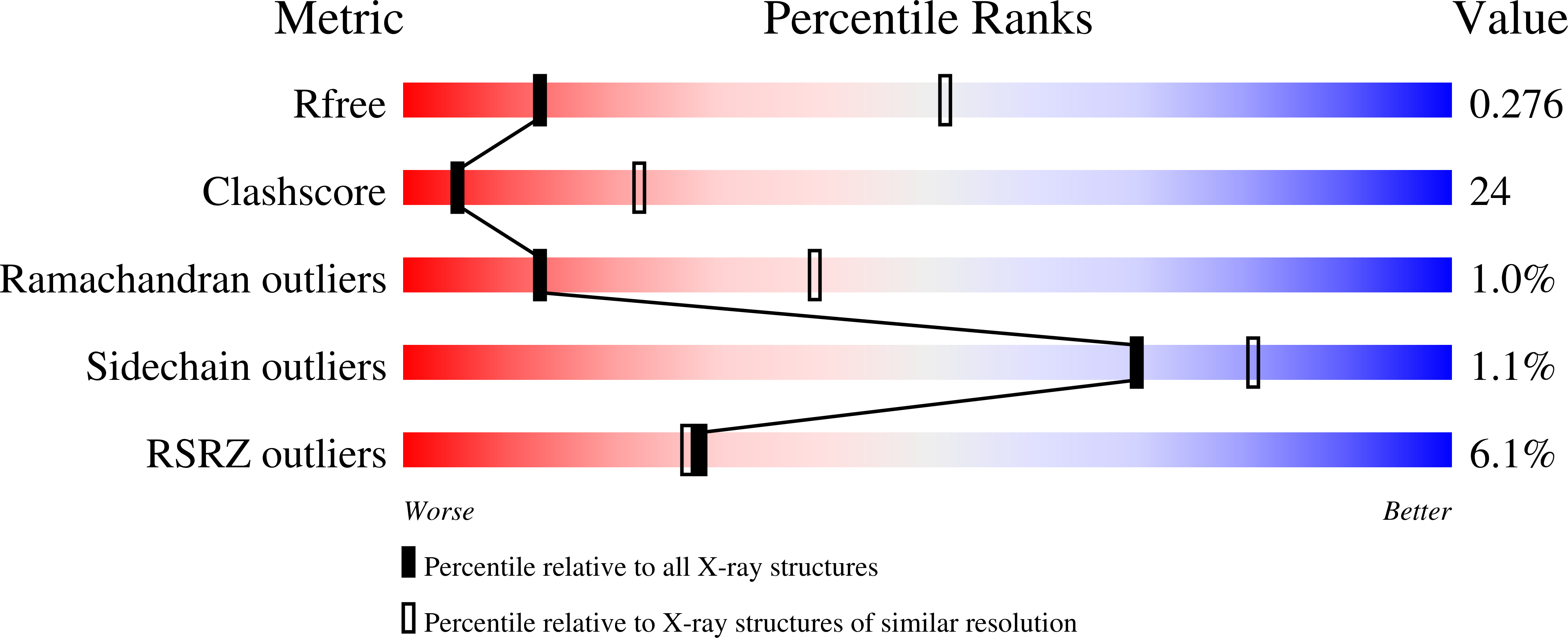
Resolution:
3.40 Å
R-Value Free:
0.27
R-Value Work:
0.27
R-Value Observed:
0.27
Space Group:
P 1 21 1