

Search Count: 20
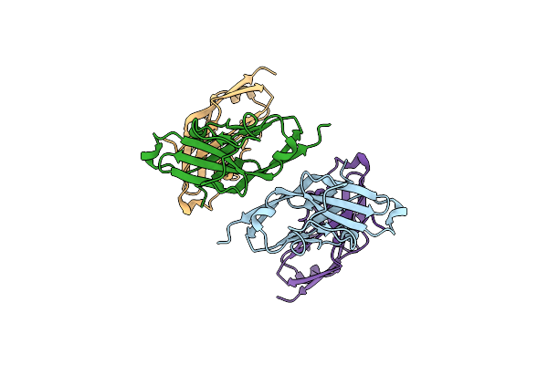 |
Cell Surface Receptor
Organism: Homo sapiens
Method: X-RAY DIFFRACTION Resolution:1.78 Å Release Date: 2019-04-17 Classification: IMMUNE SYSTEM |
Organism: Homo sapiens
Method: X-RAY DIFFRACTION
Release Date: 2019-04-17
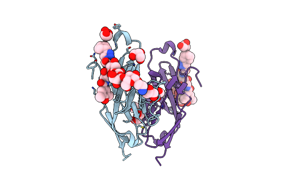 |
Cell Surface Receptor In Complex With Ligand At 1.80-A Resolution
Organism: Homo sapiens
Method: X-RAY DIFFRACTION Resolution:1.80 Å Release Date: 2019-04-17 Classification: IMMUNE SYSTEM Ligands: GOL, FVP |
Organism: Homo sapiens
Method: X-RAY DIFFRACTION
Release Date: 2019-04-17
Ligands: GOL, FVP
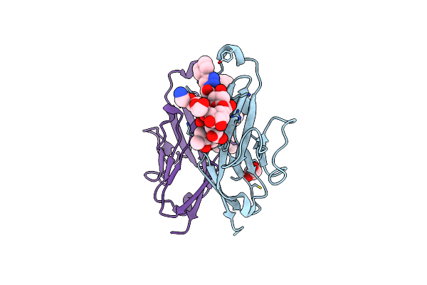 |
Cell Surface Receptor With Bound Ligand At 1.75-A Resolution
Organism: Homo sapiens
Method: X-RAY DIFFRACTION Resolution:1.75 Å Release Date: 2019-04-17 Classification: IMMUNE SYSTEM Ligands: GOL, FVP |
Organism: Homo sapiens
Method: X-RAY DIFFRACTION
Release Date: 2019-04-17
Ligands: GOL, FVP
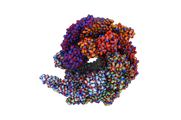 |
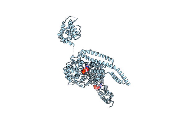
Calcarisporiella Thermophila Hsp104
Organism: Calcarisporiella thermophila
Method: ELECTRON MICROSCOPY Release Date: 2019-04-03 Classification: CHAPERONE Ligands: ADP |
Organism: Calcarisporiella thermophila
Method: ELECTRON MICROSCOPY
Release Date: 2019-04-03
Ligands: ADP
 |
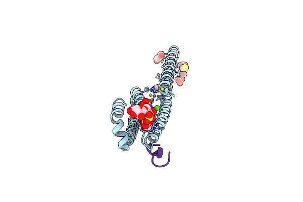
Crystal Structure Of Hsp104 R328M/R757M Mutant From Calcarisporiella Thermophila
Organism: Calcarisporiella thermophila
Method: X-RAY DIFFRACTION Resolution:2.70 Å Release Date: 2018-10-03 Classification: CHAPERONE Ligands: ADP |
Organism: Calcarisporiella thermophila
Method: X-RAY DIFFRACTION
Release Date: 2018-10-03
Ligands: ADP
 |
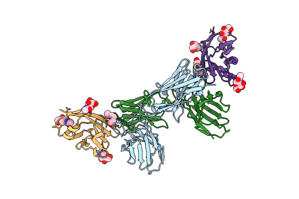
A Receptor Molecule
Organism: Homo sapiens
Method: X-RAY DIFFRACTION Resolution:2.31 Å Release Date: 2016-07-13 Classification: METAL TRANSPORT Ligands: ZN, CA, GOL, ACT, SCN, SO4, MG |
Organism: Homo sapiens
Method: X-RAY DIFFRACTION
Release Date: 2016-07-13
Ligands: ZN, CA, GOL, ACT, SCN, SO4, MG
 |
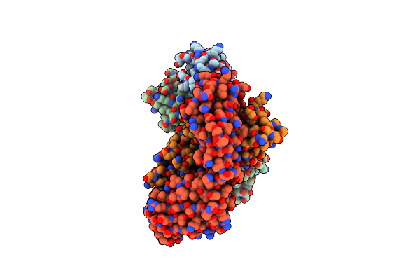
Cd47-Diabody Complex
Organism: Homo sapiens
Method: X-RAY DIFFRACTION Resolution:2.80 Å Release Date: 2016-06-29 Classification: CELL ADHESION Ligands: NAG, PGE |
Organism: Homo sapiens
Method: X-RAY DIFFRACTION
Release Date: 2016-06-29
Ligands: NAG, PGE
 |
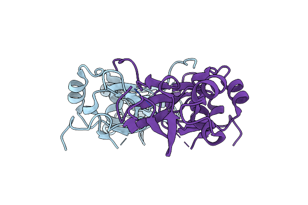
Crystal Structure Of Mid-Region Amyloid Beta Capture By Solanezumab
Organism: Homo sapiens
Method: X-RAY DIFFRACTION Resolution:2.41 Å Release Date: 2015-04-29 Classification: IMMUNE SYSTEM |
Organism: Homo sapiens
Method: X-RAY DIFFRACTION
Release Date: 2015-04-29
 |
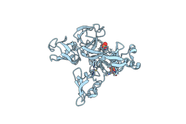
Crystal Structure Of The Mib Repeat Domain Of Mind Bomb 1
Organism: Homo sapiens
Method: X-RAY DIFFRACTION Resolution:2.06 Å Release Date: 2015-03-18 Classification: LIGASE |
Organism: Homo sapiens
Method: X-RAY DIFFRACTION
Release Date: 2015-03-18
 |
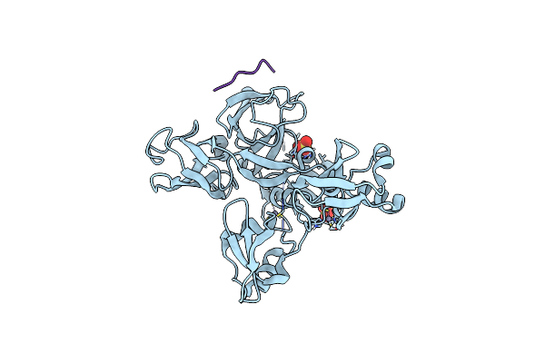
Crystal Structure Of The Mzm-Rep Domains Of Mind Bomb 1
Organism: Homo sapiens
Method: X-RAY DIFFRACTION Resolution:2.04 Å Release Date: 2015-03-18 Classification: LIGASE Ligands: ZN, SO4 |
Organism: Homo sapiens
Method: X-RAY DIFFRACTION
Release Date: 2015-03-18
Ligands: ZN, SO4
 |
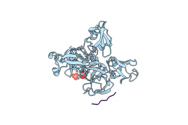
Crystal Structure Of The Mzm-Rep Domains Of Mind Bomb 1 In Complex With Jagged1 N-Box Peptide
Organism: Homo sapiens
Method: X-RAY DIFFRACTION Resolution:2.05 Å Release Date: 2015-03-18 Classification: LIGASE Ligands: ZN, SO4 |
Organism: Homo sapiens
Method: X-RAY DIFFRACTION
Release Date: 2015-03-18
Ligands: ZN, SO4
 |
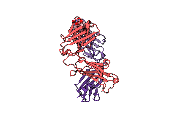
Crystal Structure Of The Mzm-Rep Domains Of Mind Bomb 1 In Complex With Fly Delta N-Box Peptide
Organism: Homo sapiens, Drosophila melanogaster
Method: X-RAY DIFFRACTION Resolution:2.15 Å Release Date: 2015-03-18 Classification: LIGASE Ligands: ZN, SO4 |
Organism: Homo sapiens, Drosophila melanogaster
Method: X-RAY DIFFRACTION
Release Date: 2015-03-18
Ligands: ZN, SO4
 |
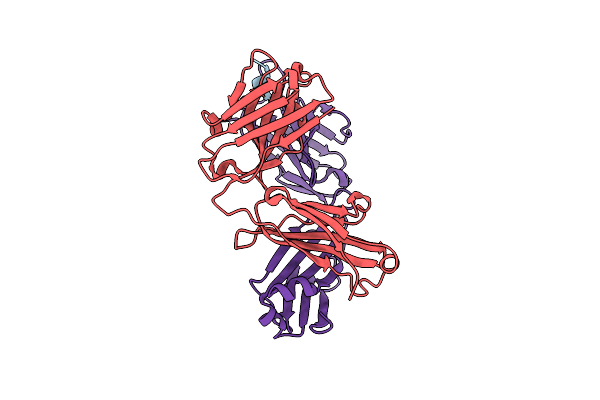
Humanised 3D6 Fab Complexed To Amyloid Beta 1-8
Organism: Mus musculus, Homo sapiens
Method: X-RAY DIFFRACTION Resolution:2.00 Å Release Date: 2015-01-28 Classification: IMMUNE SYSTEM |
Organism: Mus musculus, Homo sapiens
Method: X-RAY DIFFRACTION
Release Date: 2015-01-28
 |
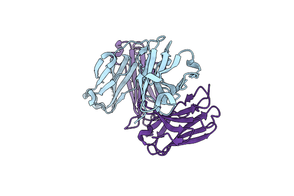
Crystal Structure Of A Humanised 3D6 Fab Bound To Amyloid Beta Peptide
Organism: Homo sapiens, mus musculus, Homo sapiens
Method: X-RAY DIFFRACTION Resolution:2.20 Å Release Date: 2013-03-13 Classification: PROTEIN FIBRIL/Immune System |
Organism: Homo sapiens, mus musculus, Homo sapiens
Method: X-RAY DIFFRACTION
Release Date: 2013-03-13
 |
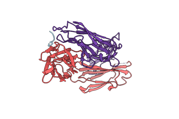
Ad Related Murine Antibody Fragment
Organism: Mus musculus, Homo sapiens
Method: X-RAY DIFFRACTION Resolution:2.00 Å Release Date: 2012-10-03 Classification: IMMUNE SYSTEM |
Organism: Mus musculus, Homo sapiens
Method: X-RAY DIFFRACTION
Release Date: 2012-10-03
 |
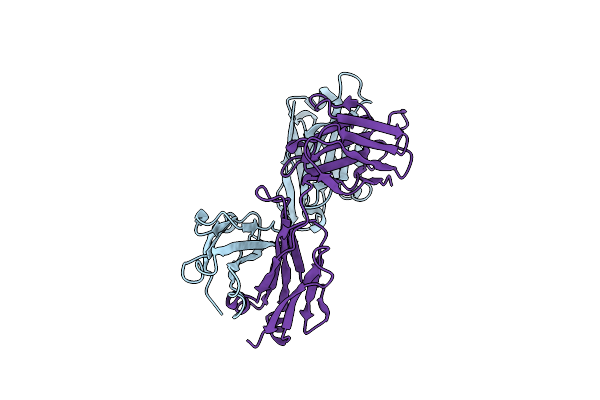
Crystal Structure Of Fab Wo2 Bound To The N Terminal Domain Of Amyloid Beta Peptide (1-28)
Organism: Mus musculus
Method: X-RAY DIFFRACTION Resolution:1.59 Å Release Date: 2008-04-15 Classification: IMMUNE SYSTEM |
Organism: Mus musculus
Method: X-RAY DIFFRACTION
Release Date: 2008-04-15
 |
Crystal Structure Of Anti-Amyloid Beta Fab Wo2 (P21, Formb)
Organism: Mus musculus
Method: X-RAY DIFFRACTION Resolution:1.90 Å Release Date: 2008-04-15 Classification: IMMUNE SYSTEM |
Organism: Mus musculus
Method: X-RAY DIFFRACTION
Release Date: 2008-04-15
 |
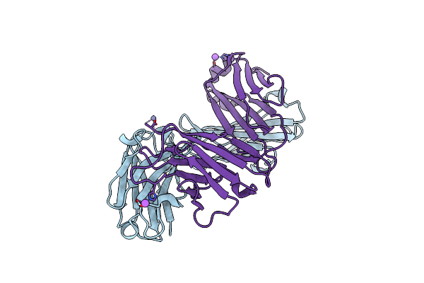
Crystal Structure Of Fab Wo2 Bound To The N Terminal Domain Of Amyloid Beta Peptide (1-16)
Organism: Mus musculus
Method: X-RAY DIFFRACTION Resolution:1.59 Å Release Date: 2008-04-15 Classification: IMMUNE SYSTEM |
Organism: Mus musculus
Method: X-RAY DIFFRACTION
Release Date: 2008-04-15
 |
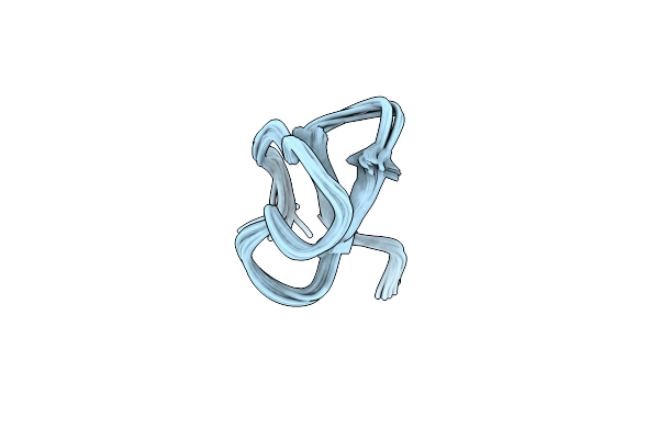
Structure Of Anti-Amyloid-Beta Fab Wo2 (Form A, P212121)
Organism: Mus musculus
Method: X-RAY DIFFRACTION Resolution:1.60 Å Release Date: 2008-04-15 Classification: IMMUNE SYSTEM |
Organism: Mus musculus
Method: X-RAY DIFFRACTION
Release Date: 2008-04-15
 |
Structure Of A Novel P-Superfamily Spasmodic Conotoxin Reveals An Inhibitory Cystine Knot Motif
|
Method: SOLUTION NMR
Release Date: 2003-01-28