

Search Count: 16
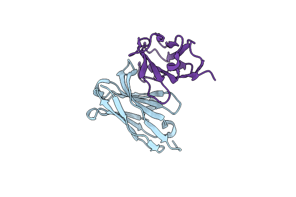 |
Engineered Single Chain Antibody C9+C14 Scfv
Organism: Mus musculus
Method: X-RAY DIFFRACTION Resolution:1.58 Å Release Date: 2020-04-15 Classification: IMMUNE SYSTEM |
Organism: Mus musculus
Method: X-RAY DIFFRACTION
Release Date: 2020-04-15
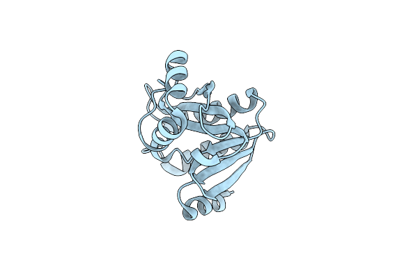 |
Crystal Structure Of The Evolved Variant Of The Computationally Designed Serine Hydrolase, Osh55.4_H1, Covalently Bound With Diisopropyl Fluorophosphate (Dfp), Northeast Structural Genomics Consortium (Nesg) Target Or273
Organism: Synthetic construct
Method: X-RAY DIFFRACTION Resolution:2.29 Å Release Date: 2013-04-24 Classification: Structural Genomics, Unknown Function |
Organism: Synthetic construct
Method: X-RAY DIFFRACTION
Release Date: 2013-04-24
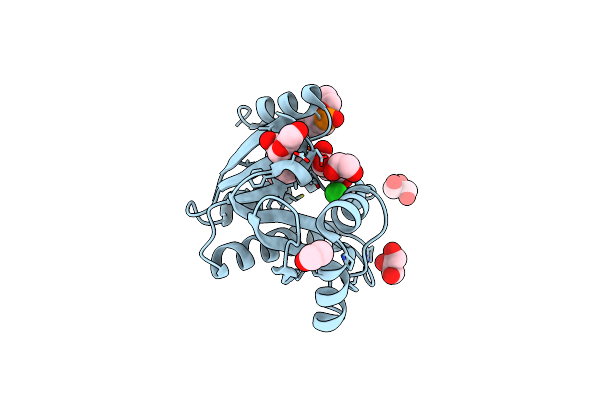 |
Crystal Structure Of The Evolved Variant Of The Computationally Designed Serine Hydrolase, Osh55.4_H1 Covalently Bound With Fp-Alkyne, Northeast Structural Genomics Consortium (Nesg) Target Or273
Organism: Synthetic construct
Method: X-RAY DIFFRACTION Resolution:1.36 Å Release Date: 2013-04-10 Classification: Structural Genomics, Unknown Function Ligands: SEF, CL, EDO, PEG, GOL |
Organism: Synthetic construct
Method: X-RAY DIFFRACTION
Release Date: 2013-04-10
Ligands: SEF, CL, EDO, PEG, GOL
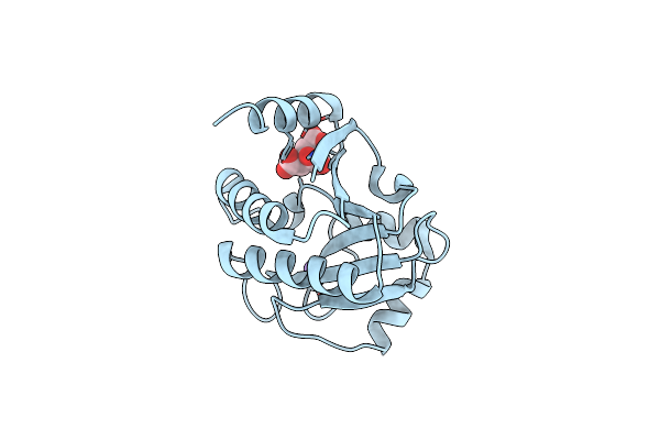 |
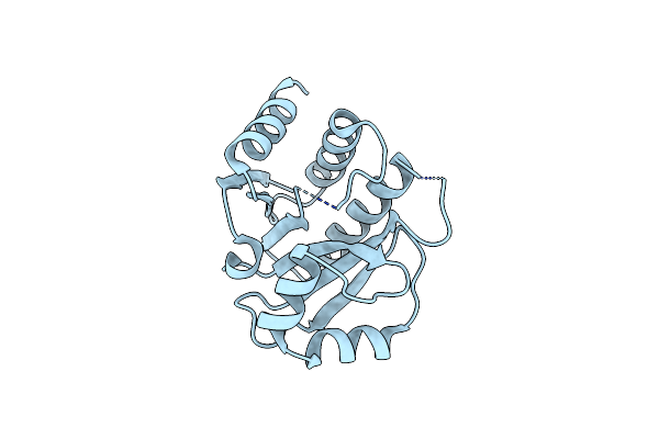
Crystal Structure Of The Apo Form Of The Evolved Variant Of The Computationally Designed Serine Hydrolase, Osh55.4_H1. Northeast Structural Genomics Consortium (Nesg) Target Or273
Organism: Artificial gene
Method: X-RAY DIFFRACTION Resolution:2.41 Å Release Date: 2013-03-20 Classification: HYDROLASE Ligands: RB, CIT |
Organism: Artificial gene
Method: X-RAY DIFFRACTION
Release Date: 2013-03-20
Ligands: RB, CIT
 |
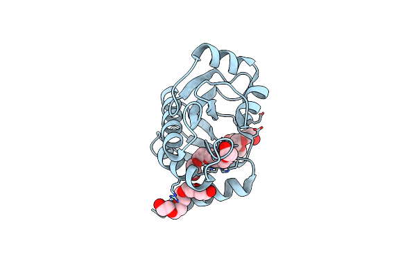
Crystal Structure Of E6D/L155R Variant Of De Novo Designed Serine Hydrolase Osh55, Northeast Structural Genomics Consortium (Nesg) Target Or187
Organism: Synthetic construct
Method: X-RAY DIFFRACTION Resolution:2.00 Å Release Date: 2012-06-13 Classification: De Novo Protein, hydrolase |
Organism: Synthetic construct
Method: X-RAY DIFFRACTION
Release Date: 2012-06-13
 |
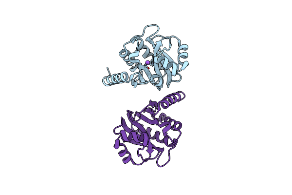
Crystal Structure Of E6H Variant Of De Novo Designed Serine Hydrolase Osh55, Northeast Structural Genomics Consortium (Nesg) Target Or185
Organism: Artificial gene
Method: X-RAY DIFFRACTION Resolution:2.20 Å Release Date: 2012-06-13 Classification: DE NOVO PROTEIN Ligands: CL, PE4, PE5, PE6 |
Organism: Artificial gene
Method: X-RAY DIFFRACTION
Release Date: 2012-06-13
Ligands: CL, PE4, PE5, PE6
 |
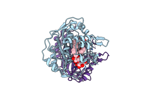
Crystal Structure Of E6A/L130D/A155H Variant Of De Novo Designed Serine Hydrolase, Northeast Structural Genomics Consortium (Nesg) Target Or186
Organism: Artificial gene
Method: X-RAY DIFFRACTION Resolution:2.70 Å Release Date: 2012-06-13 Classification: HYDROLASE |
Organism: Artificial gene
Method: X-RAY DIFFRACTION
Release Date: 2012-06-13
 |
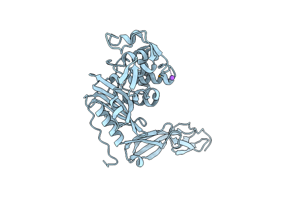
Crystal Structure Of De Novo Designed Serine Hydrolase, Northeast Structural Genomics Consortium (Nesg) Target Or165
Organism: Artificial gene
Method: X-RAY DIFFRACTION Resolution:2.49 Å Release Date: 2012-05-30 Classification: DE NOVO PROTEIN Ligands: LMU, PEG |
Organism: Artificial gene
Method: X-RAY DIFFRACTION
Release Date: 2012-05-30
Ligands: LMU, PEG
 |
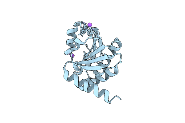
Three Dimensional Structure Of De Novo Designed Serine Hydrolase Osh26, Northeast Structural Genomics Consortium (Nesg) Target Or89
Organism: Artificial gene
Method: X-RAY DIFFRACTION Resolution:2.00 Å Release Date: 2012-04-18 Classification: DE NOVO PROTEIN |
Organism: Artificial gene
Method: X-RAY DIFFRACTION
Release Date: 2012-04-18
 |
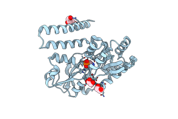
Crystal Structure Of De Novo Designed Serine Hydrolase Osh55, Northeast Structural Genomics Consortium Target Or130
Method: X-RAY DIFFRACTION
Resolution:2.60 Å Release Date: 2012-01-04 Classification: DE NOVO PROTEIN Ligands: CL, NA |
Method: X-RAY DIFFRACTION
Release Date: 2012-01-04
Ligands: CL, NA
 |
Crystal Structure Of Escherichia Coli Phnd In Complex With 2-Aminoethyl Phosphonate
Organism: Escherichia coli uti89
Method: X-RAY DIFFRACTION Resolution:1.71 Å Release Date: 2011-10-12 Classification: TRANSPORT PROTEIN Ligands: P7I, GOL |
Organism: Escherichia coli uti89
Method: X-RAY DIFFRACTION
Release Date: 2011-10-12
Ligands: P7I, GOL
 |
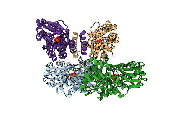
Crystal Structure Of Escherichia Coli Phnd
Organism: Escherichia coli uti89
Method: X-RAY DIFFRACTION Resolution:2.40 Å Release Date: 2011-10-12 Classification: TRANSPORT PROTEIN Ligands: UNL |
Organism: Escherichia coli uti89
Method: X-RAY DIFFRACTION
Release Date: 2011-10-12
Ligands: UNL
 |
Crystal Structure Of The Phosphonate Binding Protein, Phnd, From Escherichia Coli
Organism: Escherichia coli uti89
Method: X-RAY DIFFRACTION Resolution:2.20 Å Release Date: 2011-10-12 Classification: TRANSPORT PROTEIN Ligands: UNL |
Organism: Escherichia coli uti89
Method: X-RAY DIFFRACTION
Release Date: 2011-10-12
Ligands: UNL
 |
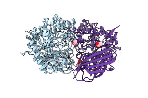
Crystal Structure Of Open, Unliganded E. Coli Phnd H157A
Organism: Escherichia coli
Method: X-RAY DIFFRACTION Resolution:3.30 Å Release Date: 2011-10-12 Classification: TRANSPORT PROTEIN |
Organism: Escherichia coli
Method: X-RAY DIFFRACTION
Release Date: 2011-10-12
 |
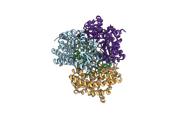
Crystal Structure Of Engineered Protein At The Resolution 1.98A, Northeast Structural Genomics Consortium Target Or128
Organism: Synthetic construct
Method: X-RAY DIFFRACTION Resolution:1.98 Å Release Date: 2011-10-05 Classification: Structural Genomics, Unknown Function Ligands: ACY, MG, PEG |
Organism: Synthetic construct
Method: X-RAY DIFFRACTION
Release Date: 2011-10-05
Ligands: ACY, MG, PEG
 |
The Crystal Structure Of A Thermophilic Trap Periplasmic Binding Protein
Organism: Thermotoga maritima
Method: X-RAY DIFFRACTION Resolution:1.90 Å Release Date: 2007-03-20 Classification: LIGAND BINDING PROTEIN |
Organism: Thermotoga maritima
Method: X-RAY DIFFRACTION
Release Date: 2007-03-20