

Search Count: 19
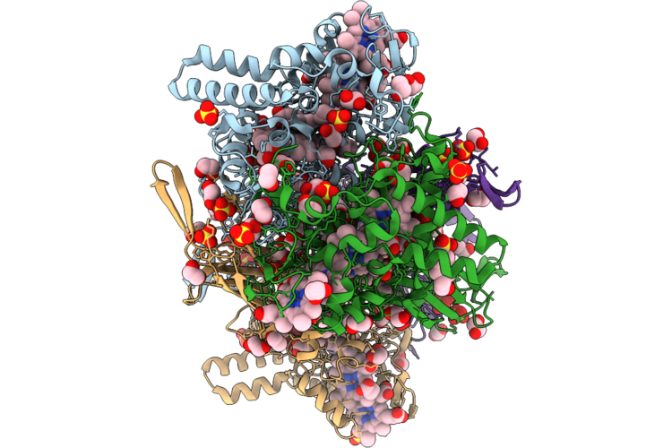 |
Crystal Structure Of A Multiheme Cytochrome C Selenoprotein (Mccsep)
Organism: Geobacter sulfurreducens pca
Method: X-RAY DIFFRACTION Resolution:1.75 Å Release Date: 2026-07-22 Classification: ELECTRON TRANSPORT Ligands: HEC, SO4, EDO, EPE, PGE |
Organism: Geobacter sulfurreducens pca
Method: X-RAY DIFFRACTION
Release Date: 2026-07-22
Ligands: HEC, SO4, EDO, EPE, PGE
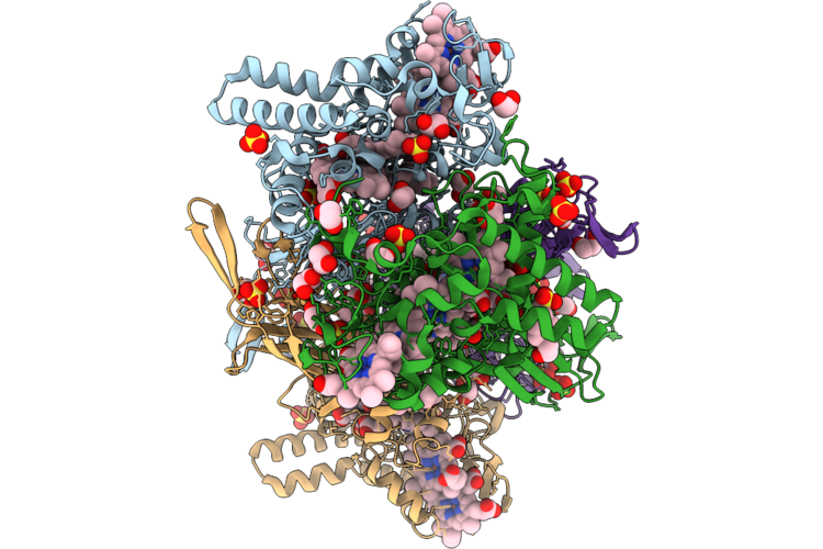 |
Crystal Structure Of A Multiheme Cytochrome C Selenoprotein Mutant (Mccsep U325A)
Organism: Geobacter sulfurreducens pca
Method: X-RAY DIFFRACTION Resolution:1.80 Å Release Date: 2026-07-22 Classification: ELECTRON TRANSPORT Ligands: HEC, SO4, EDO, EPE |
Organism: Geobacter sulfurreducens pca
Method: X-RAY DIFFRACTION
Release Date: 2026-07-22
Ligands: HEC, SO4, EDO, EPE
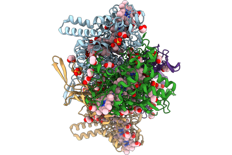 |
Crystal Structure Of A Selenite-Soaked Multiheme Cytochrome C Selenoprotein (Mccsep)
Organism: Geobacter sulfurreducens pca
Method: X-RAY DIFFRACTION Resolution:2.20 Å Release Date: 2026-07-22 Classification: ELECTRON TRANSPORT Ligands: HEC, BSY, SO4, EDO, EPE |
Organism: Geobacter sulfurreducens pca
Method: X-RAY DIFFRACTION
Release Date: 2026-07-22
Ligands: HEC, BSY, SO4, EDO, EPE
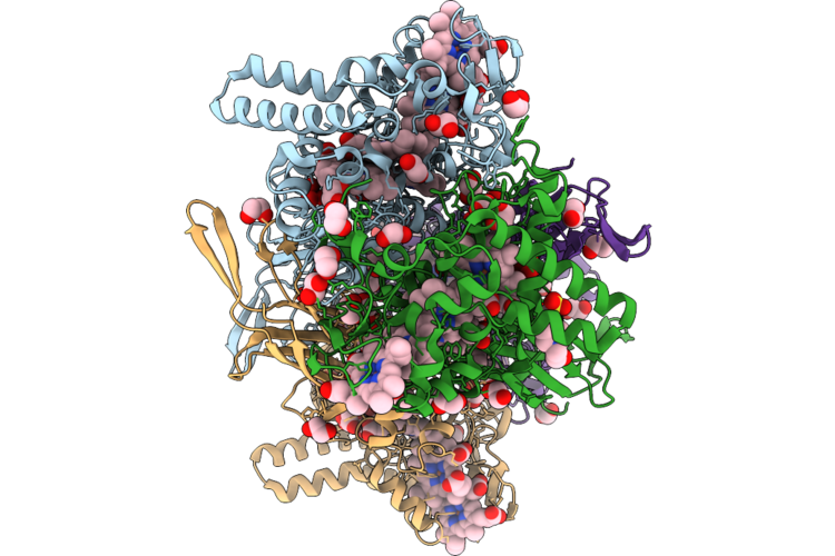 |
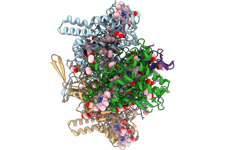
Crystal Structure Of A Selenate-Soaked Multiheme Cytochrome C Selenoprotein (Mccsep)
Organism: Geobacter sulfurreducens pca
Method: X-RAY DIFFRACTION Resolution:2.20 Å Release Date: 2026-07-22 Classification: ELECTRON TRANSPORT Ligands: HEC, SE4, EDO, EPE |
Organism: Geobacter sulfurreducens pca
Method: X-RAY DIFFRACTION
Release Date: 2026-07-22
Ligands: HEC, SE4, EDO, EPE
 |
Crystal Structure Of A Selenate-Soaked Multiheme Cytochrome C Selenoprotein Mutant (Mccsep U325A)
Organism: Geobacter sulfurreducens pca
Method: X-RAY DIFFRACTION Resolution:2.20 Å Release Date: 2026-07-22 Classification: ELECTRON TRANSPORT Ligands: HEC, SE4, EDO, EPE |
Organism: Geobacter sulfurreducens pca
Method: X-RAY DIFFRACTION
Release Date: 2026-07-22
Ligands: HEC, SE4, EDO, EPE
 |
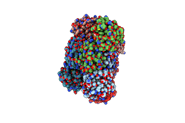
Crystal Structure Of Glyoxylate Reductase From Acetobacter Aceti In The Apo Form
Organism: Acetobacter aceti
Method: X-RAY DIFFRACTION Resolution:1.60 Å Release Date: 2024-10-23 Classification: OXIDOREDUCTASE |
Organism: Acetobacter aceti
Method: X-RAY DIFFRACTION
Release Date: 2024-10-23
 |
Crystal Structure Of Glyoxylate Reductase From Acetobacter Aceti In Complex With Nadh
Organism: Acetobacter aceti
Method: X-RAY DIFFRACTION Resolution:1.60 Å Release Date: 2024-10-23 Classification: OXIDOREDUCTASE Ligands: NAI |
Organism: Acetobacter aceti
Method: X-RAY DIFFRACTION
Release Date: 2024-10-23
Ligands: NAI
 |
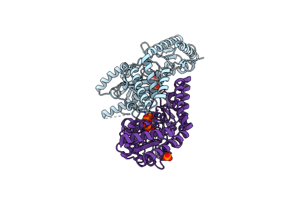
Crystal Structure Of Glyoxylate Reductase From Acetobacter Aceti In Complex With Nadph
Organism: Acetobacter aceti
Method: X-RAY DIFFRACTION Resolution:1.68 Å Release Date: 2024-10-23 Classification: OXIDOREDUCTASE Ligands: NDP |
Organism: Acetobacter aceti
Method: X-RAY DIFFRACTION
Release Date: 2024-10-23
Ligands: NDP
 |
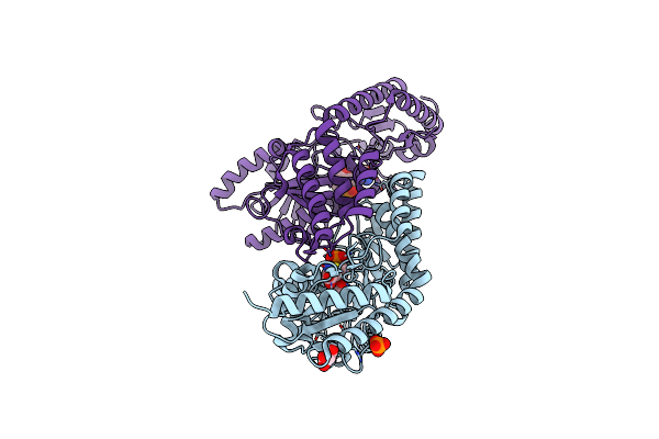
Crystal Structure Of Rat Selenocysteine Lyase
Organism: Rattus norvegicus
Method: X-RAY DIFFRACTION Resolution:2.00 Å Release Date: 2010-03-16 Classification: LYASE Ligands: PLP, PO4 |
Organism: Rattus norvegicus
Method: X-RAY DIFFRACTION
Release Date: 2010-03-16
Ligands: PLP, PO4
 |
Crystal Structure Of Rat Selenocysteine Lyase In Complex With L-Cysteine
Organism: Rattus norvegicus
Method: X-RAY DIFFRACTION Resolution:1.85 Å Release Date: 2010-03-16 Classification: LYASE Ligands: CYS, GOL, PLP, PO4 |
Organism: Rattus norvegicus
Method: X-RAY DIFFRACTION
Release Date: 2010-03-16
Ligands: CYS, GOL, PLP, PO4
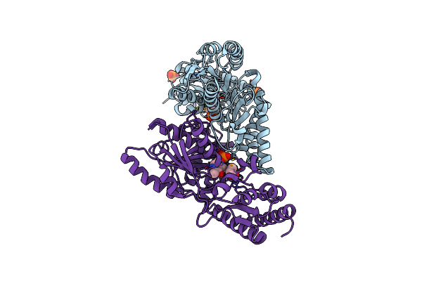 |
Crystal Structure Of Ras Selenocysteine Lyase In Complex With Selenopropionate
Organism: Rattus norvegicus
Method: X-RAY DIFFRACTION Resolution:1.55 Å Release Date: 2010-03-16 Classification: LYASE Ligands: PLP, SLP, PO4 |
Organism: Rattus norvegicus
Method: X-RAY DIFFRACTION
Release Date: 2010-03-16
Ligands: PLP, SLP, PO4
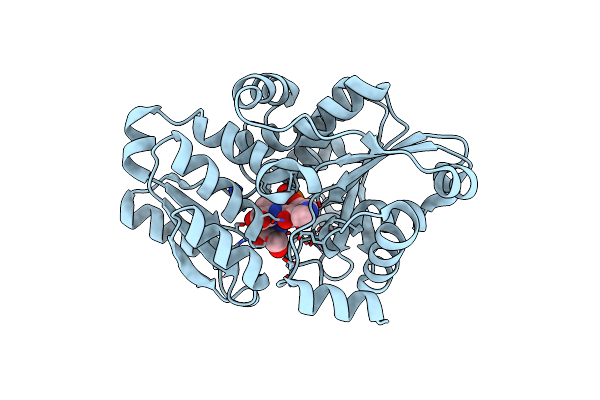 |
Crystal Structure Of Modified Serine Racemase Complexed With Serine
Organism: Schizosaccharomyces pombe
Method: X-RAY DIFFRACTION Resolution:2.20 Å Release Date: 2009-07-28 Classification: ISOMERASE Ligands: PDD, SER, MG |
Organism: Schizosaccharomyces pombe
Method: X-RAY DIFFRACTION
Release Date: 2009-07-28
Ligands: PDD, SER, MG
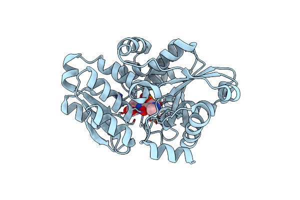 |
Crystal Structure Of Modified Serine Racemase From S.Pombe.
Organism: Schizosaccharomyces pombe
Method: X-RAY DIFFRACTION Resolution:1.70 Å Release Date: 2009-04-28 Classification: ISOMERASE Ligands: MG, PDD |
Organism: Schizosaccharomyces pombe
Method: X-RAY DIFFRACTION
Release Date: 2009-04-28
Ligands: MG, PDD
 |
Crystal Structure Of S.Pombe Serine Racemase Complex With Amppcp
Organism: Schizosaccharomyces pombe
Method: X-RAY DIFFRACTION Resolution:1.90 Å Release Date: 2005-11-01 Classification: ISOMERASE Ligands: MG, PLP, ACP |
Organism: Schizosaccharomyces pombe
Method: X-RAY DIFFRACTION
Release Date: 2005-11-01
Ligands: MG, PLP, ACP
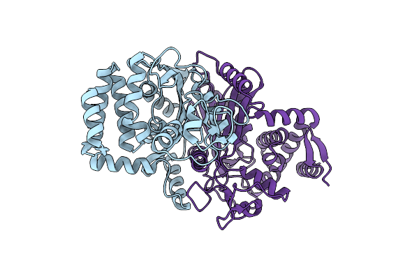 |
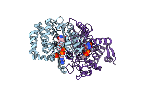
Crystal Structure Of Delta1-Piperideine-2-Carboxylate Reductase From Pseudomonas Syringae Pvar.Tomato
Organism: Pseudomonas syringae pv. tomato
Method: X-RAY DIFFRACTION Resolution:1.55 Å Release Date: 2005-10-04 Classification: OXIDOREDUCTASE |
Organism: Pseudomonas syringae pv. tomato
Method: X-RAY DIFFRACTION
Release Date: 2005-10-04
 |
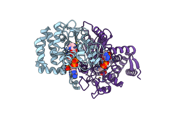
Crystal Structure Of Delta1-Piperideine-2-Carboxylate Reductase From Pseudomonas Syringae Complexed With Nadph
Organism: Pseudomonas syringae pv. tomato
Method: X-RAY DIFFRACTION Resolution:1.80 Å Release Date: 2005-10-04 Classification: OXIDOREDUCTASE Ligands: NDP |
Organism: Pseudomonas syringae pv. tomato
Method: X-RAY DIFFRACTION
Release Date: 2005-10-04
Ligands: NDP
 |
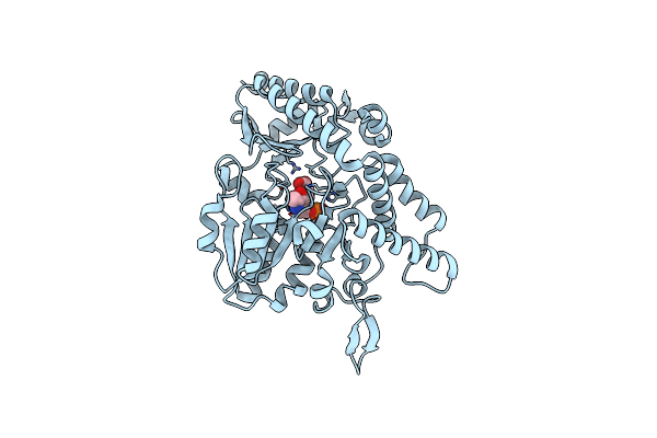
Crystal Structure Of Delta1-Piperideine-2-Carboxylate Reductase From Pseudomonas Syringae Complexed With Nadph And Pyrrole-2-Carboxylate
Organism: Pseudomonas syringae pv. tomato
Method: X-RAY DIFFRACTION Resolution:1.70 Å Release Date: 2005-10-04 Classification: OXIDOREDUCTASE Ligands: NDP, PYC |
Organism: Pseudomonas syringae pv. tomato
Method: X-RAY DIFFRACTION
Release Date: 2005-10-04
Ligands: NDP, PYC
 |
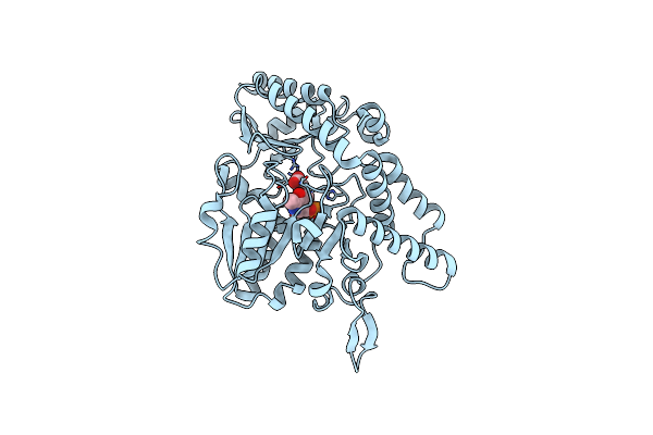
Crystal Structure Of Csdb Complexed With L-Propargylglycine
Organism: Escherichia coli
Method: X-RAY DIFFRACTION Resolution:2.80 Å Release Date: 2003-07-01 Classification: LYASE Ligands: LPG, PLP |
Organism: Escherichia coli
Method: X-RAY DIFFRACTION
Release Date: 2003-07-01
Ligands: LPG, PLP
 |
Csdb Protein, Nifs Homologue
Organism: Escherichia coli
Method: X-RAY DIFFRACTION Resolution:2.80 Å Release Date: 2000-07-17 Classification: LYASE Ligands: PLP, ACY |
Organism: Escherichia coli
Method: X-RAY DIFFRACTION
Release Date: 2000-07-17
Ligands: PLP, ACY