
Search Count: 37
All
Selected
 |
Organism: Streptococcus pneumoniae
Method: X-RAY DIFFRACTION Resolution:1.85 Å Release Date: 2026-02-11 Classification: PEPTIDE BINDING PROTEIN Ligands: GOL |
 |
Organism: Arabidopsis thaliana
Method: X-RAY DIFFRACTION Resolution:1.74 Å Release Date: 2025-05-14 Classification: LIGASE Ligands: ZN |
 |
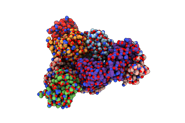
Y-Degron Fused Zz-Domain Of The Arabidopsis Thaliana E3 Ubiquitin-Protein Ligase Prt1
Organism: Arabidopsis thaliana
Method: X-RAY DIFFRACTION Resolution:1.67 Å Release Date: 2025-05-14 Classification: LIGASE Ligands: ZN, MG |
 |
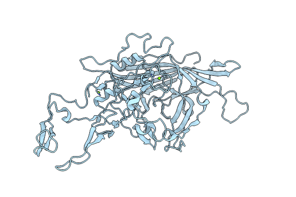
F-Degron Fused Zz-Domain Of The Arabidopsis Thaliana E3 Ubiquitin-Protein Ligase Prt1
Organism: Arabidopsis thaliana
Method: X-RAY DIFFRACTION Resolution:2.10 Å Release Date: 2025-05-14 Classification: LIGASE Ligands: ZN, MG |
 |
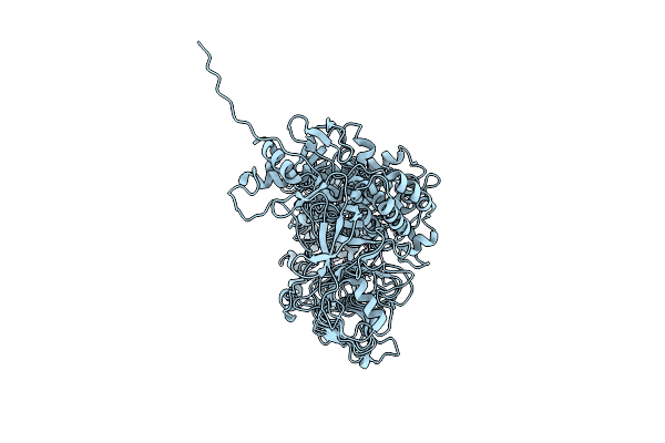
W-Degron Fused Zz-Domain Of The Arabidopsis Thaliana E3 Ubiquitin-Protein Ligase Prt1
Organism: Arabidopsis thaliana
Method: X-RAY DIFFRACTION Resolution:2.79 Å Release Date: 2025-05-14 Classification: LIGASE Ligands: ZN, SO4 |
 |
Organism: Homo sapiens, Hepatitis e virus
Method: X-RAY DIFFRACTION Resolution:1.98 Å Release Date: 2024-07-10 Classification: VIRAL PROTEIN Ligands: ACT, SO4 |
 |
Organism: Hepatitis e virus rat/r63/deu/2009, Homo sapiens
Method: X-RAY DIFFRACTION Resolution:3.92 Å Release Date: 2024-07-10 Classification: VIRAL PROTEIN |
 |
Organism: Homo sapiens, Paslahepevirus balayani
Method: X-RAY DIFFRACTION Resolution:2.41 Å Release Date: 2024-07-10 Classification: VIRAL PROTEIN Ligands: ZN |
 |
Organism: Paslahepevirus balayani, Homo sapiens
Method: X-RAY DIFFRACTION Resolution:1.91 Å Release Date: 2024-07-10 Classification: VIRAL PROTEIN Ligands: B3P |
 |
Organism: Homo sapiens, Paslahepevirus balayani
Method: X-RAY DIFFRACTION Resolution:2.07 Å Release Date: 2024-07-10 Classification: VIRAL PROTEIN |
 |
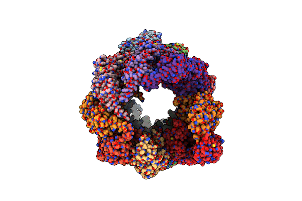
Organism: Adeno-associated virus - 4
Method: ELECTRON MICROSCOPY Release Date: 2023-01-25 Classification: VIRUS LIKE PARTICLE Ligands: MG |
 |
Organism: Gemella haemolysans
Method: ELECTRON MICROSCOPY Release Date: 2022-11-23 Classification: IMMUNE SYSTEM |
 |
Organism: Gemella haemolysans, Homo sapiens
Method: ELECTRON MICROSCOPY Release Date: 2022-11-23 Classification: IMMUNE SYSTEM |
 |
Organism: Vibrio cholerae
Method: SOLUTION NMR Release Date: 2021-10-20 Classification: TRANSCRIPTION |
 |
Organism: Homo sapiens
Method: ELECTRON MICROSCOPY Release Date: 2019-07-03 Classification: CHAPERONE Ligands: ADP |
 |
Organism: Homo sapiens, Adeno-associated virus - 2
Method: ELECTRON MICROSCOPY Release Date: 2019-06-12 Classification: VIRUS Ligands: MG |
 |
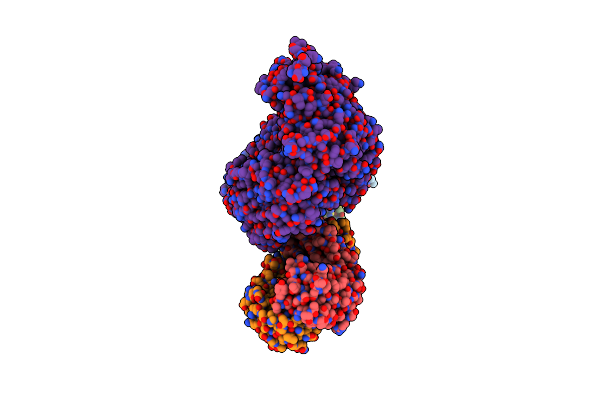
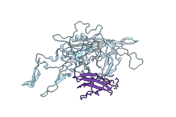
Helicobacter Pylori Adhesin Hopq Type I Bound To The N-Terminal Domain Of Human Ceacam1
Organism: Homo sapiens, Helicobacter pylori (strain g27)
Method: X-RAY DIFFRACTION Resolution:2.80 Å Release Date: 2018-06-27 Classification: CELL ADHESION Ligands: BR |
 |
Helicobacter Pylori Adhesin Hopq Type Ii Bound To The N-Terminal Domain Of Human Ceacam1
Organism: Homo sapiens, Helicobacter pylori
Method: X-RAY DIFFRACTION Resolution:2.59 Å Release Date: 2018-06-27 Classification: CELL ADHESION Ligands: SO4 |
 |
 |
The 2.8 A Electron Microscopy Structure Of Adeno-Associated Virus-Dj Bound By A Heparanoid Pentasaccharide
Organism: Adeno-associated virus
Method: ELECTRON MICROSCOPY Release Date: 2017-05-24 Classification: VIRUS LIKE PARTICLE |