

Search Count: 33
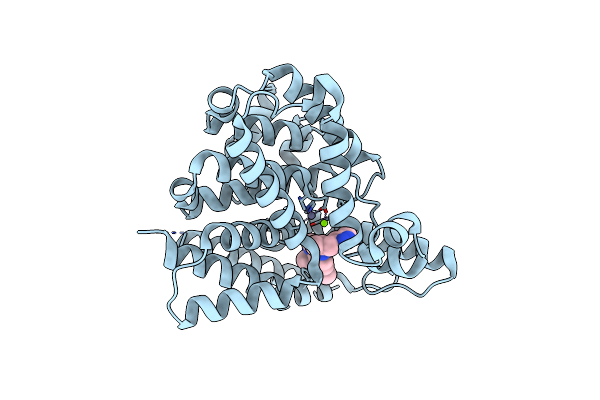 |
Pde1B Complexed With Compound 3S
Organism: Homo sapiens
Method: X-RAY DIFFRACTION Resolution:1.90 Å Release Date: 2018-05-30 Classification: Hydrolase/Hydrolase Inhibitor Ligands: ZN, MG, 0NY |
Organism: Homo sapiens
Method: X-RAY DIFFRACTION
Release Date: 2018-05-30
Ligands: ZN, MG, 0NY
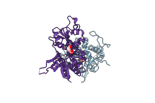 |
Glutamate- And Glycine-Bound Glun1/Glun2A Agonist Binding Domains
Organism: Rattus norvegicus
Method: X-RAY DIFFRACTION Resolution:1.70 Å Release Date: 2016-09-21 Classification: TRANSPORT PROTEIN, RECEPTOR Ligands: GLY, GLU |
Organism: Rattus norvegicus
Method: X-RAY DIFFRACTION
Release Date: 2016-09-21
Ligands: GLY, GLU
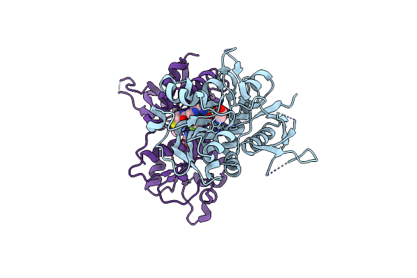 |
Glutamate- And Glycine-Bound Glun1/Glun2A Agonist Binding Domains With Mpx-004
Organism: Rattus norvegicus
Method: X-RAY DIFFRACTION Resolution:2.52 Å Release Date: 2016-09-21 Classification: TRANSPORT PROTEIN, RECEPTOR Ligands: GLY, GLU, 67R |
Organism: Rattus norvegicus
Method: X-RAY DIFFRACTION
Release Date: 2016-09-21
Ligands: GLY, GLU, 67R
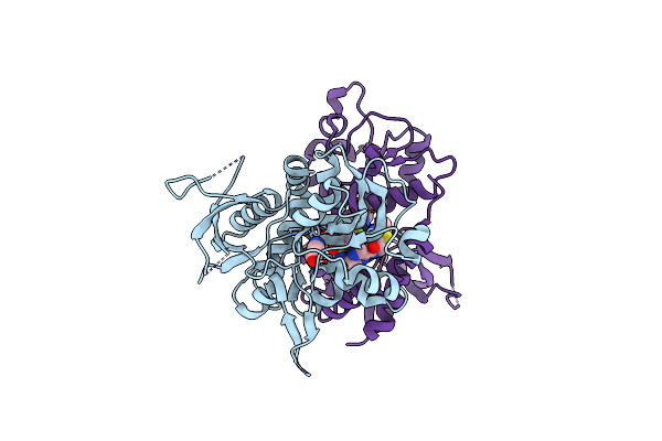 |
Glutamate- And Glycine-Bound Glun1/Glun2A Agonist Binding Domains With Mpx 007
Organism: Rattus norvegicus
Method: X-RAY DIFFRACTION Resolution:2.25 Å Release Date: 2016-09-21 Classification: TRANSPORT PROTEIN, RECEPTOR Ligands: GLY, GLU, 67Q |
Organism: Rattus norvegicus
Method: X-RAY DIFFRACTION
Release Date: 2016-09-21
Ligands: GLY, GLU, 67Q
 |
Glutamate- And Dcka-Bound Glun1/Glun2A Agonist Binding Domains With Mpx-007
Organism: Rattus norvegicus
Method: X-RAY DIFFRACTION Resolution:2.72 Å Release Date: 2016-09-21 Classification: TRANSPORT PROTEIN, RECEPTOR Ligands: 2JK, 6ND, GLU |
Organism: Rattus norvegicus
Method: X-RAY DIFFRACTION
Release Date: 2016-09-21
Ligands: 2JK, 6ND, GLU
 |
Agonist-Bound Glun1/Glun2A Agonist Binding Domains With Tcn201
Organism: Rattus norvegicus
Method: X-RAY DIFFRACTION Resolution:2.28 Å Release Date: 2016-09-07 Classification: TRANSPORT PROTEIN, RECEPTOR Ligands: GLY, GLU, 67P |
Organism: Rattus norvegicus
Method: X-RAY DIFFRACTION
Release Date: 2016-09-07
Ligands: GLY, GLU, 67P
 |
Crystal Structure Of Human Pde1B Bound To Inhibitor 7A (6,7,8-Trimethoxy-N-(Pentan-3-Yl)Quinazolin-4-Amine)
Organism: Homo sapiens
Method: X-RAY DIFFRACTION Resolution:2.40 Å Release Date: 2014-07-16 Classification: HYDROLASE/HYDROLASE INHIBITOR Ligands: 0NK, MG, ZN |
Organism: Homo sapiens
Method: X-RAY DIFFRACTION
Release Date: 2014-07-16
Ligands: 0NK, MG, ZN
 |
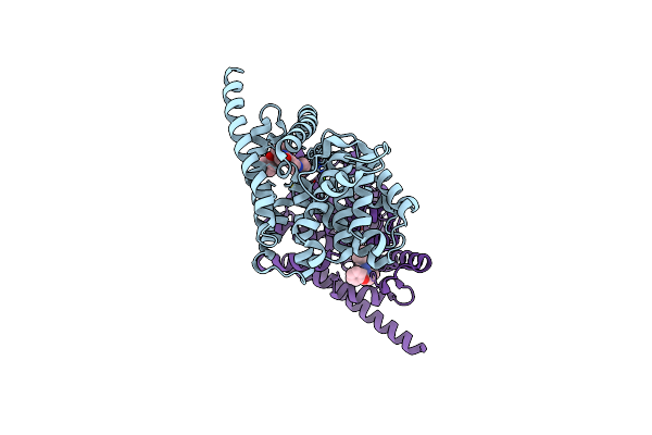
Crystal Structure Of Human Pde1B Bound To Inhibitor 19A (7,8-Dimethoxy-N-[(2S)-1-(3-Methyl-1H-Pyrazol-5-Yl)Propan-2-Yl]Quinazolin-4-Amine)
Organism: Homo sapiens
Method: X-RAY DIFFRACTION Resolution:1.90 Å Release Date: 2014-07-16 Classification: HYDROLASE/HYDROLASE INHIBITOR Ligands: ZN, MG, 0NY |
Organism: Homo sapiens
Method: X-RAY DIFFRACTION
Release Date: 2014-07-16
Ligands: ZN, MG, 0NY
 |
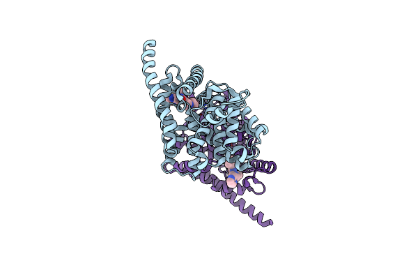
Human Pde9 In Complex With Selective Compound
Organism: Homo sapiens
Method: X-RAY DIFFRACTION Resolution:2.40 Å Release Date: 2013-05-29 Classification: HYDROLASE/HYDROLASE INHIBITOR Ligands: ZN, MG, 0WF |
Organism: Homo sapiens
Method: X-RAY DIFFRACTION
Release Date: 2013-05-29
Ligands: ZN, MG, 0WF
 |
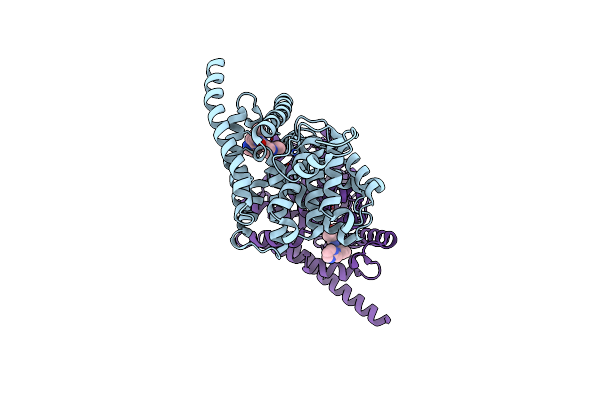
Human Pde9 In Complex With Selective Compound
Organism: Homo sapiens
Method: X-RAY DIFFRACTION Resolution:3.00 Å Release Date: 2013-05-29 Classification: HYDROLASE/HYDROLASE INHIBITOR Ligands: ZN, MG, 0WL |
Organism: Homo sapiens
Method: X-RAY DIFFRACTION
Release Date: 2013-05-29
Ligands: ZN, MG, 0WL
 |
Human Phosphodiesterase 9 In Complex With Inhibitors
Organism: Homo sapiens
Method: X-RAY DIFFRACTION Resolution:2.50 Å Release Date: 2013-02-27 Classification: Hydrolase/Hydrolase Inhibitor Ligands: ZN, MG, 7RG |
Organism: Homo sapiens
Method: X-RAY DIFFRACTION
Release Date: 2013-02-27
Ligands: ZN, MG, 7RG
 |
Structure Of Pde10-Inhibitor Complex
Organism: Rattus norvegicus
Method: X-RAY DIFFRACTION Resolution:2.00 Å Release Date: 2011-06-15 Classification: HYDROLASE/HYDROLASE INHIBITOR Ligands: SO4, ZN, MG, PFK |
Organism: Rattus norvegicus
Method: X-RAY DIFFRACTION
Release Date: 2011-06-15
Ligands: SO4, ZN, MG, PFK
 |
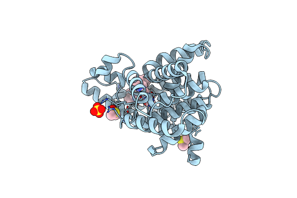
Structure Of Pde10-Inhibitor Complex
Organism: Rattus norvegicus
Method: X-RAY DIFFRACTION Resolution:1.80 Å Release Date: 2011-06-15 Classification: HYDROLASE/HYDROLASE INHIBITOR Ligands: SO4, ZN, MG, PFR |
Organism: Rattus norvegicus
Method: X-RAY DIFFRACTION
Release Date: 2011-06-15
Ligands: SO4, ZN, MG, PFR
 |
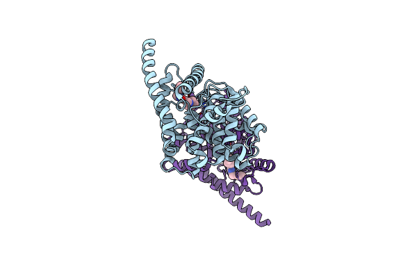
Structure Of Pde10-Inhibitor Complex
Organism: Rattus norvegicus
Method: X-RAY DIFFRACTION Resolution:1.80 Å Release Date: 2011-06-15 Classification: HYDROLASE/HYDROLASE INHIBITOR Ligands: ZN, MG, SO4, DMS, PFW |
Organism: Rattus norvegicus
Method: X-RAY DIFFRACTION
Release Date: 2011-06-15
Ligands: ZN, MG, SO4, DMS, PFW
 |
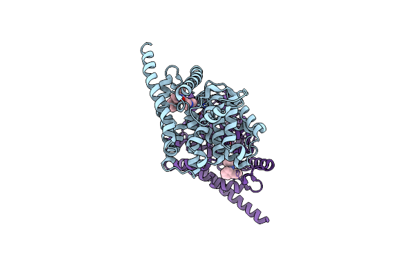
Human Phosphodiesterase 9 In Complex With Inhibitor
Organism: Homo sapiens
Method: X-RAY DIFFRACTION Resolution:2.72 Å Release Date: 2009-12-01 Classification: HYDROLASE Ligands: ZN, MG, WTC |
Organism: Homo sapiens
Method: X-RAY DIFFRACTION
Release Date: 2009-12-01
Ligands: ZN, MG, WTC
 |
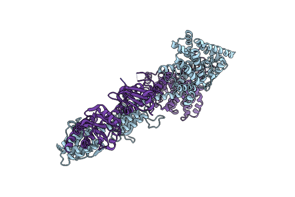
Human Pde9 In Complex With Selective Inhibitor
Organism: Homo sapiens
Method: X-RAY DIFFRACTION Resolution:2.30 Å Release Date: 2009-12-01 Classification: HYDROLASE Ligands: ZN, MG, JAR |
Organism: Homo sapiens
Method: X-RAY DIFFRACTION
Release Date: 2009-12-01
Ligands: ZN, MG, JAR
 |
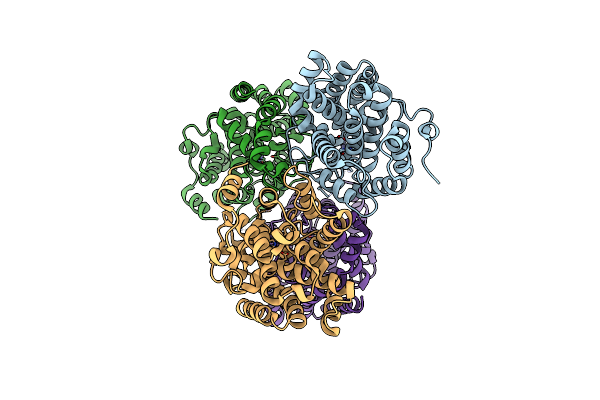
X-Ray Structure Of Pde2A
Organism: Homo sapiens
Method: X-RAY DIFFRACTION Resolution:3.02 Å Release Date: 2009-10-27 Classification: HYDROLASE Ligands: ZN, MG |
Organism: Homo sapiens
Method: X-RAY DIFFRACTION
Release Date: 2009-10-27
Ligands: ZN, MG
 |
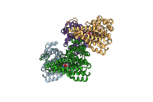
Catalytic Domain Of Hpde2A
Organism: Homo sapiens
Method: X-RAY DIFFRACTION Resolution:2.49 Å Release Date: 2009-10-27 Classification: HYDROLASE Ligands: ZN |
Organism: Homo sapiens
Method: X-RAY DIFFRACTION
Release Date: 2009-10-27
Ligands: ZN
 |
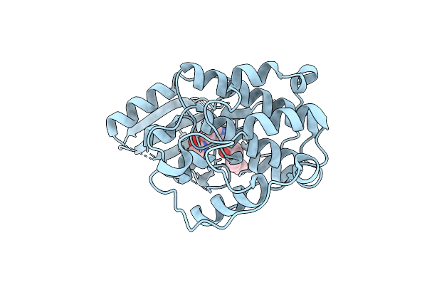
Hpde2A Catalytic Domain Complexed With Ibmx
Organism: Homo sapiens
Method: X-RAY DIFFRACTION Resolution:1.58 Å Release Date: 2009-10-27 Classification: HYDROLASE Ligands: ZN, MG, IBM |
Organism: Homo sapiens
Method: X-RAY DIFFRACTION
Release Date: 2009-10-27
Ligands: ZN, MG, IBM
 |
Novel Cdk-5 Inhibitors - Crystal Structure Of Inhibitor Efp With Cdk-2
Organism: Homo sapiens
Method: X-RAY DIFFRACTION Resolution:1.80 Å Release Date: 2009-09-08 Classification: TRANSFERASE Ligands: EFP |
Organism: Homo sapiens
Method: X-RAY DIFFRACTION
Release Date: 2009-09-08
Ligands: EFP