

Search Count: 54
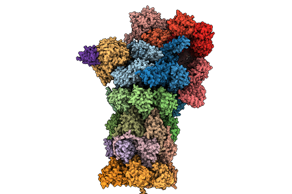 |
Co-Chaperone Bag1-Bound Human 26S Proteasome In Sbag1 State
Organism: Homo sapiens
Method: ELECTRON MICROSCOPY Release Date: 2025-12-31 Classification: HYDROLASE Ligands: ATP, ADP |
Organism: Homo sapiens
Method: ELECTRON MICROSCOPY
Release Date: 2025-12-31
Ligands: ATP, ADP
 |
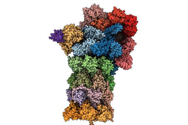
Co-Chaperone Bag1-Bound Human 26S Proteasome In Sbag1.2 State
Organism: Homo sapiens
Method: ELECTRON MICROSCOPY Release Date: 2025-12-31 Classification: HYDROLASE Ligands: ATP, ADP |
Organism: Homo sapiens
Method: ELECTRON MICROSCOPY
Release Date: 2025-12-31
Ligands: ATP, ADP
 |
Co-Chaperone Bag1-Bound Human 26S Proteasome In Sbag1.3 State
Organism: Homo sapiens
Method: ELECTRON MICROSCOPY Release Date: 2025-12-31 Classification: HYDROLASE Ligands: ATP, ADP |
Organism: Homo sapiens
Method: ELECTRON MICROSCOPY
Release Date: 2025-12-31
Ligands: ATP, ADP
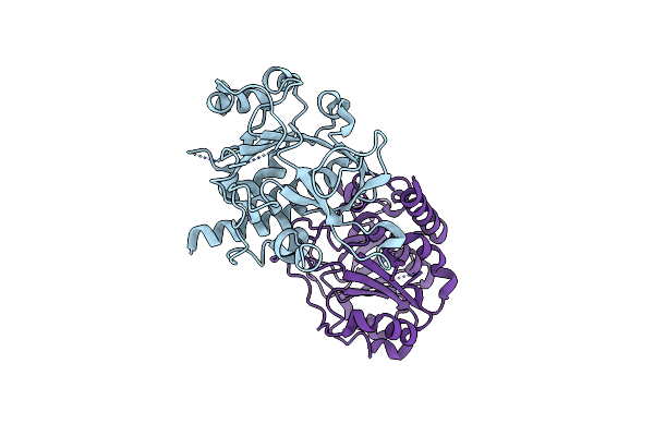 |
Crystal Structure Of Eccc5 Duf Domain Of Mycobacterium Tuberculosis
Organism: Mycobacterium tuberculosis h37rv
Method: X-RAY DIFFRACTION Resolution:2.05 Å Release Date: 2024-11-27 Classification: UNKNOWN FUNCTION |
Organism: Mycobacterium tuberculosis h37rv
Method: X-RAY DIFFRACTION
Release Date: 2024-11-27
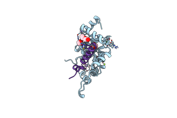 |
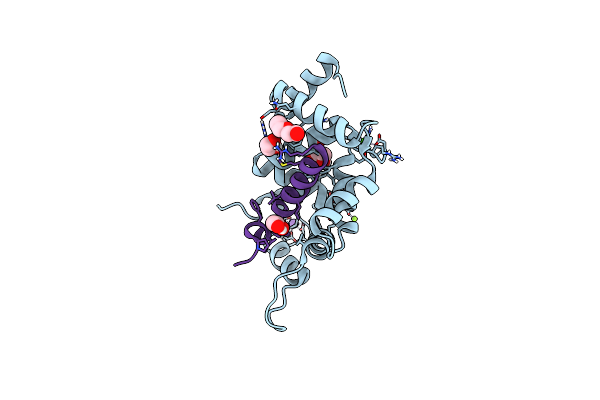
X-Ray Structure Of Human Ncs-1 Bound To Ric-8A
Organism: Homo sapiens, Rattus norvegicus
Method: X-RAY DIFFRACTION Resolution:1.70 Å Release Date: 2023-12-13 Classification: STRUCTURAL PROTEIN Ligands: CA, NA, MG, 1PE, GOL, CL |
Organism: Homo sapiens, Rattus norvegicus
Method: X-RAY DIFFRACTION
Release Date: 2023-12-13
Ligands: CA, NA, MG, 1PE, GOL, CL
 |
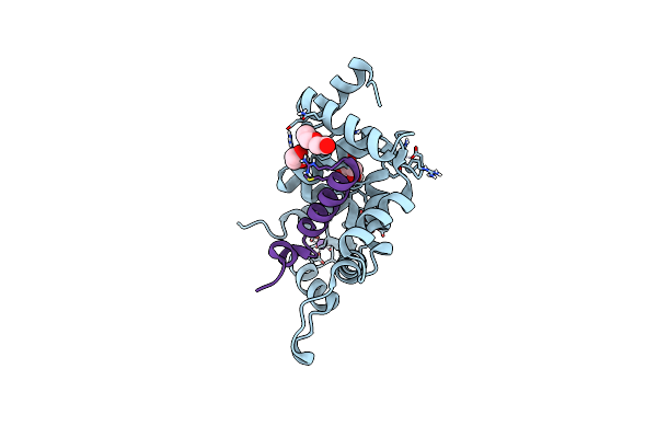
X-Ray Structure Of Human Ncs-1 Bound To Ric-8A
Organism: Homo sapiens, Rattus norvegicus
Method: X-RAY DIFFRACTION Resolution:1.86 Å Release Date: 2023-12-13 Classification: STRUCTURAL PROTEIN Ligands: CA, NA, MG, 1PE, GOL, CL |
Organism: Homo sapiens, Rattus norvegicus
Method: X-RAY DIFFRACTION
Release Date: 2023-12-13
Ligands: CA, NA, MG, 1PE, GOL, CL
 |
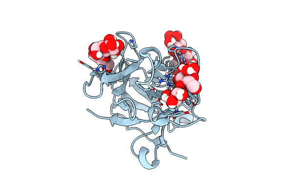
X-Ray Structure Of Human Ncs-1 Bound To Ric-8A
Organism: Homo sapiens, Rattus norvegicus
Method: X-RAY DIFFRACTION Resolution:1.85 Å Release Date: 2023-12-13 Classification: STRUCTURAL PROTEIN Ligands: CA, NA, 1PE, GOL, CL |
Organism: Homo sapiens, Rattus norvegicus
Method: X-RAY DIFFRACTION
Release Date: 2023-12-13
Ligands: CA, NA, 1PE, GOL, CL
 |
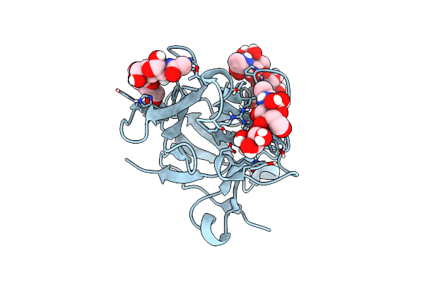
Atomic Structure Of The Beta-Trefoil Domain Of The Laccaria Bicolor Lectin Lbl In Complex With Lactose
Organism: Laccaria bicolor s238n-h53
Method: X-RAY DIFFRACTION Resolution:1.00 Å Release Date: 2023-02-22 Classification: SUGAR BINDING PROTEIN |
Organism: Laccaria bicolor s238n-h53
Method: X-RAY DIFFRACTION
Release Date: 2023-02-22
 |
N-Terminal Beta-Trefoil Lectin Domain Of The Laccaria Bicolor Lectin In Complex With N-Acetyl-Lactosamine
Organism: Laccaria bicolor s238n-h53
Method: X-RAY DIFFRACTION Resolution:0.97 Å Release Date: 2023-02-22 Classification: SUGAR BINDING PROTEIN |
Organism: Laccaria bicolor s238n-h53
Method: X-RAY DIFFRACTION
Release Date: 2023-02-22
 |
Cell Wall Binding Domain Of Endolysin From Listeria Phage A500.
Organism: Listeria phage a500
Method: X-RAY DIFFRACTION Resolution:1.59 Å Release Date: 2019-10-30 Classification: VIRAL PROTEIN |
Organism: Listeria phage a500
Method: X-RAY DIFFRACTION
Release Date: 2019-10-30
 |
Structure Of The Distal Domain Of Mouse Adenovirus 2 Fibre, Methylmercury Chloride Derivative
Organism: Murine adenovirus 2
Method: X-RAY DIFFRACTION Resolution:2.76 Å Release Date: 2018-03-14 Classification: STRUCTURAL PROTEIN Ligands: HG |
Organism: Murine adenovirus 2
Method: X-RAY DIFFRACTION
Release Date: 2018-03-14
Ligands: HG
 |
Structure Of The Distal Domain Of Mouse Adenovirus 2 Fibre, P21 Native
Organism: Murine adenovirus 2
Method: X-RAY DIFFRACTION Resolution:1.80 Å Release Date: 2018-03-14 Classification: VIRAL PROTEIN Ligands: GOL, CL |
Organism: Murine adenovirus 2
Method: X-RAY DIFFRACTION
Release Date: 2018-03-14
Ligands: GOL, CL
 |
Structure Of The Distal Domain Of Mouse Adenovirus 2 Fibre, P212121 Native
Organism: Murine adenovirus 2
Method: X-RAY DIFFRACTION Resolution:1.70 Å Release Date: 2018-03-14 Classification: VIRAL PROTEIN Ligands: GOL |
Organism: Murine adenovirus 2
Method: X-RAY DIFFRACTION
Release Date: 2018-03-14
Ligands: GOL
 |
Structure Of The Distal Domain Of Mouse Adenovirus 2 Fibre Bound To N-Acetyl-Glucosamine
Organism: Murine adenovirus 2
Method: X-RAY DIFFRACTION Resolution:2.00 Å Release Date: 2018-03-14 Classification: VIRAL PROTEIN Ligands: NAG, SO4, GOL |
Organism: Murine adenovirus 2
Method: X-RAY DIFFRACTION
Release Date: 2018-03-14
Ligands: NAG, SO4, GOL
 |
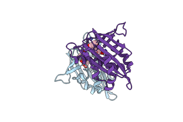
Aba Receptor From Citrus, Cspyl1
Organism: Citrus sinensis
Method: X-RAY DIFFRACTION Resolution:1.80 Å Release Date: 2017-08-02 Classification: SIGNALING PROTEIN |
Organism: Citrus sinensis
Method: X-RAY DIFFRACTION
Release Date: 2017-08-02
 |
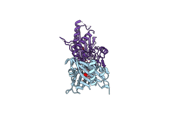
Aba Receptor From Citrus, Cspyl1
Organism: Citrus sinensis
Method: X-RAY DIFFRACTION Resolution:2.88 Å Release Date: 2017-08-02 Classification: SIGNALING PROTEIN Ligands: A8S |
Organism: Citrus sinensis
Method: X-RAY DIFFRACTION
Release Date: 2017-08-02
Ligands: A8S
 |
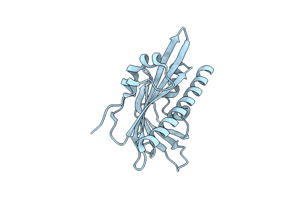
Aba Receptor From Citrus, Cspyl1
Organism: Citrus sinensis, Arabidopsis thaliana
Method: X-RAY DIFFRACTION Resolution:2.00 Å Release Date: 2017-08-02 Classification: SIGNALING PROTEIN Ligands: A8S, GOL, MN, CL |
Organism: Citrus sinensis, Arabidopsis thaliana
Method: X-RAY DIFFRACTION
Release Date: 2017-08-02
Ligands: A8S, GOL, MN, CL
 |
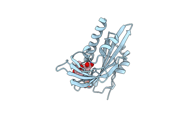
Aba Receptor From Tomato, Slpyl1
Organism: Solanum lycopersicum
Method: X-RAY DIFFRACTION Resolution:1.65 Å Release Date: 2017-08-02 Classification: SIGNALING PROTEIN |
Organism: Solanum lycopersicum
Method: X-RAY DIFFRACTION
Release Date: 2017-08-02
 |
Aba Receptor From Tomato, Slpyl1
Organism: Solanum lycopersicum
Method: X-RAY DIFFRACTION Resolution:1.67 Å Release Date: 2017-08-02 Classification: SIGNALING PROTEIN Ligands: A8S, SO4 |
Organism: Solanum lycopersicum
Method: X-RAY DIFFRACTION
Release Date: 2017-08-02
Ligands: A8S, SO4
 |
Crystal Structure Of The Full-Length Cell Wall-Binding Module Of Cpl7 Mutant R223A
Organism: Streptococcus phage cp-7
Method: X-RAY DIFFRACTION Resolution:2.80 Å Release Date: 2017-03-08 Classification: HYDROLASE Ligands: GOL |
Organism: Streptococcus phage cp-7
Method: X-RAY DIFFRACTION
Release Date: 2017-03-08
Ligands: GOL