

Search Count: 16
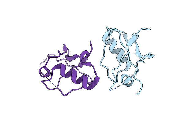 |
Structure Of The Ubl Domain Of Sacsin
Organism: Homo sapiens
Method: X-RAY DIFFRACTION Resolution:2.10 Å Release Date: 2017-05-31 Classification: CHAPERONE |
Organism: Homo sapiens
Method: X-RAY DIFFRACTION
Release Date: 2017-05-31
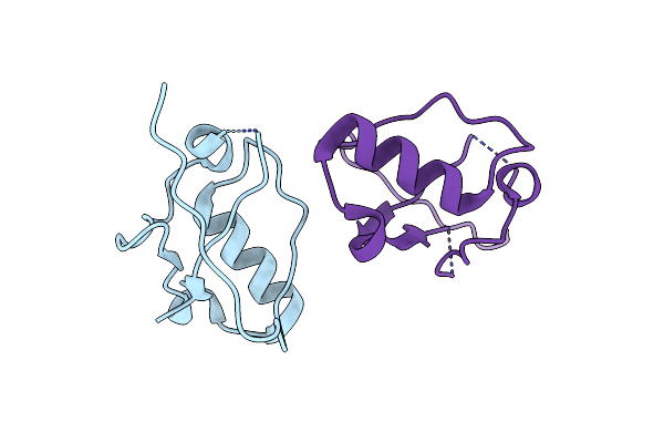 |
Structure Of The Ubl Domain Of Sacsin Mutant L78M
Organism: Homo sapiens
Method: X-RAY DIFFRACTION Resolution:2.40 Å Release Date: 2017-05-31 Classification: CHAPERONE |
Organism: Homo sapiens
Method: X-RAY DIFFRACTION
Release Date: 2017-05-31
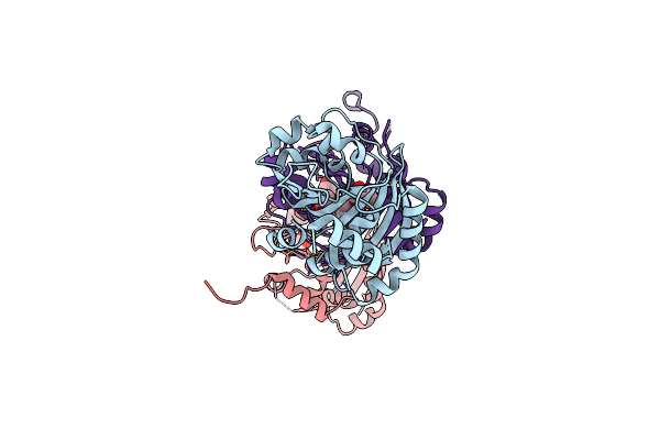 |
Crystal Structure Of The Sr1 Domain Of Human Sacsin
Organism: Homo sapiens
Method: X-RAY DIFFRACTION Resolution:1.56 Å Release Date: 2017-05-24 Classification: CHAPERONE Ligands: GOL |
Organism: Homo sapiens
Method: X-RAY DIFFRACTION
Release Date: 2017-05-24
Ligands: GOL
 |
Crystal Structure Of The F270M, K291M, L318M Mutant Of Sr1 Domain Of Human Sacsin
Organism: Homo sapiens
Method: X-RAY DIFFRACTION Resolution:1.91 Å Release Date: 2017-05-24 Classification: CHAPERONE |
Organism: Homo sapiens
Method: X-RAY DIFFRACTION
Release Date: 2017-05-24
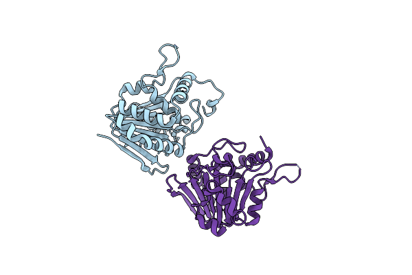 |
Crystal Structure Of The I113M, F270M, K291M, L308M Mutant Of Sr1 Domain Of Human Sacsin
Organism: Homo sapiens
Method: X-RAY DIFFRACTION Resolution:1.80 Å Release Date: 2017-05-24 Classification: CHAPERONE |
Organism: Homo sapiens
Method: X-RAY DIFFRACTION
Release Date: 2017-05-24
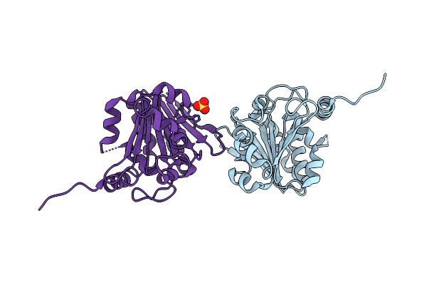 |
Crystal Structure Of The Sr1 Domain Of Lizard Sacsin
Organism: Anolis carolinensis
Method: X-RAY DIFFRACTION Resolution:1.84 Å Release Date: 2017-05-24 Classification: CHAPERONE Ligands: SO4 |
Organism: Anolis carolinensis
Method: X-RAY DIFFRACTION
Release Date: 2017-05-24
Ligands: SO4
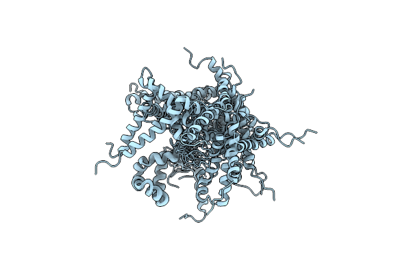 |
Solution Structure Of The Yeast Ddi1 Hdd Domain
Organism: Saccharomyces cerevisiae
Method: SOLUTION NMR Release Date: 2016-10-05 Classification: UNKNOWN FUNCTION |
Organism: Saccharomyces cerevisiae
Method: SOLUTION NMR
Release Date: 2016-10-05
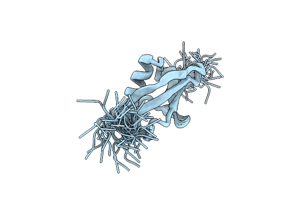 |
Solution Structure Of The Ubl Domain Of Yeast Ddi1
Organism: Saccharomyces cerevisiae
Method: SOLUTION NMR Release Date: 2016-07-20 Classification: UNKNOWN FUNCTION |
Organism: Saccharomyces cerevisiae
Method: SOLUTION NMR
Release Date: 2016-07-20
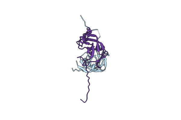 |
New Crystal Structure Of Yeast Ddi1 Aspartyl Protease Reveals Substrate Engagement Mode
Organism: Saccharomyces cerevisiae
Method: X-RAY DIFFRACTION Resolution:1.80 Å Release Date: 2016-04-13 Classification: HYDROLASE |
Organism: Saccharomyces cerevisiae
Method: X-RAY DIFFRACTION
Release Date: 2016-04-13
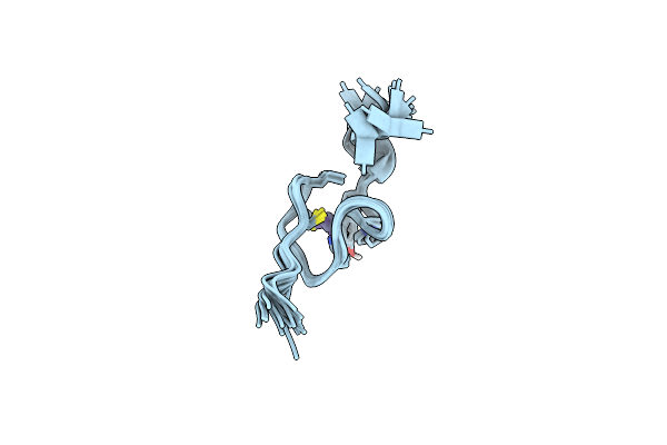 |
Zn-Binding Domain Of Eukaryotic Translation Initiation Factor 3, Subunit G
Organism: Homo sapiens
Method: SOLUTION NMR Release Date: 2015-01-07 Classification: METAL BINDING PROTEIN Ligands: ZN |
Organism: Homo sapiens
Method: SOLUTION NMR
Release Date: 2015-01-07
Ligands: ZN
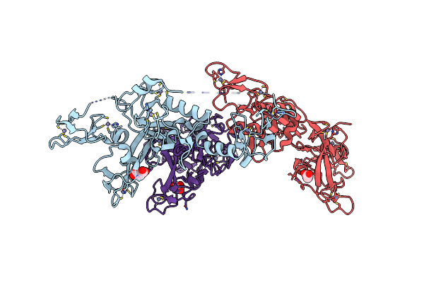 |
Crystal Structure Of Parkin C-Terminal Ring Domains
Organism: Rattus norvegicus
Method: X-RAY DIFFRACTION Resolution:2.80 Å Release Date: 2013-05-15 Classification: LIGASE Ligands: ZN, MLI, CL |
Organism: Rattus norvegicus
Method: X-RAY DIFFRACTION
Release Date: 2013-05-15
Ligands: ZN, MLI, CL
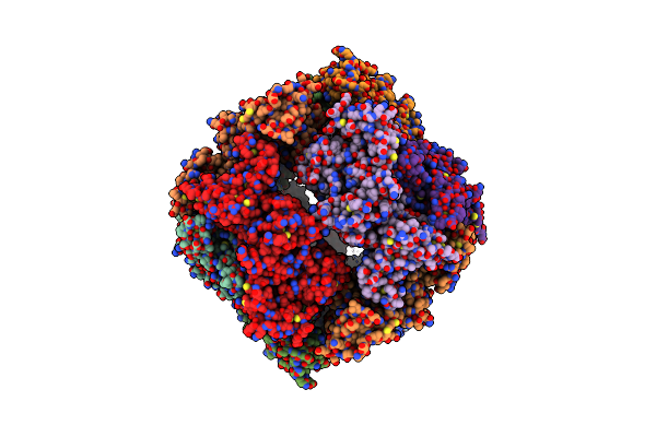 |
Crystal Structure Of Parkin
Organism: Rattus norvegicus
Method: X-RAY DIFFRACTION Resolution:6.50 Å Release Date: 2013-05-15 Classification: LIGASE Ligands: ZN |
Organism: Rattus norvegicus
Method: X-RAY DIFFRACTION
Release Date: 2013-05-15
Ligands: ZN
 |
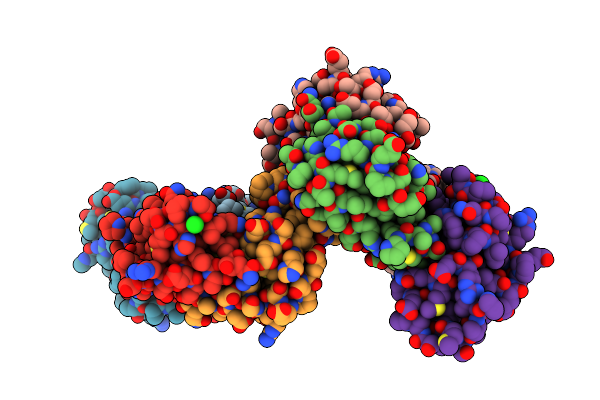
Crystal Structure Of The C-Terminal Lobe Of The Human Ubr5 Hect Domain
Organism: Homo sapiens
Method: X-RAY DIFFRACTION Resolution:1.97 Å Release Date: 2012-01-25 Classification: LIGASE |
Organism: Homo sapiens
Method: X-RAY DIFFRACTION
Release Date: 2012-01-25
 |
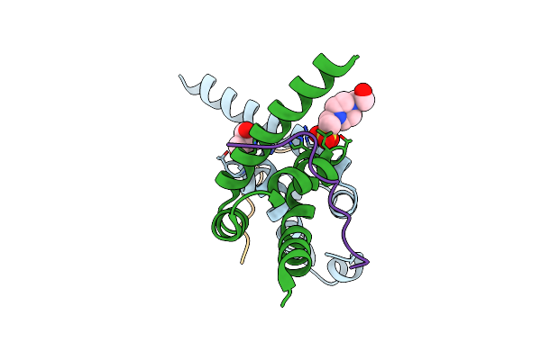
Crystal Structure Of The Mlle Domain Of Poly(A)-Binding Protein
Organism: Homo sapiens
Method: X-RAY DIFFRACTION Resolution:2.50 Å Release Date: 2010-02-09 Classification: RNA BINDING PROTEIN Ligands: CL |
Organism: Homo sapiens
Method: X-RAY DIFFRACTION
Release Date: 2010-02-09
Ligands: CL
 |
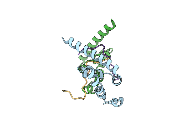
Crystal Structure Of The Mlle Domain Of Poly(A)-Binding Protein In Complex With The Binding Region Of Paip2
Organism: Homo sapiens
Method: X-RAY DIFFRACTION Resolution:1.40 Å Release Date: 2010-02-09 Classification: PROTEIN BINDING Ligands: GOL, EPE |
Organism: Homo sapiens
Method: X-RAY DIFFRACTION
Release Date: 2010-02-09
Ligands: GOL, EPE
 |
Crystal Structure Of The Mlle Domain Of Poly(A)-Binding Protein In Complex With The Binding Region Of Paip2
Organism: Homo sapiens
Method: X-RAY DIFFRACTION Resolution:1.50 Å Release Date: 2010-02-09 Classification: PROTEIN BINDING Ligands: CL |
Organism: Homo sapiens
Method: X-RAY DIFFRACTION
Release Date: 2010-02-09
Ligands: CL