

Search Count: 39
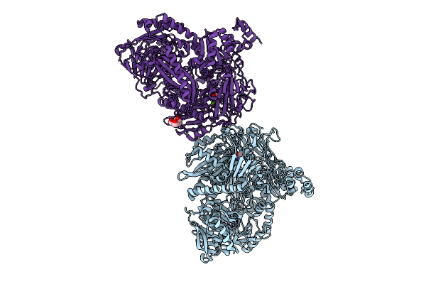 |
Crystal Structure Of N-Methylhydantoinase In Complex With 1-Methylimidazolidine-2,4-Dione
Organism: Glutamicibacter protophormiae
Method: X-RAY DIFFRACTION Resolution:2.07 Å Release Date: 2026-04-15 Classification: HYDROLASE Ligands: CA, A1BC1, NH4, BTB |
Organism: Glutamicibacter protophormiae
Method: X-RAY DIFFRACTION
Release Date: 2026-04-15
Ligands: CA, A1BC1, NH4, BTB
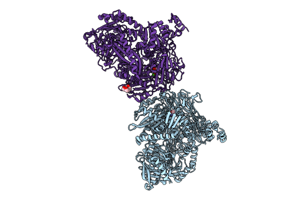 |
Crystal Structure Of N-Methylhydantoinase In Complex With 1-Methylimidazolidine-2,4-Dione, Iodide Soak
Organism: Glutamicibacter protophormiae
Method: X-RAY DIFFRACTION Resolution:2.62 Å Release Date: 2026-04-15 Classification: HYDROLASE Ligands: CA, A1BC1, NH4, IOD, BTB |
Organism: Glutamicibacter protophormiae
Method: X-RAY DIFFRACTION
Release Date: 2026-04-15
Ligands: CA, A1BC1, NH4, IOD, BTB
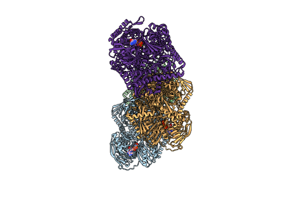 |
Crystal Structure Of N-Methylhydantoinase In Complex With Mg-Adpnp
Organism: Glutamicibacter protophormiae
Method: X-RAY DIFFRACTION Resolution:3.13 Å Release Date: 2026-04-15 Classification: HYDROLASE Ligands: ANP, MG, NH4 |
Organism: Glutamicibacter protophormiae
Method: X-RAY DIFFRACTION
Release Date: 2026-04-15
Ligands: ANP, MG, NH4
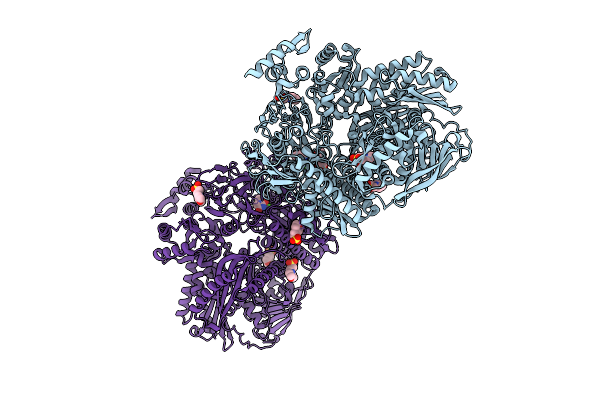 |
Crystal Structure Of N-Methylhydantoinase In Complex With 1-Methylimidazolidine-2,4-Dione, C2221 Form
Organism: Glutamicibacter protophormiae
Method: X-RAY DIFFRACTION Resolution:2.80 Å Release Date: 2026-04-15 Classification: HYDROLASE Ligands: CA, A1BC1, NH4, MES, SO4 |
Organism: Glutamicibacter protophormiae
Method: X-RAY DIFFRACTION
Release Date: 2026-04-15
Ligands: CA, A1BC1, NH4, MES, SO4
 |
Crystal Structure Of N-Methylhydantoinase In Complex With Zn2+ And Adpnp
Organism: Glutamicibacter protophormiae
Method: X-RAY DIFFRACTION Resolution:2.58 Å Release Date: 2026-04-15 Classification: HYDROLASE Ligands: ANP, ZN, NH4, ACT |
Organism: Glutamicibacter protophormiae
Method: X-RAY DIFFRACTION
Release Date: 2026-04-15
Ligands: ANP, ZN, NH4, ACT
 |
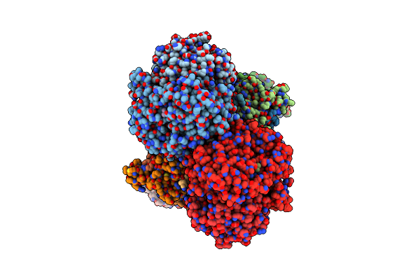
Crystal Structure Of N-Methylhydantoinase In Complex With 1-Methylimidazolidine-2,4-Dione, C-Terminal Residues Visible
Organism: Glutamicibacter protophormiae
Method: X-RAY DIFFRACTION Resolution:2.07 Å Release Date: 2026-04-15 Classification: HYDROLASE Ligands: CA, A1BC1, BTB, NH4, SO4 |
Organism: Glutamicibacter protophormiae
Method: X-RAY DIFFRACTION
Release Date: 2026-04-15
Ligands: CA, A1BC1, BTB, NH4, SO4
 |
Cytochrome Bc1 Complex (Bos Taurus)
Organism: Bos taurus
Method: ELECTRON MICROSCOPY Release Date: 2024-07-03 Classification: MEMBRANE PROTEIN Ligands: HEM, HEC, FES |
Organism: Bos taurus
Method: ELECTRON MICROSCOPY
Release Date: 2024-07-03
Ligands: HEM, HEC, FES
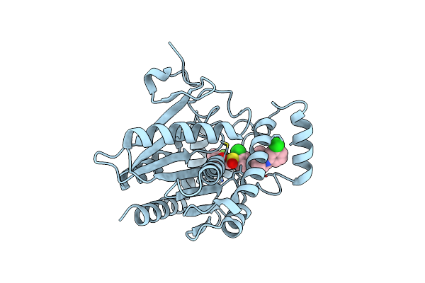 |
Crystal Structure Of Human Monoglyceride Lipase With Compound Lei-515
Organism: Homo sapiens
Method: X-RAY DIFFRACTION Resolution:1.55 Å Release Date: 2023-08-23 Classification: HYDROLASE Ligands: NUX |
Organism: Homo sapiens
Method: X-RAY DIFFRACTION
Release Date: 2023-08-23
Ligands: NUX
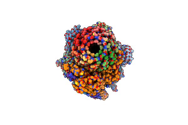 |
F1Fo-Atp Synthase From Acinetobacter Baumannii (State 1)
Organism: Acinetobacter baumannii (strain atcc 17978 / cip 53.77 / lmg 1025 / ncdc kc755 / 5377)
Method: ELECTRON MICROSCOPY Release Date: 2022-02-02 Classification: MEMBRANE PROTEIN Ligands: ATP, MG, ADP, PO4 |
Organism: Acinetobacter baumannii (strain atcc 17978 / cip 53.77 / lmg 1025 / ncdc kc755 / 5377)
Method: ELECTRON MICROSCOPY
Release Date: 2022-02-02
Ligands: ATP, MG, ADP, PO4
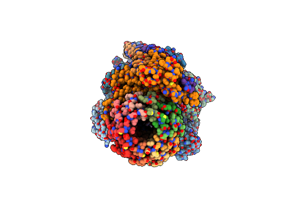 |
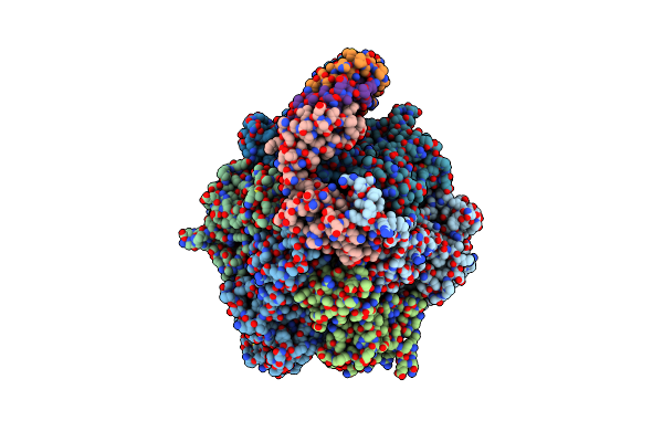
F1Fo-Atp Synthase From Acinetobacter Baumannii (State 2)
Organism: Acinetobacter baumannii atcc 17978
Method: ELECTRON MICROSCOPY Release Date: 2022-02-02 Classification: MEMBRANE PROTEIN Ligands: ATP, MG, ADP |
Organism: Acinetobacter baumannii atcc 17978
Method: ELECTRON MICROSCOPY
Release Date: 2022-02-02
Ligands: ATP, MG, ADP
 |
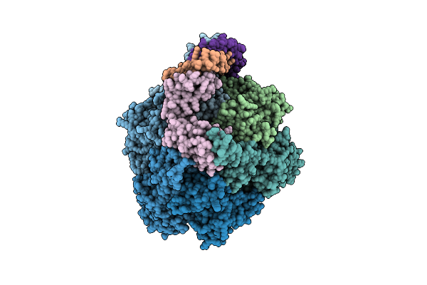
F1Fo-Atp Synthase From Acinetobacter Baumannii (State 3)
Organism: Acinetobacter baumannii (strain atcc 17978 / cip 53.77 / lmg 1025 / ncdc kc755 / 5377)
Method: ELECTRON MICROSCOPY Release Date: 2022-02-02 Classification: MEMBRANE PROTEIN Ligands: ATP, MG, ADP |
Organism: Acinetobacter baumannii (strain atcc 17978 / cip 53.77 / lmg 1025 / ncdc kc755 / 5377)
Method: ELECTRON MICROSCOPY
Release Date: 2022-02-02
Ligands: ATP, MG, ADP
 |
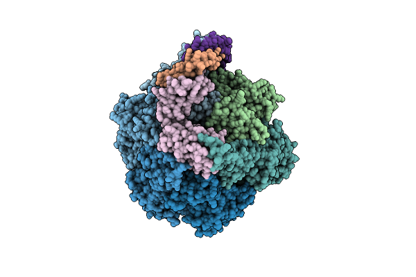
Chloroplast F1Fo Conformation 1
Organism: Spinacia oleracea
Method: ELECTRON MICROSCOPY Resolution:3.15 Å Release Date: 2018-05-23 Classification: MEMBRANE PROTEIN Ligands: ATP, MG, ADP |
Organism: Spinacia oleracea
Method: ELECTRON MICROSCOPY
Release Date: 2018-05-23
Ligands: ATP, MG, ADP
 |
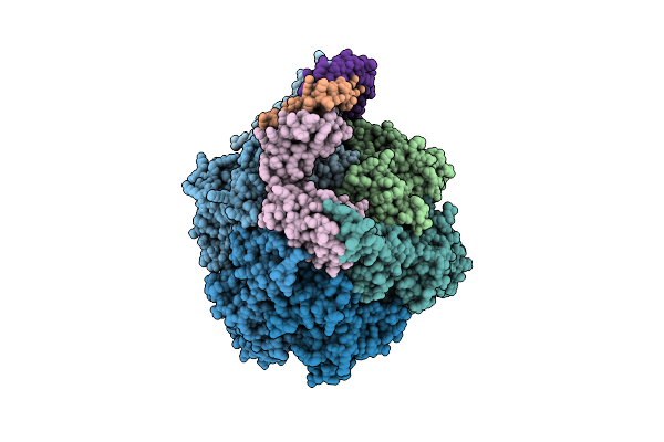
Chloroplast F1Fo Conformation 2
Organism: Spinacia oleracea
Method: ELECTRON MICROSCOPY Release Date: 2018-05-23 Classification: MEMBRANE PROTEIN Ligands: MG, ATP, ADP |
Organism: Spinacia oleracea
Method: ELECTRON MICROSCOPY
Release Date: 2018-05-23
Ligands: MG, ATP, ADP
 |
Chloroplast F1Fo Conformation 3
Organism: Spinacia oleracea
Method: ELECTRON MICROSCOPY Release Date: 2018-05-23 Classification: MEMBRANE PROTEIN Ligands: ATP, MG, ADP |
Organism: Spinacia oleracea
Method: ELECTRON MICROSCOPY
Release Date: 2018-05-23
Ligands: ATP, MG, ADP
 |
Crystal Structure Of Aicarft Bound To An Antifolate
Organism: Homo sapiens
Method: X-RAY DIFFRACTION Resolution:2.39 Å Release Date: 2018-01-10 Classification: TRANSFERASE, HYDROLASE Ligands: 8UM, AMZ, MG |
Organism: Homo sapiens
Method: X-RAY DIFFRACTION
Release Date: 2018-01-10
Ligands: 8UM, AMZ, MG
 |
Crystal Structure Of Aicarft Bound To An Antifolate
Organism: Homo sapiens
Method: X-RAY DIFFRACTION Resolution:1.79 Å Release Date: 2018-01-10 Classification: TRANSFERASE, HYDROLASE Ligands: AMZ, MG, 8US |
Organism: Homo sapiens
Method: X-RAY DIFFRACTION
Release Date: 2018-01-10
Ligands: AMZ, MG, 8US
 |
High Resolution X-Ray Crystal Structure Of Synthetic Insulin Lispro
Organism: Homo sapiens
Method: X-RAY DIFFRACTION Resolution:1.35 Å Release Date: 2017-01-11 Classification: HORMONE Ligands: IPH, ZN, CL, NA |
Organism: Homo sapiens
Method: X-RAY DIFFRACTION
Release Date: 2017-01-11
Ligands: IPH, ZN, CL, NA
 |
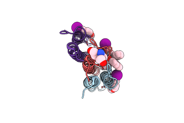
Structure Of The F1C10 Complex From Yarrowia Lipolytica Atp Synthase
Organism: Yarrowia lipolytica
Method: X-RAY DIFFRACTION Resolution:3.50 Å Release Date: 2016-06-29 Classification: HYDROLASE Ligands: ATP, MG, ADP |
Organism: Yarrowia lipolytica
Method: X-RAY DIFFRACTION
Release Date: 2016-06-29
Ligands: ATP, MG, ADP
 |
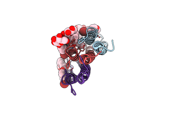
Crystal Structure Of A Mycobacterial Atp Synthase Rotor Ring In Complex With Iodo-Bedaquiline
Organism: Mycobacterium phlei
Method: X-RAY DIFFRACTION Resolution:1.80 Å Release Date: 2016-01-20 Classification: HYDROLASE Ligands: IBQ, TAM |
Organism: Mycobacterium phlei
Method: X-RAY DIFFRACTION
Release Date: 2016-01-20
Ligands: IBQ, TAM
 |
Crystal Structure Of A Mycobacterial Atp Synthase Rotor Ring In Complex With Bedaquiline
Organism: Mycobacterium phlei
Method: X-RAY DIFFRACTION Resolution:1.70 Å Release Date: 2015-05-20 Classification: HYDROLASE Ligands: BQ1, BOG |
Organism: Mycobacterium phlei
Method: X-RAY DIFFRACTION
Release Date: 2015-05-20
Ligands: BQ1, BOG