

Search Count: 70
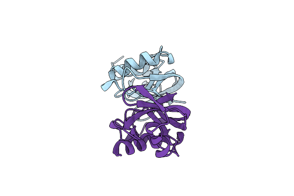 |
X-Ray Structure Of Ss-Rnase-2
Organism: Salmo salar
Method: X-RAY DIFFRACTION Resolution:1.89 Å Release Date: 2021-04-28 Classification: HYDROLASE |
Organism: Salmo salar
Method: X-RAY DIFFRACTION
Release Date: 2021-04-28
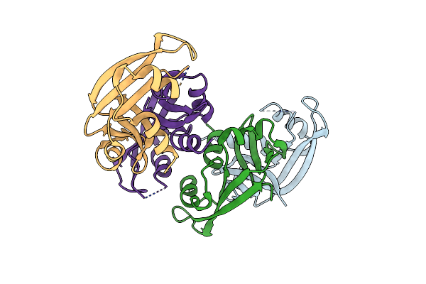 |
X-Ray Structure Of Ss-Rnase-2 Des116-120
Organism: Salmo salar
Method: X-RAY DIFFRACTION Resolution:2.88 Å Release Date: 2021-04-28 Classification: HYDROLASE |
Organism: Salmo salar
Method: X-RAY DIFFRACTION
Release Date: 2021-04-28
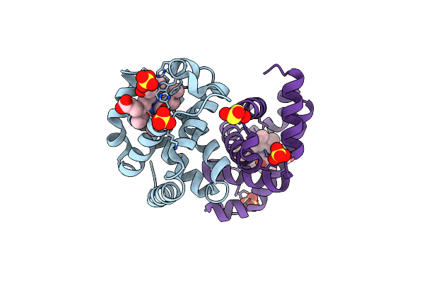 |
Crystal Structure Of Monocarboxylated Hemoglobin From The Sub-Antarctic Fish Eleginops Maclovinus
Organism: Eleginops maclovinus
Method: X-RAY DIFFRACTION Resolution:1.49 Å Release Date: 2019-12-25 Classification: OXYGEN TRANSPORT Ligands: CMO, HEM, SO4, DTN |
Organism: Eleginops maclovinus
Method: X-RAY DIFFRACTION
Release Date: 2019-12-25
Ligands: CMO, HEM, SO4, DTN
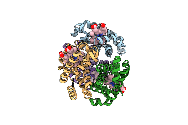 |
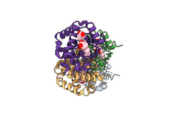
The X-Ray Structure Of Ferric-Beta4 Human Hemoglobin
Organism: Homo sapiens
Method: X-RAY DIFFRACTION Resolution:2.10 Å Release Date: 2018-06-13 Classification: OXYGEN TRANSPORT Ligands: HEM |
Organism: Homo sapiens
Method: X-RAY DIFFRACTION
Release Date: 2018-06-13
Ligands: HEM
 |
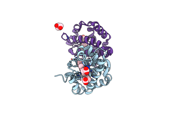
X-Ray Structure Of A New Fully Ligated Carbomonoxy Form Of Trematomus Newnesi Hemoglobin (Hb1Tnco).
Organism: Trematomus newnesi
Method: X-RAY DIFFRACTION Resolution:1.94 Å Release Date: 2017-08-09 Classification: OXYGEN TRANSPORT Ligands: CMO, HEM |
Organism: Trematomus newnesi
Method: X-RAY DIFFRACTION
Release Date: 2017-08-09
Ligands: CMO, HEM
 |
Crystal Structure Of Trematomus Bernacchii Hemoglobin In A Partially Cyanided State
Organism: Trematomus bernacchii
Method: X-RAY DIFFRACTION Resolution:1.54 Å Release Date: 2014-12-24 Classification: OXYGEN TRANSPORT/PROTEIN BINDING Ligands: CYN, HEM, GOL |
Organism: Trematomus bernacchii
Method: X-RAY DIFFRACTION
Release Date: 2014-12-24
Ligands: CYN, HEM, GOL
 |
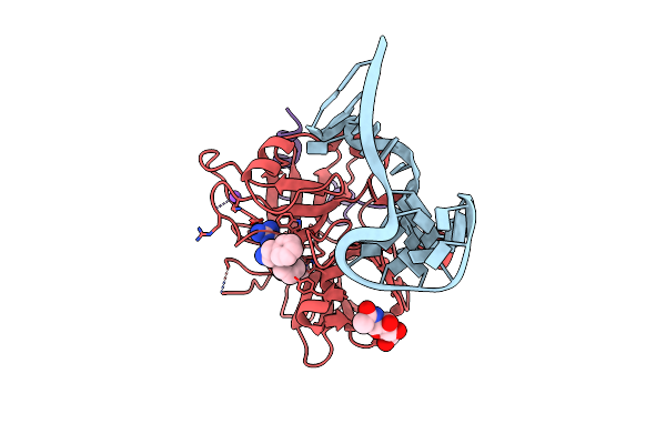
X-Ray Structure Of An All Lna Quadruplex
Method: X-RAY DIFFRACTION
Resolution:1.70 Å Release Date: 2014-03-05 Classification: DNA, RNA Ligands: K, GOL |
Method: X-RAY DIFFRACTION
Release Date: 2014-03-05
Ligands: K, GOL
 |
Crystal Structure Of Human Alpha Thrombin In Complex With A 27-Mer Aptamer Bound To Exosite Ii
Organism: Synthetic dna, Homo sapiens
Method: X-RAY DIFFRACTION Resolution:2.40 Å Release Date: 2013-10-16 Classification: HYDROLASE/HYDROLASE INHIBITOR/DNA Ligands: 0G6, NA, NAG |
Organism: Synthetic dna, Homo sapiens
Method: X-RAY DIFFRACTION
Release Date: 2013-10-16
Ligands: 0G6, NA, NAG
 |
The X-Ray Crystal Structure Of A Dimeric Variant Of Human Pancreatic Ribonuclease
Organism: Homo sapiens
Method: X-RAY DIFFRACTION Resolution:2.70 Å Release Date: 2013-10-02 Classification: HYDROLASE Ligands: SO4, NA, CL |
Organism: Homo sapiens
Method: X-RAY DIFFRACTION
Release Date: 2013-10-02
Ligands: SO4, NA, CL
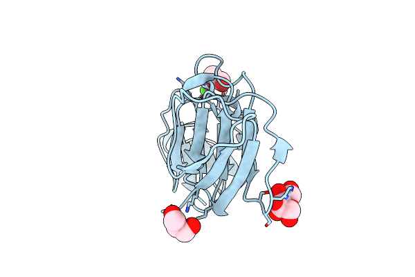 |
Atomic Resolution Structure Of Geodin, A Beta-Gamma Crystallin From Geodia Cydonium
Organism: Geodia cydonium
Method: X-RAY DIFFRACTION Resolution:0.99 Å Release Date: 2013-06-05 Classification: UNKNOWN FUNCTION Ligands: CA, GOL |
Organism: Geodia cydonium
Method: X-RAY DIFFRACTION
Release Date: 2013-06-05
Ligands: CA, GOL
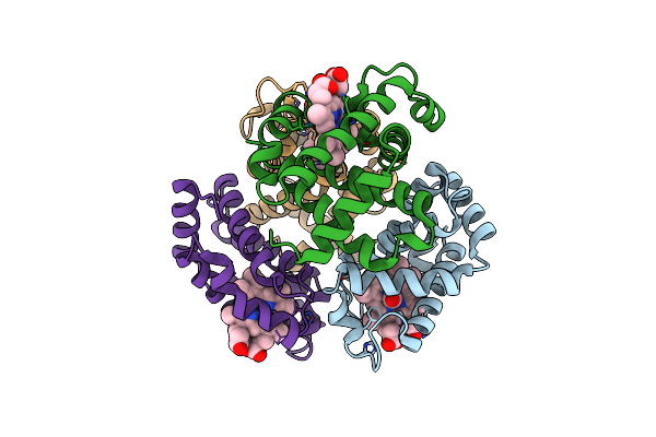 |
Crystal Structure Of T-State Carbonmonoxy Hemoglobin From Trematomus Bernacchii At Ph 8.4
Organism: Trematomus bernacchii
Method: X-RAY DIFFRACTION Resolution:2.20 Å Release Date: 2013-02-20 Classification: OXYGEN TRANSPORT Ligands: CMO, HEM |
Organism: Trematomus bernacchii
Method: X-RAY DIFFRACTION
Release Date: 2013-02-20
Ligands: CMO, HEM
 |
Crystallographic Analysis Of The Interaction Of Nitric Oxide With Hemoglobin From Trematomus Bernacchii In The T Quaternary Structure (Fully Ligated State).
Organism: Trematomus bernacchii
Method: X-RAY DIFFRACTION Resolution:2.50 Å Release Date: 2013-01-16 Classification: OXYGEN TRANSPORT Ligands: NO, HEM |
Organism: Trematomus bernacchii
Method: X-RAY DIFFRACTION
Release Date: 2013-01-16
Ligands: NO, HEM
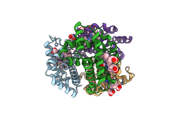 |
X-Ray Structure Of Carbonmonoxy Hemoglobin Of Eleginops Maclovinus
Organism: Eleginops maclovinus
Method: X-RAY DIFFRACTION Resolution:1.45 Å Release Date: 2012-11-07 Classification: OXYGEN TRANSPORT Ligands: CMO, HEM, GOL |
Organism: Eleginops maclovinus
Method: X-RAY DIFFRACTION
Release Date: 2012-11-07
Ligands: CMO, HEM, GOL
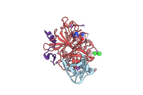 |
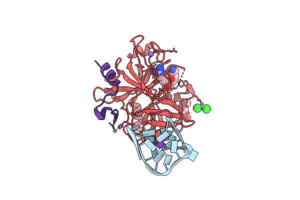
X-Ray Structure Of The Complex Between Human Alpha Thrombin And Thrombin Binding Aptamer In The Presence Of Sodium Ions
Organism: Synthetic dna, Homo sapiens
Method: X-RAY DIFFRACTION Resolution:1.80 Å Release Date: 2012-07-18 Classification: HYDROLASE/HYDROLASE INHIBITOR/DNA Ligands: ZN, 0G6, NAG, NA, CL |
Organism: Synthetic dna, Homo sapiens
Method: X-RAY DIFFRACTION
Release Date: 2012-07-18
Ligands: ZN, 0G6, NAG, NA, CL
 |
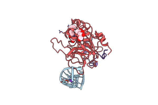
X-Ray Structure Of The Complex Between Human Alpha Thrombin And Thrombin Binding Aptamer In The Presence Of Potassium Ions
Organism: Synthetic dna, Homo sapiens
Method: X-RAY DIFFRACTION Resolution:2.05 Å Release Date: 2012-07-18 Classification: HYDROLASE/HYDROLASE INHIBITOR/DNA Ligands: ZN, 0G6, NAG, NA, CL, K |
Organism: Synthetic dna, Homo sapiens
Method: X-RAY DIFFRACTION
Release Date: 2012-07-18
Ligands: ZN, 0G6, NAG, NA, CL, K
 |
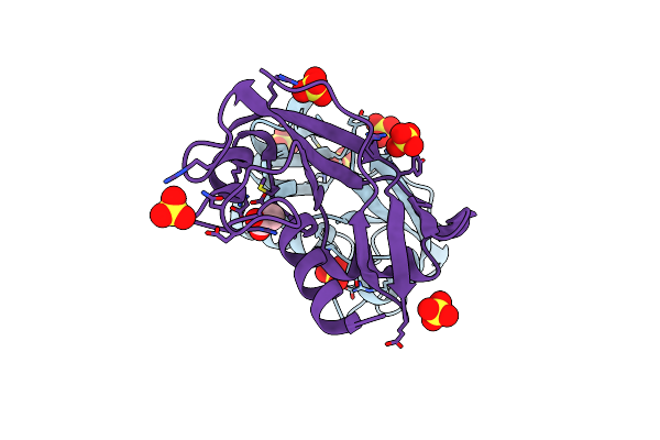
X-Ray Structure Of The Complex Between Human Alpha Thrombin And A Modified Thrombin Binding Aptamer (Mtba)
Organism: Homo sapiens
Method: X-RAY DIFFRACTION Resolution:2.14 Å Release Date: 2011-10-19 Classification: HYDROLASE/HYDROLASE INHIBITOR/DNA Ligands: 0G6, NAG, NA, K |
Organism: Homo sapiens
Method: X-RAY DIFFRACTION
Release Date: 2011-10-19
Ligands: 0G6, NAG, NA, K
 |
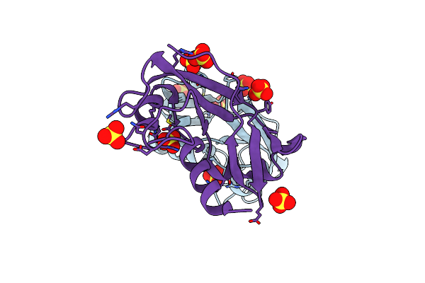
The X-Ray Structure Of Zebrafish Rnase1 From A New Crystal Form At Ph 4.5
Organism: Danio rerio
Method: X-RAY DIFFRACTION Resolution:1.38 Å Release Date: 2010-12-08 Classification: HYDROLASE Ligands: SO4, ACT |
Organism: Danio rerio
Method: X-RAY DIFFRACTION
Release Date: 2010-12-08
Ligands: SO4, ACT
 |
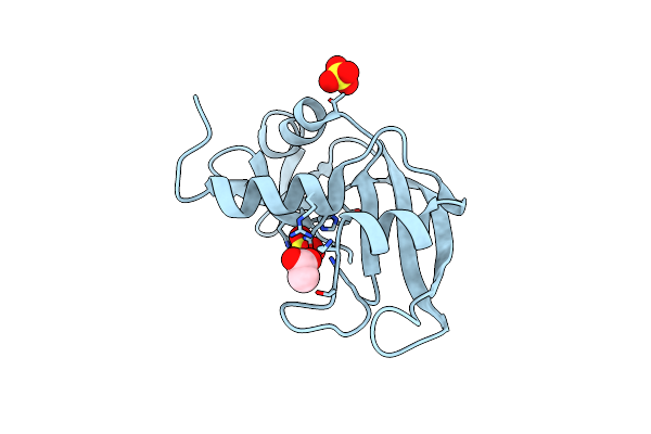
The X-Ray Structure Of Zf-Rnase-1 From A New Crystal Form At Ph 7.3
Organism: Danio rerio
Method: X-RAY DIFFRACTION Resolution:1.61 Å Release Date: 2010-12-08 Classification: HYDROLASE Ligands: SO4 |
Organism: Danio rerio
Method: X-RAY DIFFRACTION
Release Date: 2010-12-08
Ligands: SO4
 |
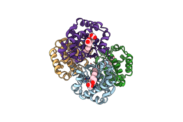
The X-Ray Structure Of Zebrafish Rnase5
Organism: Danio rerio
Method: X-RAY DIFFRACTION Resolution:1.80 Å Release Date: 2010-11-24 Classification: HYDROLASE Ligands: ACT, SO4 |
Organism: Danio rerio
Method: X-RAY DIFFRACTION
Release Date: 2010-11-24
Ligands: ACT, SO4
 |
The Crystal Structure Of Hemoglobin I From Trematomus Newnesi In Deoxygenated State Obtained Through An Oxidation/Reduction Cycle In Which Potassium Hexacyanoferrate And Sodium Dithionite Were Alternatively Added
Organism: Trematomus newnesi
Method: X-RAY DIFFRACTION Resolution:2.20 Å Release Date: 2010-07-28 Classification: OXYGEN TRANSPORT Ligands: HEM |
Organism: Trematomus newnesi
Method: X-RAY DIFFRACTION
Release Date: 2010-07-28
Ligands: HEM