

Search Count: 19
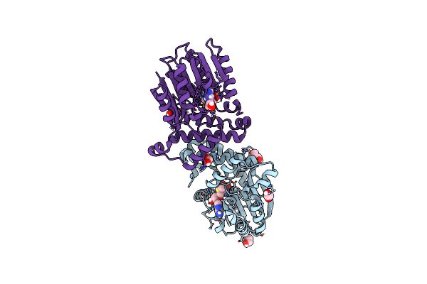 |
Crystal Structure Of Mycobacterium Tuberculosis Mmaa1 With S-Adenosyl Methionine (Sam)
Organism: Mycobacterium tuberculosis h37rv
Method: X-RAY DIFFRACTION Resolution:1.40 Å Release Date: 2024-12-11 Classification: TRANSFERASE Ligands: SAM, GOL, EDO, NA |
Organism: Mycobacterium tuberculosis h37rv
Method: X-RAY DIFFRACTION
Release Date: 2024-12-11
Ligands: SAM, GOL, EDO, NA
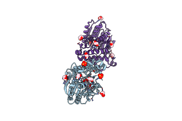 |
Crystal Structure Of Mycobacterium Tuberculosis Mmaa1 With Sinefungin (Sfg)
Organism: Mycobacterium tuberculosis
Method: X-RAY DIFFRACTION Resolution:1.50 Å Release Date: 2024-12-11 Classification: TRANSFERASE Ligands: SFG, EDO, SO4 |
Organism: Mycobacterium tuberculosis
Method: X-RAY DIFFRACTION
Release Date: 2024-12-11
Ligands: SFG, EDO, SO4
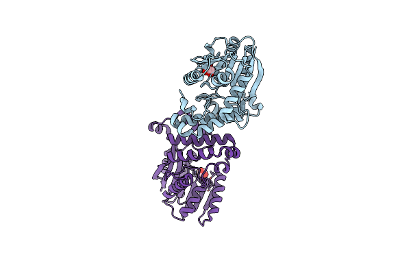 |
Crystal Structure Of Mycobacterium Tuberculosis Mmaa1 In Apo Form
Organism: Mycobacterium tuberculosis
Method: X-RAY DIFFRACTION Resolution:1.90 Å Release Date: 2024-12-11 Classification: TRANSFERASE Ligands: GOL |
Organism: Mycobacterium tuberculosis
Method: X-RAY DIFFRACTION
Release Date: 2024-12-11
Ligands: GOL
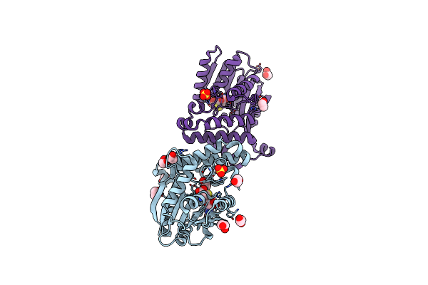 |
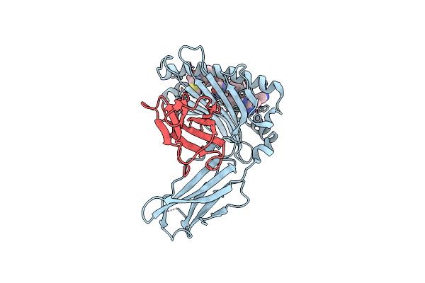
Crystal Structure Of Mycobacterium Tuberculosis Mmaa1 With S-Adenosyl Homocysteine
Organism: Mycobacterium tuberculosis
Method: X-RAY DIFFRACTION Resolution:1.55 Å Release Date: 2024-12-11 Classification: TRANSFERASE Ligands: SAH, EDO, SO4 |
Organism: Mycobacterium tuberculosis
Method: X-RAY DIFFRACTION
Release Date: 2024-12-11
Ligands: SAH, EDO, SO4
 |
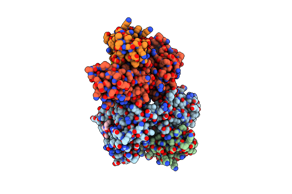
Crystal Structure Of The Murine Class I Major Histocompatibility Complex H-2Db In Complex With Lcmv-Derived Gp33 Altered Peptide Ligand V3P
Organism: Mus musculus
Method: X-RAY DIFFRACTION Resolution:2.60 Å Release Date: 2014-01-01 Classification: Immune system/peptide |
Organism: Mus musculus
Method: X-RAY DIFFRACTION
Release Date: 2014-01-01
 |
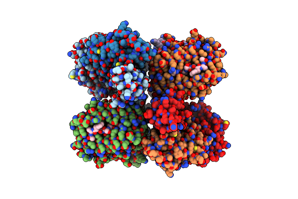
Crystal Structure Of The Murine Class I Major Histocompatibility Complex H-2Db In Complex The With Lcmv-Derived Gp33 Altered Peptide Ligand (V3P,Y4A)
Organism: Mus musculus, Lymphocytic choriomeningitis virus
Method: X-RAY DIFFRACTION Resolution:2.49 Å Release Date: 2012-08-08 Classification: Immune system/agonist Ligands: SO4, GOL |
Organism: Mus musculus, Lymphocytic choriomeningitis virus
Method: X-RAY DIFFRACTION
Release Date: 2012-08-08
Ligands: SO4, GOL
 |
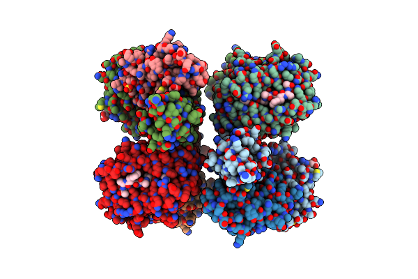
Crystal Structure Of The Murine Class I Major Histocompatibility Complex H-2Db In Complex With The Lcmv-Derived Gp33 Altered Peptide Ligand (V3P, Y4S)
Organism: Mus musculus, Lymphocytic choriomeningitis virus
Method: X-RAY DIFFRACTION Resolution:2.30 Å Release Date: 2012-08-08 Classification: Immune system/agonist |
Organism: Mus musculus, Lymphocytic choriomeningitis virus
Method: X-RAY DIFFRACTION
Release Date: 2012-08-08
 |
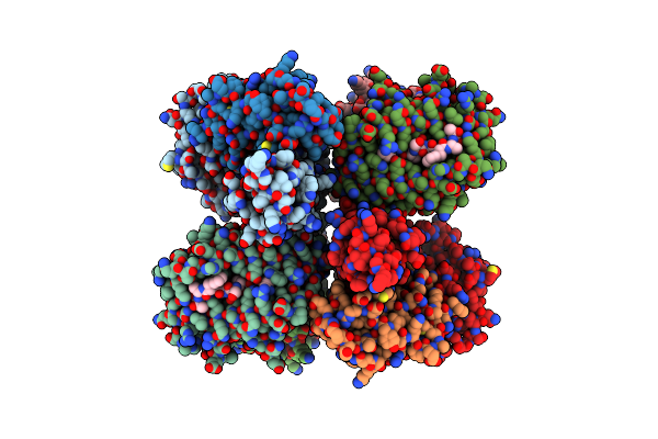
Crystal Structure Of The Murine Class I Major Histocompatibility Complex H-2Db In Complex With The Lcmv-Derived Gp33 Altered Peptide Ligand (A2G,V3P,Y4A)
Organism: Mus musculus, Lymphocytic choriomeningitis virus
Method: X-RAY DIFFRACTION Resolution:2.10 Å Release Date: 2012-08-08 Classification: Immune system/agonist Ligands: SO4, GOL |
Organism: Mus musculus, Lymphocytic choriomeningitis virus
Method: X-RAY DIFFRACTION
Release Date: 2012-08-08
Ligands: SO4, GOL
 |
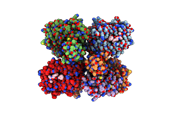
Crystal Structure Of The Murine Class I Major Histocompatibility Complex H-2Db In Complex With The Lcmv-Derived Gp33 Altered Peptide Ligand (A2G, V3P, Y4S)
Organism: Mus musculus, Lymphocytic choriomeningitis virus
Method: X-RAY DIFFRACTION Resolution:2.15 Å Release Date: 2012-08-08 Classification: Immune system/agonist |
Organism: Mus musculus, Lymphocytic choriomeningitis virus
Method: X-RAY DIFFRACTION
Release Date: 2012-08-08
 |
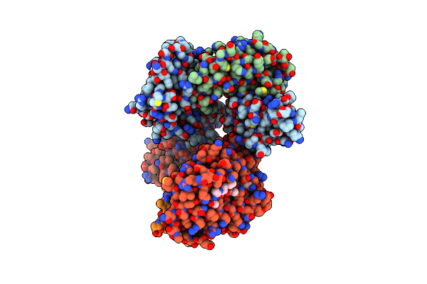
Crystal Structure Of The Murine Class I Major Histocompatibility Complex H-2Db In Complex With The Lcmv-Derived Gp33 Altered Peptide Ligand (V3P, Y4F)
Organism: Mus musculus, Lymphocytic choriomeningitis virus
Method: X-RAY DIFFRACTION Resolution:2.50 Å Release Date: 2012-08-08 Classification: Immune system/agonist |
Organism: Mus musculus, Lymphocytic choriomeningitis virus
Method: X-RAY DIFFRACTION
Release Date: 2012-08-08
 |
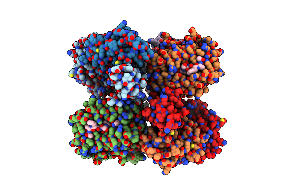
Crystal Structures Of The Murine Class I Major Histocompatibility Complex H-2Db In Complex With Lcmv-Derived Gp33 Altered Peptide Ligand (Y4A)
Organism: Mus musculus, Lymphocytic choriomeningitis virus
Method: X-RAY DIFFRACTION Resolution:2.41 Å Release Date: 2012-03-21 Classification: IMMUNE SYSTEM |
Organism: Mus musculus, Lymphocytic choriomeningitis virus
Method: X-RAY DIFFRACTION
Release Date: 2012-03-21
 |
Crystal Structures Of The Murine Class I Major Histocompatibility Complex H-2Db In Complex With Lcmv-Derived Gp33 Altered Peptide Ligand (Y4S)
Organism: Mus musculus, Lymphocytic choriomeningitis virus
Method: X-RAY DIFFRACTION Resolution:2.00 Å Release Date: 2012-03-21 Classification: IMMUNE SYSTEM |
Organism: Mus musculus, Lymphocytic choriomeningitis virus
Method: X-RAY DIFFRACTION
Release Date: 2012-03-21
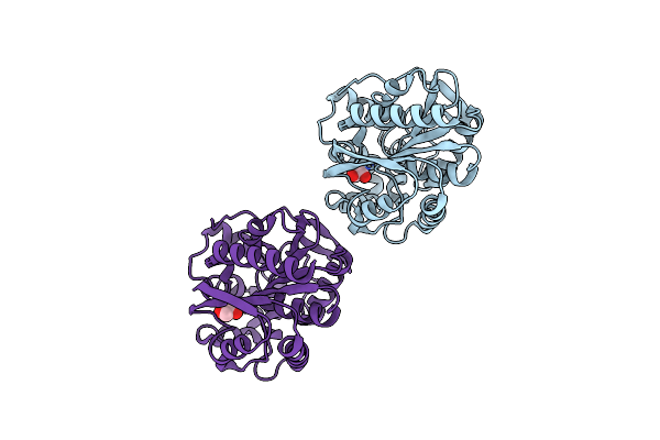 |
Crystal Structure Of Haloalkane Dehalogenase Rv2579 From Mycobacterium Tuberculosis
Organism: Mycobacterium tuberculosis
Method: X-RAY DIFFRACTION Resolution:1.19 Å Release Date: 2008-02-12 Classification: HYDROLASE Ligands: CL, EDO |
Organism: Mycobacterium tuberculosis
Method: X-RAY DIFFRACTION
Release Date: 2008-02-12
Ligands: CL, EDO
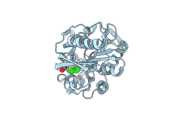 |
Crystal Structure Of Haloalkane Dehalogenase Rv2579 From Mycobacterium Tuberculosis Complexed With 1,2-Dichloroethane
Organism: Mycobacterium tuberculosis
Method: X-RAY DIFFRACTION Resolution:1.60 Å Release Date: 2007-11-13 Classification: HYDROLASE Ligands: CL, ACT, DCE |
Organism: Mycobacterium tuberculosis
Method: X-RAY DIFFRACTION
Release Date: 2007-11-13
Ligands: CL, ACT, DCE
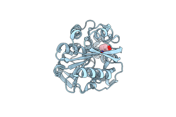 |
Crystal Structure Of Haloalkane Dehalogenase Rv2579 From Mycobacterium Tuberculosis Complexed With 1,3-Propandiol
Organism: Mycobacterium tuberculosis
Method: X-RAY DIFFRACTION Resolution:1.50 Å Release Date: 2007-11-13 Classification: HYDROLASE Ligands: BR, PDO |
Organism: Mycobacterium tuberculosis
Method: X-RAY DIFFRACTION
Release Date: 2007-11-13
Ligands: BR, PDO
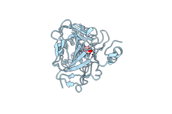 |
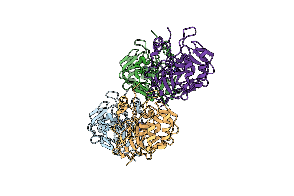
Crystal Structure Of Human Carbonic Anhydrase Ii Complexed With An Acetate Ion
Organism: Homo sapiens
Method: X-RAY DIFFRACTION Resolution:1.81 Å Release Date: 2005-09-27 Classification: LYASE Ligands: ACT, ZN |
Organism: Homo sapiens
Method: X-RAY DIFFRACTION
Release Date: 2005-09-27
Ligands: ACT, ZN
 |
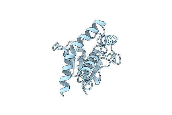
Crystal Structure Of Human Carbonic Anhydrase Ii In A New Crystal Form
Organism: Homo sapiens
Method: X-RAY DIFFRACTION Resolution:2.20 Å Release Date: 2005-09-20 Classification: LYASE Ligands: ZN |
Organism: Homo sapiens
Method: X-RAY DIFFRACTION
Release Date: 2005-09-20
Ligands: ZN
 |
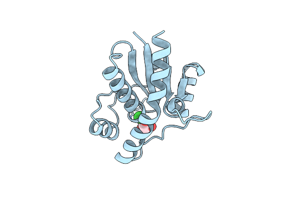
Crystal Structure Of Mycobacterium Tuberculosis Low Molecular Protein Tyrosine Phosphatase (Mptpa) At 1.9A Resolution
Organism: Mycobacterium tuberculosis
Method: X-RAY DIFFRACTION Resolution:1.90 Å Release Date: 2005-03-22 Classification: HYDROLASE Ligands: CL |
Organism: Mycobacterium tuberculosis
Method: X-RAY DIFFRACTION
Release Date: 2005-03-22
Ligands: CL
 |
Crystal Structure Of Mycobacterium Tuberculosis Low Molecular Weight Protein Tyrosine Phosphatase (Mptpa) At 2.5A Resolution With Glycerol In The Active Site
Organism: Mycobacterium tuberculosis
Method: X-RAY DIFFRACTION Resolution:2.50 Å Release Date: 2005-03-22 Classification: HYDROLASE Ligands: CL, GOL |
Organism: Mycobacterium tuberculosis
Method: X-RAY DIFFRACTION
Release Date: 2005-03-22
Ligands: CL, GOL